. 2024 Oct 18;10(42):eado7024.
doi: 10.1126/sciadv.ado7024. Epub 2024 Oct 16.

2'3'-cGAMP interactome identifies 2'3'-cGAMP/Rab18/FosB signaling in cell migration control independent of innate immunity

Affiliations

2'3'-cGAMP interactome identifies 2'3'-cGAMP/Rab18/FosB signaling in cell migration control independent of innate immunity

Yu Deng et al. Sci Adv. .

Erratum in

Abstract

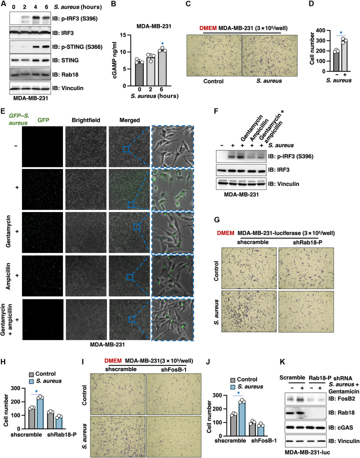
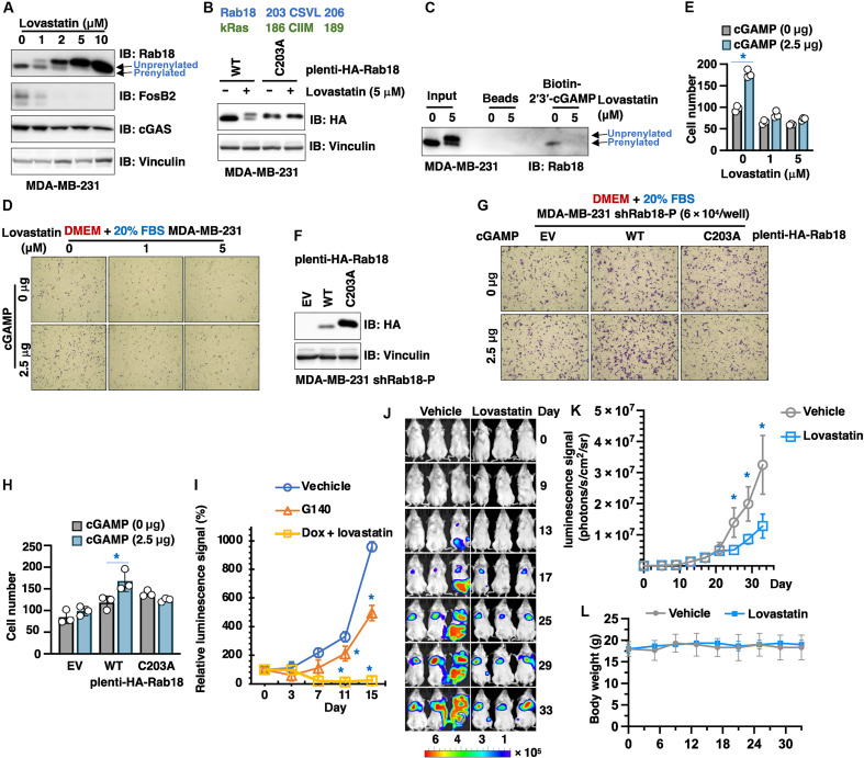
c-di-GAMP was first identified in bacteria to promote colonization, while mammalian 2'3'-cGAMP is synthesized by cGAS to activate STING for innate immune stimulation. However, 2'3'-cGAMP function beyond innate immunity remains elusive. Here, we report that 2'3'-cGAMP promotes cell migration independent of innate immunity. 2'3'-cGAMP interactome analysis identifies the small GTPase Rab18 as a 2'3'-cGAMP binding partner and effector in cell migration control. Mechanistically, 2'3'-cGAMP binds Rab18 to facilitate GTP loading and subsequent Rab18 activation, which further promotes FosB transcription in facilitating cell migration. Induced synthesis of endogenous 2'3'-cGAMP by intrabreast tumor bacterium S. aureus infection or low-dose doxorubicin treatment facilitates cell migration depending on the cGAS/cGAMP/Rab18/FosB signaling. We find that lovastatin induces Rab18 deprenylation that abolishes 2'3'-cGAMP recognition therefore suppressing cell migration. Together, our study reveals a previously unidentified 2'3'-cGAMP function in cell migration control via the 2'3'-cGAMP/Rab18/FosB signaling that provides additional insights into clinical applications of 2'3'-cGAMP.


Figures

Fig. 1.
Fig. 1.. 2′3′-cGAMP treatment promotes cell migration independent of STING.
(A, B, F, G, I, J, O, P, Q, and R) Representative images of transwell assays using indicated numbers of indicated MDA-MB-231 cells under indicated conditions for 24 hours. Triplicates of (A), (F), (I), (Q), and (P) are quantified in (B), (G), (J), (P), and (R). Error bars were calculated as mean ± SD, n = 3 biological replicates. *P < 0.05 [one-way analysis of variance (ANOVA) test]. Where indicated, the amount of 2′3′-cGAMP is used in 300 μl of serum-free media in transwells. (C) 2′3′-cGAMP enzyme-linked immunosorbent assays (ELISAs) using 2 u 105 MDA-MB-231 cells from (A). (D and E) Representative images of transwell assays using 3 × 10⁴ of indicated cells under indicated conditions for 24 hours and quantified in (E). Error bars were calculated as mean ± SD, n = 3 biological replicates. *P < 0.05 (one-way ANOVA test). Where indicated, 2.5 μg of 2′3′-cGAMP is used in 300 μl of serum-free media in transwells. (H) Immunoblot (IB) analysis of whole cell lysates (WCL) derived from indicated MDA-MB-231 cells. MDA-MB-231 cells were infected with either shscramble or shSTING viruses and selected in puromycin (1romyci to eliminate noninfected cells for 72 hours before cell collection. (K) 2′3′-cGAMP ELISA measurements using 106 indicated cells. (L and M) Representative images of transwell assays using 3 a 104 indicated cells treated with 10 M G140 for 24 hours and quantified in (M). Error bars were calculated as mean ± SD, n = 3. *P < 0.05 (one-way ANOVA test). (N) 2′3′-cGAMP ELISA measurements using 2 × 10⁵ MDA-MB-231 cells treated with indicated doses of Dox for 24 hours. DMSO, dimethyl sulfoxide; ns, not significant.
Fig. 2.
Fig. 2.. 2′3′-cGAMP binds Rab18 to promote cell migration.
(A) A cartoon illustration of the pipeline for the chemical-proteomic approaches to identify 2′3-cGAMP binding partners. This model image is generated using BioRender. (B) A volcano plot from (A) showing significantly enriched (log2 fold change equals or larger than 1.5) 2′3′-cGAMP binding proteins. (C) A list of GTPases, GEFs, and GAPs from (B). (D and E) Representative images of transwell assays using 3 × 10⁴ indicated MDA-MB-231 cells under indicated conditions for 24 hours. Where indicated, MDA-MB-231 cells were infected with indicated shRNA viruses and selected in puromycin (1romyci to eliminate noninfected cells for 72 hours before cell collection. Transwell assays data are quantified in (E). Where indicated, 2.5 g of 2′3′-cGAMP is used in 300 liters of serum-free media in transwells. (F) IB analysis of WCL derived from indicated MDA-MB-231 cells. (G and H) Representative images of transwell assays using 3 × 10⁵ indicated MDA-MB-231 cells treated with 2.5 g of 2′3′-cGAMP (in 300 liters of serum-free media in transwells) for 24 hours. Error bars were calculated as mean ± SD, n = 3 biological replicates. *P < 0.05 (one-way ANOVA test). (I) IB analysis of WCL derived from indicated MDA-MB-231 cells. (J and K) Representative images of transwell assays using 3 × 10⁴ indicated MDA-MB-231 cells under indicated conditions for 24 hours and quantified in (K). Error bars were calculated as mean ± SD, n = 3 biological replicates. *P < 0.05 (one-way ANOVA test). Where indicated, 2.5 g of 2′3′-cGAMP is used in 300 liters of serum-free media in transwells.
Fig. 3.
Fig. 3.. 2′3′-cGAMP binds Rab18-E68, R69, R71, and K98 residues to facilitate Rab18 activation.
(A and B) Immunoblot (IB) analysis of biotin-2′3′-cGAMP and biotin-GTP pull-downs using bacterially purified GST-Rab18 (A) or tag-free Rab18 (B) proteins. (C) 2′3′-cGAMP ELISA analysis of endogenous Rab18-IPs (immunoprecipitations) using MDA-MB-231 lysates. (D) IB analysis of biotin-2′3′-cGAMP pull-downs from MDA-MB-231 whole cell lysates (WCL). (E) An illustration of a simulated 2′3′-cGAMP/Rab18 complex structure by MedusaDock, and interacting Rab18 residues were identified by Prankweb. (F to H, M, and N) In vitro Rab18 GTPases activity assays using bacterially purified GST-Rab18 proteins under indicated conditions. Error bars were calculated as mean ± SD, n = 6 biological replicates. *P < 0.05 (one-way ANOVA test). (I) IB analysis of biotin-2′3′-cGAMP pull-downs using GST-Rab18 proteins with GTP. The cartoon illustration is generated using BioRender. (J) An illustration of Rab18 residues interacting with 2′3′-cGAMP from the structural simulation by Prankweb. (K and L) IB analyses of biotin-2′3′-cGAMP (K) or biotin-GTP (L) pull-downs using indicated GST-Rab18 proteins. (O) In vitro Rab18 GTPases activity assays by incubating indicated GST-Rab18 proteins with or without 2′3′-cGAMP for indicated periods. (P) IB analysis of WCL derived from indicated MDA-MB-231 cells. EV, empty vector control. (Q and R) Representative images of transwell assays using indicated numbers of indicated MDA-MB-231 cells under indicated conditions for 24 hours. Error bars were calculated as mean ± SD, n = 3 biological replicates. *P < 0.05 (one-way ANOVA test). Where indicated, 2.5 μg of 2′3′-cGAMP is used in 300 liters of serum-free media in transwells.
Fig. 4.
Fig. 4.. 2′3′-cGAMP/Rab18 signaling promotes FosB transcription and expression to facilitate cell migration.
(A) A volcano plot from RNA-seq data from THP1STING−/− cells comparing 2′3′-cGAMP–induced gene transcriptional changes in THP1-STING−/− cells. (B) RT-PCR analyses of expression changes of indicated genes from MDA-MB-231 cells treated with indicated of 2′3′-cGAMP (2.5MP edan for 2 hours. (C) A representative heatmap for genes with indicated changes using RT2 Profiler PCR arrays by comparing MDA-MB-231-shSTING cells treated with 2′3′-cGAMP (2.5MP ays for 2 hours compared with vehicle treatment control cells. (D) IB analysis of WCL from MDA-MB-231 cells treated with indicated doses of 2′3′-cGAMP for 24 hours. (E and F) IB analysis of WCL from indicated MDA-MB-231 cells treated with indicated doses of 2′3′-cGAMP for 24 hours. (G) IB analysis of WCL from indicated MDA-MB-231 cells. Where indicated, MDA-MB-231 cells were infected with indicated shRNA viruses and selected in puromycin (1romyci to eliminate noninfected cells for 72 hours before cell collection. (H and I) Representative images of transwell assays using 3 × 10⁴ control or FosB-depleted MDA-MB-231 cells treated with indicated doses of 2′3′-cGAMP for 24 hours. Error bars were calculated as mean ± SD, n = 3 biological replicates. *P < 0.05 (one-way ANOVA test). Where indicated, 2.5 g of 2′3′-cGAMP is used in 300 liters of serum-free media in transwells. (J and K) IB analysis of WCL derived from indicated MDA-MB-231 cells treated with indicated doses of Dox for 24 hours before cell collection. (L) Quantifications of transwell assays using indicated numbers of control or FosB-depleted MDA-MB-231 cells treated with indicated doses of Dox for 24 hours. Error bars were calculated as mean ± SD, n = 3 biological replicates. *P < 0.05 (one-way ANOVA test). (M and N) Representative images of transwell assays using indicated cells under indicated conditions for 24 hours (M) and quantified in (N). Error bars were calculated as mean ± SD, n = 3 biological replicates. *P < 0.05 (one-way ANOVA test).
Fig. 5.
Fig. 5.. S. aureus infection induces 2′3′-cGAMP synthesis in cells to promote MDA-MB-231 cell migration.
(A) IB analysis of WCL derived from MDA-MB-231 cells infected with HG003 MSSA S. aureus for indicated time periods. (B) 2′3′-cGAMP ELISA quantifications of intracellular 2′3′-cGAMP levels in 106 MDA-MB-231 cells obtained from (A). (C and D) Representative images of transwell assays using 3 × 10⁵ MDA-MB-231 cells infected with control or S. aureus for 6 hours followed by transwell assays and quantified in (D). Error bars were calculated as mean ± SD, n = 3 biological replicates. *P < 0.05 (one-way ANOVA test). (E) Representative immunofluorescent images of MDA-MB-231 cells infected with BC47 LAC-GFP S. aureus for 6 hours followed by treatment with indicated antibiotics for 1 hour. Concentrations of antibiotics are as below: gentamicin, 200 μg/ml; ampicillin, 200 μg/ml; doxycycline, 20 g/ml. (F) IB analysis of WCL from MDA-MB-231 cells obtained from (E). (G to J) Representative images of transwell assays using 3 × 10⁵ indicated MDA-MB-231 cells infected with control or S. aureus for 6 hours in DMEM media and quantified in (H) and (J). Error bars were calculated as mean ± SD, n = 3 biological replicates. *P < 0.05 (one-way ANOVA test). (K) IB analysis of WCL from MDA-MB-231 cells obtained from (G).
Fig. 6.
Fig. 6.. S. aureus infection or HSV-1 infection induces MDA-MB-231 cell migration.
(A) A cartoon illustration for the design of the NSG mouse tail vein injection assays. The cartoon illustration is generated using BioRender. (B and C) MDA-MB-231 cells stably expressing firefly luciferase were preinfected with vehicle or HG003 MSSA S. aureus (S. a) for 6 hours followed by treatment with gentamicin for 1 hour to remove noninfected bacteria. A total of 3 × 10⁵ resulting cells were injected into tail veins of NSG mice, and noninvasive imaging was taken on indicated days postinjection (B) and quantified in (C). (D) Indicated number of MDA-MB-231 cells were infected with HSV-1 by indicated MOI, and supernatants were collected to perform transwell assays under indicated conditions and quantified. (E) Before HSV-1 infection, MDA-MB-231 cells were treated with G140 (10 mM) for 2 hours, and supernatants were similarly collected and used for transwell assays as in (D) and quantified in (F).
Fig. 7.
Fig. 7.. Statin treatments induce Rab18 deprenylation to attenuate 2′3′-cGAMP binding.
(A) IB analysis of WCL derived from MDA-MB-231 cells treated with indicated doses of lovastatin for 24 hours. Both prenylated and unprenylated species are labeled as indicated. (B) IB analysis of WCL derived from WT or C203A-Rab18 expressing MDA-MB-231 cells treated with 5 M lovastatin for 24 hours. (C) IB analysis of biotin-2′3′-cGAMP pull-downs from MDA-MB-231 cells treated with indicated doses of lovastatin for 24 hours. (D and E) Representative images of transwell assays using 3 × 10⁴ MDA-MB-231 cells treated with indicated doses of 2′3′-cGAMP and lovastatin for 24 hours. Error bars were calculated as mean ± SD, n = 3 biological replicates. *P < 0.05 (one-way ANOVA test). (F) IB analysis of MDA-MB-231 cells stably expressing indicated Rab18 by lentiviral infection depleted of endogenous Rab18. (G and H) Representative images of transwell assays using 6 a 104 indicated MDA-MB-231 cells treated with indicated doses of 2′3′-cGAMP for 24 hours. Error bars were calculated as mean ± SD, n = 3 biological replicates. *P < 0.05 (one-way ANOVA test). (I) MDA-MB-231 cells stably expressing firefly luciferase were pretreated with vehicle, G140 (10 M), or Dox (0.05 M) with lovastatin (5 M) for 24 hours. A total of 5 × 10⁵ resulting cells were injected into tail veins of NSG mice, and noninvasive imaging was performed on indicated days postinjection and quantified. (J to L) MDA-MB-231 cells stably expressing firefly luciferase were pretreated with vehicle or lovastatin (5 μM) for 24 hours. A total of 5 × 10⁵ resulting cells were injected into tail veins of NSG mice, and noninvasive imaging was performed on indicated days postinjection (J) and quantified in (K). Animal body weights are shown in (L).

References

    1. Gomelsky M., cAMP, c-di-GMP, c-di-AMP and now cGMP: Bacteria use them all! Mol. Microbiol. 79, 562–565 (2011). - PMC - PubMed
    1. Romling U., Galperin M. Y., Gomelsky M., Cyclic di-GMP: The first 25 years of a universal bacterial second messenger. Microbiol. Mol. Biol. Rev. 77, 1–52 (2013). - PMC - PubMed
    1. Cohen D., Melamed S., Millman A., Shulman G., Oppenheimer-Shaanan Y., Kacen A., Doron S., Amitai G., Sorek R., Cyclic GMP-AMP signalling protects bacteria against viral infection. Nature 574, 691–695 (2019). - PubMed
    1. Ren A., Wang X. C., Kellenberger C. A., Rajashankar K. R., Jones R. A., Hammond M. C., Patel D. J., Structural basis for molecular discrimination by a 3′,3'-cGAMP sensing riboswitch. Cell Rep. 11, 1–12 (2015). - PMC - PubMed
    1. Zhang X., Shi H., Wu J., Zhang X., Sun L., Chen C., Chen Z. J., Cyclic GMP-AMP containing mixed phosphodiester linkages is an endogenous high-affinity ligand for STING. Mol. Cell 51, 226–235 (2013). - PMC - PubMed