

Reference formulas for chest CT-derived lobar volumes in the lung-healthy general population
- PMID: 39414656
- PMCID: PMC12021944
- DOI: 10.1007/s00330-024-11123-6
Reference formulas for chest CT-derived lobar volumes in the lung-healthy general population
Abstract
Introduction: Lung hyperinflation, a key contributor to dyspnea in chronic obstructive pulmonary disease (COPD), can be quantified via chest computed tomography (CT). Establishing reference equations for lobar volumes and total lung volume (TLV) can aid in evaluating lobar hyperinflation, especially for targeted lung volume reduction therapies.
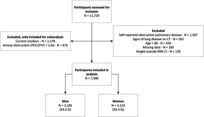
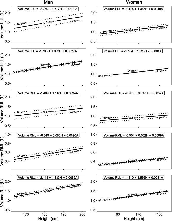
Methods: The Imaging in Lifelines study (ImaLife) comprises 11,729 participants aged 45 and above with analyzed inspiratory low-dose thoracic CT scans. Lung and lobar volumes were measured using an automatic AI-based segmentation algorithm (LungSeg). For the main analysis, participants were excluded if they had self-reported COPD/asthma, lung disease on CT, airflow obstruction on lung function testing, were currently smoking, aged over 80 years, or had height outside the 99% confidence interval. Reference equations for TLV and lobar volumes were determined using linear regression considering age and height, stratified by sex. For the subanalysis, participants who were currently smoking or experiencing airflow obstruction were compared to the group of the main analysis.
Results: The study included 7306 lung-healthy participants, 97.5% Caucasian, 43.6% men, with mean age of 60.3 ± 9.5 years. Lung and lobar volumes generally increased with age and height. Men consistently had higher volumes than women when adjusted for height. R2 values ranged from 7.8 to 19.9%. In smokers and those with airway obstruction, volumes were larger than in lung-healthy groups, with the largest increases measured in the upper lobes.
Conclusion: The established reference equations for CT-derived TLV and lobar volumes provide a standardized interpretation for individuals aged 45 to 80 of Northern European descent.
Key points: Question Lobar lung volumes can be derived from inspiratory CT scans, but healthy-lung reference values are lacking. Findings Lung and lobar volumes generally increased with age and height. Reference equations for lung/lobar volumes were derived from a sizeable lung-healthy population. Clinical relevance This study provides reference equations for inspiratory CT-derived lung and lobar volumes in a lung-healthy population, potentially useful for assessing candidates for lung volume reduction therapies, for lobe removal in lung cancer patients, and in case of restrictive pulmonary diseases.
Keywords: Lung; Lung volume measurements; Population; Pulmonary disease (chronic obstructive); Tomography (X-ray computed).
© 2024. The Author(s).
Conflict of interest statement
Compliance with ethical standards. Guarantor: The scientific guarantor of this publication is R.V. Conflict of interest: J.T.B., I.D., S.A.R., J.M.V., K.K. and M.d.B. declare no relationships with any companies whose products or services may be related to the subject matter of the article. D.J.S. is an advisor to Thirona B.V., Nijmegen, The Netherlands. R.V. is supported by institutional research grants from Siemens Healthineers and received an honorarium for lectures from Siemens Healthineers. Statistics and biometry: J.M.V. has significant statistical expertise. Informed consent: Written informed consent was obtained from all participants in this study. Ethical approval: This study has been approved by the medical ethics committee of the University Medical Center Groningen, the Netherlands, and has been registered with the Dutch Central Committee on Research involving Human Subjects ( https://www.toetsingonline.nl , Identifier: NL58592.042.16). Study subjects or cohorts overlap: Participants have been previously reported in other ImaLife studies, studying different topics related to chest CT ( https://wiki.lifelines.nl/doku.php?id=imal ). In a small subcohort from ImaLife (N = 290 middle-aged participants), we studied the relationship of CT-derived TLV to the GLI-2021 equation for total lung capacity (PMID 37327210). Methodology: Retrospective Cross-sectional study Performed at one institution
Figures

References
MeSH terms
LinkOut - more resources
Full Text Sources
Medical
