. 2024 Oct 16;15(10):750.
doi: 10.1038/s41419-024-07140-4.

Venetoclax triggers sublethal apoptotic signaling in venetoclax-resistant acute myeloid leukemia cells and induces vulnerability to PARP inhibition and azacitidine

Affiliations

Venetoclax triggers sublethal apoptotic signaling in venetoclax-resistant acute myeloid leukemia cells and induces vulnerability to PARP inhibition and azacitidine

Mahesh Tambe et al. Cell Death Dis. .

Abstract

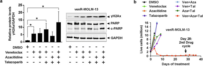
Venetoclax plus azacitidine treatment is clinically beneficial for elderly and unfit acute myeloid leukemia (AML) patients. However, the treatment is rarely curative, and relapse due to resistant disease eventually emerges. Since no current clinically feasible treatments are known to be effective at the state of acquired venetoclax resistance, this is becoming a major challenge in AML treatment. Studying venetoclax-resistant AML cell lines, we observed that venetoclax induced sublethal apoptotic signaling and DNA damage even though cell survival and growth were unaffected. This effect could be due to venetoclax inducing a sublethal degree of mitochondrial outer membrane permeabilization. Based on these results, we hypothesized that the sublethal apoptotic signaling induced by venetoclax could constitute a vulnerability in venetoclax-resistant AML cells. This was supported by screens with a broad collection of drugs, where we observed a synergistic effect between venetoclax and PARP inhibition in venetoclax-resistant cells. Additionally, the venetoclax-PARP inhibitor combination prevented the acquisition of venetoclax resistance in treatment naïve AML cell lines. Furthermore, the addition of azacitidine to the venetoclax-PARP inhibitor combination enhanced venetoclax induced DNA damage and exhibited exceptional sensitivity and long-term responses in the venetoclax-resistant AML cell lines and samples from AML patients that had clinically relapsed under venetoclax-azacitidine therapy. In conclusion, we mechanistically identify a new vulnerability in acquired venetoclax-resistant AML cells and identify PARP inhibition as a potential therapeutic approach to overcome acquired venetoclax resistance in AML.


Conflict of interest statement

MKo reports personal fees from Astellas Pharma, AbbVie, Bristol-Myers Squibb, Faron, Jazz, Pharmaceuticals, Novartis, and Pfizer and research funding from AbbVie outside the submitted work. CAH has received honoraria from Amgen, personal fees from Autolus, and research funding from Celgene/BMS, Kronos Bio, Novartis, Oncopeptides, Orion Pharma, WntReseach, Zentalis Pharmaceuticals, and the IMI2 projects HARMONY and HARMONY PLUS unrelated to this study. MCK is currently an employee of Ascendis Pharma.

Figures

Fig. 1
Fig. 1. Characterization of venetoclax-resistant AML cell lines.
Percentage cell viability of a MOLM-13 cells (parental and venR), b Kasumi-1 cells (parental and venR), and c MV4-11 cells (parental and venR) in response to venetoclax (BCL2i), S63845 (MCL1i) and A-1331852 (BCL-xLi) treated at the indicated concentrations. Cell viability was measured after 72 h of drug treatment using the CellTiter-Glo assay. The data is mean ± s.d. from 2 to 3 independent experiments. df The immunoblots showing BCL2, MCL1, and BCL-xL proteins in d MOLM-13 (parental and venR cells), e Kasumi-1 (parental and venR cells), and f MV4-11 (parental and venR cells) normalized to β-actin.
Fig. 2
Fig. 2. Venetoclax induces sublethal MOMP and caspase-dependent sublethal apoptotic signaling and DNA damage in venetoclax-resistant AML cells.
a Percentage of Annexin V+/DRAQ7− and DRAQ7+ (Annexin V+/DRAQ7+ and Annexin V−/DRAQ7+) cells. The parental MOLM-13 and venR-MOLM-13 cells were treated with venetoclax (100 nM) for the indicated times. The data are visualized as mean ± s.d. from independent experiments (parental cells = 2 experiments, venR cells = 3 experiments). b Immunoblots and the corresponding quantification of percentage of cytochrome C (Cyt-C) in cytoplasmic and mitochondrial cell fractions of parental MOLM-13 and venR-MOLM-13 cells treated with DMSO or 100 nM venetoclax (Ven) for 4 h. HeLa cell lysate served as a positive control for the detection of mitochondrial markers CV-α and PDHE1-α, and cytoplasmic marker GAPDH. The bands indicated with * are non-specific bands. The data is mean ± s.d. from two independent experiments, and one corresponding immunoblot is shown. c The parental MOLM-13 and venR-MOLM-13 cells were treated with DMSO or 100 nM venetoclax for 24 h. Immunoblots showing the expression of γH2Ax, cleaved-caspase3 (c-Casp3), PARP (non-cleaved), c-PARP (cleaved-PARP), and GAPDH in parental MOLM-13 and venR-MOLM-13 cells. The graph shows a fold change of relative protein levels normalized to GAPDH and DMSO control (dotted line). d, e The venR-cells were treated with venetoclax and Q-VD-OPh as single agents and in combination for 24 h. d The graph shows percentage of Annexin V+/DRAQ7− and DRAQ7+ (Annexin V+/DRAQ7+ and Annexin V-/DRAQ7+) cells. Data were compared using a student’s t-test (two-tailed unpaired) to determine statistical significance such that p ≤ 0.05 = * and p ≤ 0.01 = **. e The immunoblot shows expression of γH2Ax, PARP, cPARP (cleaved-PARP), and GAPDH. The non-specific bands are indicated with *. The data is mean ± s.d. from three independent experiments.
Fig. 3
Fig. 3. PARP inhibition delays AML cells acquired resistance to venetoclax.
The total number of live cells (in millions) at indicated timepoints in a parental cell lines and b venR-cell lines. The cells were treated with DMSO, 100 nM venetoclax, and 1 µM olaparib as single agents and in combination, with one exception that parental MOLM-13 and venR-MOLM-13 were treated with 50 nM venetoclax for the first 3 days. The total number of live cells was counted after every 2–3 days using trypan blue staining and a Countess II automated cell counter.
Fig. 4
Fig. 4. Triple combination of venetoclax, azacitidine, and PARPi induces increased DNA damage and cell death in venetoclax-resistant AML cells.
a Quantification of γH2Ax when normalized to GAPDH. The immunoblot showing the expression of γH2Ax, PARP (non-cleaved), c-PARP (cleaved-PARP), and GAPDH in the venR-MOLM-13 cells analyzed at 96 h post-drug treatment. The quantification is mean ± s.d. from three independent experiments, and one representative immunoblot is shown. Data were compared using a student’s t-test (two-tailed unpaired) to determine statistical significance such that p ≤ 0.05 = *. b The total number of live cells (in millions) at indicated timepoints. The venR-MOLM-13 cells were treated with DMSO, 100 nM venetoclax (Ven), 100 nM azacitidine (Aza), and 20 nM talazoparib (Tal) as single agents and in combination. The 28-day treatment cycle included daily treatment with all these drugs for the first 7 days, after which azacitidine was discontinued while venetoclax and talazoparib were continued 5 days a week.
Fig. 5
Fig. 5. The triple combination of venetoclax, azacitidine, and PARPi more effectively kills cells of AML patients than healthy bone marrow cells from healthy donors.
a, b The total number of live cells (in millions) at indicated timepoints in a primary AML samples obtained from patients relapsing under venetoclax-azacitidine treatment and b bone marrow samples obtained from healthy donors. The cells were treated with DMSO, 100 nM venetoclax, 100 nM azacitidine, and 20 nM talazoparib as single agents and in combination. The cells were treated with venetoclax and PARPi every other day, while azacitidine was added daily for the first 5 days, after which azacitidine was discontinued.
Fig. 6
Fig. 6. The illustration outlines the proposed mechanistic model.
In response to venetoclax, the venetoclax-sensitive cells undergo widespread MOMP, apoptosis, and cell death. In contrast, the venetoclax-resistant cells undergo sublethal MOMP, sublethal apoptotic signaling, and low-level DNA damage, with the majority of these cells surviving. We speculate that, in venR cells, (i) venetoclax alone induces DNA damage that can be repaired by PARP-dependent base excision repair (BER) pathway resulting in cell survival, (ii) combining PARPi with venetoclax inhibits PARP-mediated DNA repair and increases venetoclax-induced DNA damage, however, the cells survive likely due to DNA repair by homologous recombination (HR) pathway, (iii) the addition of azacitidine to venetoclax/PARPi combination amplifies the venetoclax-induced DNA damage likely due to trapping of PARP1 and azacitidine at the DNA damage sites, which results in induction of cytotoxic double-stranded DNA breaks and cell death.

References

    1. DiNardo CD, Jonas BA, Pullarkat V, Thirman MJ, Garcia JS, Wei AH, et al. Azacitidine and venetoclax in previously untreated acute myeloid leukemia. N. Engl J Med. 2020;383:617–29. - PubMed
    1. DiNardo CD, Maiti A, Rausch CR, Pemmaraju N, Naqvi K, Daver NG, et al. 10-day decitabine with venetoclax for newly diagnosed intensive chemotherapy ineligible, and relapsed or refractory acute myeloid leukaemia: a single-centre, phase 2 trial. Lancet Haematol. 2020;7:e724–736. - PMC - PubMed
    1. Abhishek M, Caitlin RR, Jorge EC, Naveen P, Naval GD, Farhad R, et al. Outcomes of relapsed or refractory acute myeloid leukemia after frontline hypomethylating agent and venetoclax regimens. Haematologica. 2020;106:894–8. - PMC - PubMed
    1. Bock FJ, Tait SWG. Mitochondria as multifaceted regulators of cell death. Nat Rev Mol Cell Biol. 2020;21:85–100. - PubMed
    1. Ichim G, Lopez J, Ahmed SU, Muthalagu N, Giampazolias E, Delgado ME, et al. Limited mitochondrial permeabilization causes DNA damage and genomic instability in the absence of cell death. Mol Cell. 2015;57:860–72. - PMC - PubMed

MeSH terms

LinkOut - more resources