. 2024 Oct 16;15(1):8914.
doi: 10.1038/s41467-024-53269-3.

Sensory neurons regulate stimulus-dependent humoral immunity in mouse models of bacterial infection and asthma

Affiliations

Sensory neurons regulate stimulus-dependent humoral immunity in mouse models of bacterial infection and asthma

Diane Aguilar et al. Nat Commun. .

Abstract

Sensory neurons sense pathogenic infiltration to drive innate immune responses, but their role in humoral immunity is unclear. Here, using mouse models of Streptococcus pneumoniae infection and Alternaria alternata asthma, we show that sensory neurons are required for B cell recruitment and antibody production. In response to S. pneumoniae, sensory neuron depletion increases bacterial burden and reduces B cell numbers, IgG release, and neutrophil stimulation. Meanwhile, during A. alternata-induced airway inflammation, sensory neuron depletion decreases B cell population sizes, IgE levels, and asthmatic characteristics. Mechanistically, during bacterial infection, sensory neurons preferentially release vasoactive intestinal polypeptide (VIP). In response to asthma, sensory neurons release substance P. Administration of VIP into sensory neuron-depleted mice suppresses bacterial burden, while VIPR1 deficiency increases infection. Similarly, exogenous substance P delivery aggravates asthma in sensory neuron-depleted mice, while substance P deficiency ameliorates asthma. Our data, thus demonstrate that sensory neurons release select neuropeptides which target B cells dependent on the immunogen.


Conflict of interest statement

D.A., M.S., and N.J. declare the following competing interests. U.S. Patent Application Serial Number 63/492,846. Methods of using sensory neuron neurotransmitters to enhance humoral immunity. The remaining authors do not declare competing interests.

Figures

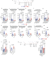
Fig. 1
Fig. 1. Sensory neurons are required for successful clearance of S. pneumoniae following pre-exposure and infection.
a Model of S. pneumoniae pre-exposure and infection, created in BioRender. Aguilar, D. (2022) BioRender.com/h64o602. Mice received escalating doses of resiniferatoxin (RTX) or Vehicle (Veh) starting 21 days prior to inoculation. 104 CFU of S. pneumoniae (Serotype 19F ATCC49619) in 50 µl PBS was delivered on day 0 to expose mice, then 108 CFU in 50 µl PBS was delivered on Day 9. Bacterial burden was enumerated on Days 10, 11 and 15. The red asterisk and dotted line denote the separate set of experiments assessing cross-protection between serotypes: 104 CFU of S. pneumoniae (Serotype 19F ATCC49619) was delivered on day 0 to expose mice, then 106 CFU (Serotype 3, ATCC6303) in 50 µl PBS was delivered on Day 9, survival was assessed as was bacterial burden on Day 11. b Bacterial recovery from lungs in untreated sensory neuron intact mice (Naïve), untreated sensory neuron ablated mice (Naive RTX), pre-exposed and infected sensory neuron intact (Veh), or pre-exposed and infected sensory neuron ablated mice (RTX). Naïve n = 6, Naïve RTX: n = 5; Day 1: Veh: n = 4, RTX: n = 4; Day 8: Veh: n = 5, RTX: n = 5; Day 10: Veh: n = 13, RTX: n = 5; Day 11: Veh: n = 13, RTX: n = 5; Day 15: Veh: n = 5, RTX: n = 5. Two-way ANOVA with Newman-Keuls post hoc test, mean ± sd. Data were pooled from three independent experiments. Gram stain shows S. pneumoniae infection (purple and green arrows point to example of gram-positive space) was greater in RTX compared to Veh. Scale bar = 20 µm. c Veh mice had reduced bacterial burden in response to primary inoculation with serotype 19 F and infection with serotype 3 (Veh n = 7, RTX n = 6). Data were compared with a two-sided t test, mean ± sd. d 90% of Veh mice survived infection with serotype 3 compared to 20% of RTX mice after inoculation with 19 F (Veh n = 8, RTX n = 7). Data were compared with the Log-rank (Mantel-Cox) test, mean ± sd. e Quantification of B cell activating factor (BAFF), APRIL13 (TNFSF13), TNF, IL-1β, IL-2, IL-6, IL-5, CXCL13, CCL5, CCL7 in sensory neuron intact (Veh n = 7) and sensory neuron ablated (RTX, n = 6) mice 16 h following the final 108 CFU dose of 19F. Samples were analyzed with Luminex bead assay. IL-6 was re-run with ELISA. Two-sided t test, mean ± sd. Data were pooled from 2 independent experiments.
Fig. 2
Fig. 2. Sensory neuron ablation reduces immunoglobulins, B cells, and disrupts neutrophil effector function after S. pneumoniae.
a In response to our pre-exposure and infection model (as in Fig. 1a), the image created in BioRender. Aguilar, D. (2022) BioRender.com/h64o602. b IgA, IgM, IgG1, IgG2a, IgG2b, and IgG3 were reduced with sensory neuron ablation 16 h (Day 10) after the final infection. Veh: n = 7, RTX: n = 7. Isotyping Luminex was used to analyze lung homogenates. Two-sided T test, mean ± sd. IgG specific to S. pneumoniae was tested with crude Sp extract. Serial dilutions were used to model the IC50 with a 4-point log model to determine differences between Veh (n = 8) and RTX (n = 6). Two-sided T test, mean ± sd. Data were collected from two independent experiments. c B memory B220+CD19+CD44+CD38+ cells, d B resident memory B220+CD19+CD44+CD38+IgD-CD62l- cells, (e) Isotype switched B220+CD19+IgM-IgD- cells, (f) Plasma CD138+ cells and, (g) Plasmablast CD138+B220lowCD19- cells were downregulated by sensory neuron ablation at 16 and 48 h after infection. Only B memory and plasma cells were different between Veh and RTX on Day 8. Two-way ANOVA with Holm-Sidak test, mean ± sd. Day 0: Veh: n = 5, RTX: n = 5; Day 8: Veh: n = 5, RTX: n = 5; Day 10: Veh: n = 6, RTX: n = 6; Day 11: Veh: n = 6, RTX: n = 6. h µMT mice had reduced burden at 16 h in comparison to sensory neuron intact mice (Veh). Sensory neuron ablated (RTX) mice demonstrated a bacterial burden similar to mice with immature B cells (µMT) 48 h after infection. Sensory neuron intact mice (Veh) reduced their bacterial burden at 48 h. Veh n = 13, RTX n = 5, µMT infected n = 7. Data from four independent experiments. Two-way ANOVA with Holm-Sidak test, mean ± sd. i Ly6G A18 neutrophil depletion antibody or isotype control was delivered before, during, and after the final infection dose of S. pneumoniae; the image was created in BioRender. Aguilar, D. (2022) BioRender.com/h64o602. Bacterial burden (Log CFU/g lung mass) from lung homogenates was greater with neutrophil depletion in sensory neuron intact infected mice compared to isotype control 48 h (Day 11) after infection. Sensory neuron-depleted mice (RTX) did not further increase bacterial burden with neutrophil depletion, demonstrating that B-cell neutrophil interaction was disrupted. One-way ANOVA with Holm-Sidak post-hoc test, mean ± sd. Veh (Iso): n = 12, Veh (Ly6g): n = 8, RTX (iso): n = 10, RTX(Ly6g): n = 9. Data were pooled from two independent experiments. j Naïve neutrophils were incubated with decomplemented serum harvested from naïve, Veh, or RTX mice (pre-exposed and infected) and plated in S. pneumoniae 19 F coated wells (1000 CFU). Neutrophils were lysed 30 min later and plated on blood agar plates, and CFU was enumerated 24 h later. One-way ANOVA with Holm-Sidak post-hoc test, mean ± sd. n = 6 per group.
Fig. 3
Fig. 3. Vasoactive intestinal peptide attracts B cells and regulates immunoglobulin production, and release.
a B220+ cells clustered around sensory nerves from the lungs of sensory neuron intact mice (Veh). In RTX sensory neuron-depleted mice, B220+ cells are further dispersed. 30 cells (10 cells per sample) from n = 3 mice for Veh & RTX. Two-sided t-test, mean ± sd. be Prominent sensory neuron neuropeptides were increased in lung homogenates 16 h after pre-exposure and infection with S. pneumoniae compared to naïve mice. b VIP was increased with infection and suppressed with sensory neuron depletion. c NPY was increased with infection and suppressed with sensory neuron depletion. d Substance P did not significantly increase following infection with S. pneumoniae. e CGRP did not significantly increase following infection with S. pneumoniae. Lung homogenates were analyzed with ELISA. be PBS: n = 6, Veh: n = 7, RTX: n = 7. One-way ANOVA with Tukey’s post hoc test, mean ± sd. Data from 2 independent experiments. fi B cells were isolated from spleens of naïve mice and cultured in media with IL4 + LPS and supplemented with VIP or NPY. Cells and media were sampled after 96 h of incubation. f VIP did not increase IgG bound to B220+CD19+ cells. g VIP significantly increased bound IgG in CD138+ plasma cells. h Proliferation, as assessed by intracellular Ki67 staining, was not affected by neuropeptides (from CD138+ gate). n = 10 all conditions. i Secreted IgG was increased by VIP and NPY. Control: n = 4; VIP, NPY: n = 5. One-way ANOVA and Tukey’s post-hoc test, mean ± sd. Data from 2 independent experiments. jl B cells were isolated from the spleens of VIP1 receptor knockout (VIPR1–/–) and WT littermates and cultured for 96 h. j B220+CD19+ cells were not significantly affected by VIPR1 deletion. k VIPR1–/–CD138+ cells did not significantly upregulate IgG in response to VIP and other neuropeptides. WT: n = 7; VIPR1–/–: n = 8. l IgG released into the media was not increased with VIP or other peptides with VIPR1 genetic deletion, VIP increased WT B cell IgG release. WT: n = 4; VIPR1–/–: n = 4. Two-way ANOVA with Holm-Sidak post-hoc test, mean ± sd. mp B cells were isolated from the lungs of naïve or S. pneumoniae pre-exposed and infected mice. m, n VIP increased bound IgG in B220+CD19+and CD138+ cells. This effect was greater in naïve cells. Naïve: n = 8, S. pneumoniae: n = 8. o released IgG was increased with VIP in both naïve and pre-exposed and infected cells. Naïve: n = 4, S. pneumoniae: n = 5. p, q pre-exposed and infected cells show higher markers of B cell exhaustion after additional stimulation in culture. Two-way ANOVA with Holm-Sidak post-hoc test, mean ± sd. Naïve: n = 4, S. pneumoniae: n = 8.
Fig. 4
Fig. 4. Vasoactive intestinal peptide supplementation improves bacterial clearance, memory B cells, plasma cells, and IgG in response to pre-exposure and infection with S. pneumoniae.
a Vasoactive intestinal peptide or vehicle (PBS) was delivered as per the schematic in addition to the model of S. pneumoniae pre-exposure and infection with serotype 19 F, image created in BioRender. Aguilar, D. (2022) BioRender.com/h64o602. Cells were gated on Live CD45+ cells, and total populations were assessed with counting beads. b B memory cells, (c) B resident memory, (d) Isotype switched, (e) Plasma cells, (f) plasmablasts were reduced with sensory neuron ablation. Supplementation with VIP increased B cell populations. VIP supplementation did not further increase B cell populations in sensory neuron intact mice. g IgG was reduced with RTX as previously but increased with the supplementation of VIP to RTX mice. One-way ANOVA with Holm Sidak post hoc test, mean ± sd. Veh (PBS): n = 8, Veh (VIP): n = 8, RTX(PBS): n = 7, RTX(VIP): n = 7. h VIP1 receptor knockout (VIPR1–/–) mice had reduced B memory cells compared WT mice,(i) B resident memory and, (j) Isotype switched B cells were similar in VIPR1–/–and WT mice, (k) Plasma cells, and (l) plasmablasts were reduced in VIPR1–/–vs WT mice. Two-sided T test. WT n = 5, VIPR1-/- n = 5. m VIPR1–/–mice had reduced IgG following pre-exposure and infection compared to WT littermates. n VIP supplementation suppressed bacterial burden in RTX mice. No effect of VIP supplementation was observed in Veh mice. Veh (PBS): n = 8, Veh (VIP): n = 7-8, RTX(PBS): n = 6-7, RTX(VIP): n = 6-7. o VIPR1–/–mice have increased bacterial burden following pre-exposure and infection with S. pneumoniae compared to WT littermates. One-way ANOVA with Holm Sidak post hoc test, mean ± sd. Data from 3 independent experiments. WT: n = 11, VIPR1–/– n = 7. Two-sided t-test, mean ± sd. Data from 3 independent experiments. p Ly6g neutralizing antibody or isotype was given to WT or VIPR1–/–mice; imagecreated in BioRender. Aguilar, D. (2022) BioRender.com/h64o602. Bacterial burden (Log CFU/ g tissue mass) from lung homogenates was greater with neutrophil depletion in WT mice compared to isotype control. VIPR1–/–mice did not further increase bacterial burden with neutrophil depletion, demonstrating that VIP significantly stimulates B cells to increase neutrophil-mediated bacterial clearance. One-way ANOVA with Holm-Sidak post-hoc test, mean ± sd. WT(Iso): n = 6, WT(Ly6g): n = 5, VIPR1–/–Iso): n = 4, VIPR1–/– (Ly6g): n = 5. Data were pooled from two independent experiments.
Fig. 5
Fig. 5. Sensory neuron ablation reduces B lymphocytes, immunoglobulins, and mast cells in A. alternata-treated mice.
a A. alternata induced asthma was elicited as per Cavagnero et al. 25 µg of A. alternata extract in 50 µl PBS was delivered as per the schematic image created in BioRender. Aguilar, D. (2022) BioRender.com/h64o602. Experiments took place 16 hrs later. b IgE, IgG1, and IgG3 (n = 6, RTX: n = 7) were reduced with sensory neuron ablation after the final A. alternata dose. Isotyping Luminex was used to analyze lung homogenates. A. alternata extract with lung homogenates and IC50 modeling was used to determine IgE specific to A. alternata (Veh n = 6, RTX n = 4) Two-sided t test. Data were collected from two independent experiments. c B memory B220+CD19+CD44+CD38+ cells, (d) B resident memory B220+CD19+CD44+CD38+IgD-CD62l- cells, (e) Isotype switched B220+CD19+IgM-IgD- cells, (f) Plasma CD138+ cells and, (g) Plasmablasts CD138+B220lowCD19- cells and (h) mast cells CD117+ were downregulated by sensory neuron ablation 16h after A. alternata asthma induction. Cells were gated on Live CD45+ cells, and counting bead calculation determined the total cell population. Veh: n = 10, RTX: n = 7. Two-sided t test, mean ± sd. Data from 3 independent experiments.
Fig. 6
Fig. 6. Sensory neuron ablation reduces airway hyperresponsiveness and goblet cell metaplasia in A. alternata-induced asthma.
a A. alternata-induced asthma was elicited as per Cavagnero et al. 25 µg of A. alternata extract in 50 µl PBS (PBS only for veh) was delivered as per the schematic image created in BioRender. Aguilar, D. (2022) BioRender.com/h64o602. Experiments took place 16hrs later. b RTX treated mice had a response to doubling doses of methacholine similar to PBS control mice and reduced compared to sensory neuron intact mice treated with A. alternata. PBS: n = 5, Veh: n = 6, RTX: n = 5. Two-way ANOVA with Tukey’s post hoc test. * Indicates a difference between Veh and RTX @8 mg/ml p = 0.0228. ** indicates difference from Veh and RTX 16&32 mg/ml: p = 0.0001, Veh and PBS 16 mg/ml: p = 0.0067, 32 mg/ml: p = 0.0001. Data from 2 independent experiments. c Correlation of IgE (from Fig. 5b) and Rn @ MCh (32 mg/ml) (d) Goblet cell metaplasia (Cells/µm) are reduced in RTX compared to sensory neuron intact A alternata treated mice. Veh: n = 6, RTX: n = 7.Two-sided t test, mean ± sd. Data from 2 independent experiments. e Representative images of periodic acid Schiff’s reagent stain to visualize mucous producing Goblet cells. Scalebar = 20 µm.
Fig. 7
Fig. 7. A. alternata increases Substance P which augments IgE production.
A. alternata increased lung concentrations of Substance P (a) and NPY (b). CGRP (c) and VIP (d) were not significantly increased following A. alternata induction. Substance P (a) and VIP (d) were significantly reduced with RTX treatment. Analyzed from lung homogenates with ELISA. PBS: n = 6, Veh: n = 6, RTX: n = 6. One-way ANOVA with Tukey’s post-hoc test. Data from 3 independent experiments. eh B cells were isolated from spleens of naïve mice and cultured in media with IL4 + LPS and supplemented with Substance P (SubP) or NPY. Cells and media were sampled after 96 h of incubation. e Substance P did not increase bound IgE on B220+CD19+ cells. f Substance P significantly increased bound IgE in CD138+ plasma cells. g Proliferation, as assessed by intracellular Ki67 staining, was not affected by neuropeptides (from CD138+ cells). h Secreted IgE was increased by Substance P preferentially. IL4 + LPS: n = 4, +SubP: n = 5, + NPY: n = 5. One-way ANOVA and Tukey’s post-hoc test, mean ± sd. Data from 2 independent experiments.
Fig. 8
Fig. 8. Substance P increases B cells and IgE in mice treated with A. alternata.
a Substance P or vehicle (PBS) was delivered in conjunction with A. alternata extract as per the schematic, in accordance with our A. alternata model of asthma, the image created in BioRender. Aguilar, D. (2022) BioRender.com/h64o602. Cells were gated on Live/CD45 + cells, and total populations were assessed with counting beads. b B memory cells, (c) B resident memory, (d) Isotype switched, (e) Plasma cells, (f) plasmablasts were reduced with sensory neuron ablation. Supplementation with Substance P increased B cell populations. Substance P supplementation did not further increase B cell populations in sensory neuron intact mice. Veh (PBS): n = 5, Veh (Sub P): n = 6, RTX(PBS): n = 6, RTX(SubP): n = 6. One-way ANOVA with Holm sidak post hoc test, mean ± sd. Data from 3 independent experiments. Tac1–/–(lack Tac1 gene, unable to produce substance P, have receptors) had similar B memory (g), but reduced B Resident Memory (h), Isotype switched (i), plasma cells (j), and plasmablast cell (k) compared to WT mice receiving A. alternata induction of asthma. WT n = 6, Tac1–/– n = 8, Two-sided t test, mean ± sd. Data from 2 independent experiments. l Supplementation with substance P increased IgE in A. alternata mice treated with RTX to a level consistent with Veh. Substance P did not further increase IgE in Veh mice (All groups n = 6). One-way ANOVA with Holm sidak post hoc test, mean ± sd. Data from 2 independent experiments. m Tac1–/– (n = 7) had reduced IgE compared to WT A. alternata mice (n = 8). Two-sided t test, mean ± sd. Data from 2 independent experiments.
Fig. 9
Fig. 9. Graphical summary. Sensory neurons stimulate B cells by the release of select neuropeptides, which enhance immunoglobulin production.
Sensory neuron neuropeptide release assists with infection but, exacerbates allergy. Image created in BioRender. Aguilar, D. (2022) BioRender.com/h64o602.

Update of

References

    1. Chiu, I. M. et al. Bacteria activate sensory neurons that modulate pain and inflammation. Nature501, 52–57 (2013). - PMC - PubMed
    1. Baral, P. et al. Nociceptor sensory neurons suppress neutrophil and γδ T cell responses in bacterial lung infections and lethal pneumonia. Nat. Med.24, 417–426 (2018). - PMC - PubMed
    1. Pinho-Ribeiro, F. A. et al. Blocking neuronal signaling to immune cells treats streptococcal invasive infection. Cell173, 1083–1097 (2018). - PMC - PubMed
    1. Riol-Blanco, L. et al. Nociceptive sensory neurons drive interleukin-23-mediated psoriasiform skin inflammation. Nature510, 157–161 (2014). - PMC - PubMed
    1. Ramirez, V. T. et al. Sensory nociceptive neurons contribute to host protection during enteric infection with citrobacter rodentium. J. Infect. Dis.221, 1978–1988 (2020). - PMC - PubMed

Publication types

MeSH terms

LinkOut - more resources