. 2024 Oct 17;15(1):8947.
doi: 10.1038/s41467-024-53280-8.

Microorganism microneedle micro-engine depth drug delivery

Affiliations

Microorganism microneedle micro-engine depth drug delivery

Bin Zheng et al. Nat Commun. .

Abstract

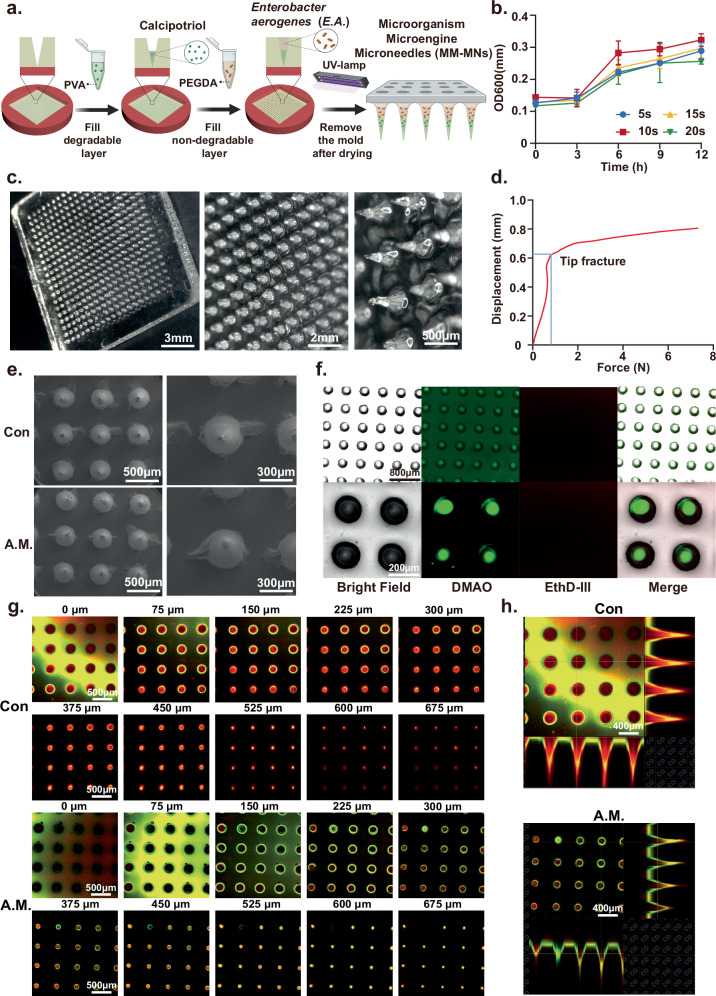
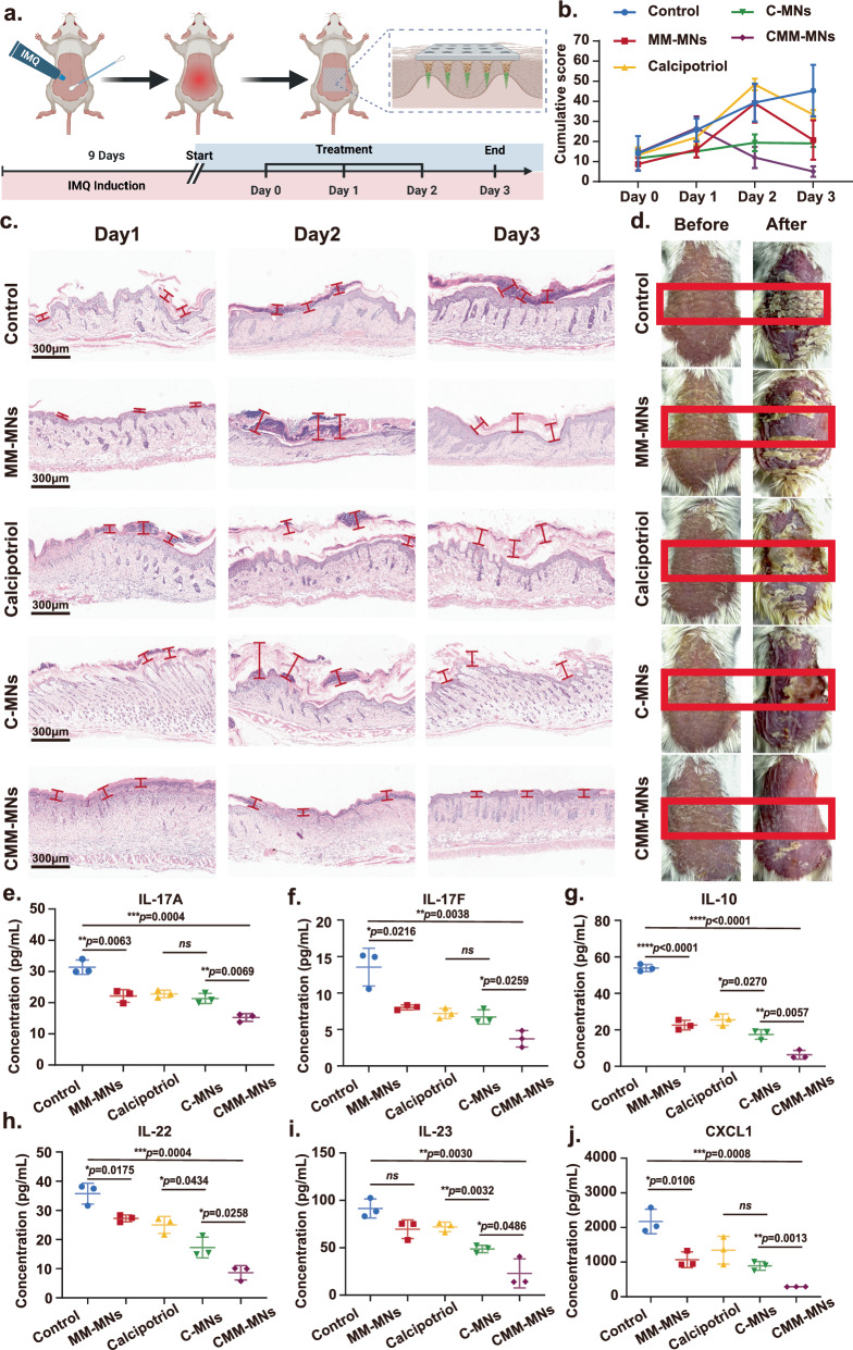
As a transdermal drug delivery method, microneedles offer minimal invasiveness, painlessness, and precise in-situ treatment. However, current microneedles rely on passive diffusion, leading to uncontrollable drug penetration. To overcome this, we developed a pneumatic microneedle patch that uses live Enterobacter aerogenes as microengines to actively control drug delivery. These microbes generate gas, driving drugs into deeper tissues, with adjustable glucose concentration allowing precise control over the process. Our results showed that this microorganism-powered system increases drug delivery depth by over 200%, reaching up to 1000 μm below the skin. In a psoriasis animal model, the technology effectively delivered calcitriol into subcutaneous tissues, offering rapid symptom relief. This innovation addresses the limitations of conventional microneedles, enhancing drug efficiency, transdermal permeability, and introducing a creative paradigm for on-demand controlled drug delivery.


Conflict of interest statement

The authors declare no competing interest.

Figures

Fig. 1
Fig. 1. Preparation, application principles, and processes of microorganism micro-engine microneedles (MM-MNs).
a The preparation process of double-layer microbiota-assisted gas dynamic microneedles. b Enterobacter aerogenes (E.A.) propels the release of drugs from the detachable layer. c The pathogenic mechanism of psoriasis. d The mechanism of microbiota-assisted gas dynamic microneedles propelling calcipotriol for deep tissue release in the treatment of psoriasis. Created with BioRender.com/w74e852.
Fig. 2
Fig. 2. Microorganisms have the potential to serve as a source of aerodynamic force.
a E.A. decomposes substrate glucose to produce H2. Created with BioRender.com/l25i234. b The results of gas chromatographic detection of the mixed gas produced by the E.A.. ce Changes in H2 production by the microorganism under different inoculation levels (c), substrate concentrations (d), and cultivation times (e). Mean ± SD of n = 3 independent samples. Two-sided Student’s t test. f The experimental diagram of different masses of agar blocks being pushed by the gas produced by E.A.. g Effect of E.A. produced gas on agar displacement distance of different masses. Mean ± SD of n = 3 independent samples. Two-sided Student’s t test. h Changes in gas production over time under different substrate concentrations. i Changes in gas production rate over time under different substrate concentrations. j Changes in maximum gas production under different substrate concentrations. k Changes in maximum gas production rate under different substrate concentrations. Mean ± SD of n = 20 independent samples. Two-sided Student’s t test. l The time to reach the maximum gas production and gas production rate. Mean ± SD of n = 3 independent samples.
Fig. 3
Fig. 3. Preparation and bacterial activity validation of microorganism micro-engine microneedles (MM-MNs).
aThe preparation process of MM-MNs. Created with BioRender.com/e35z291. b The effect of ultraviolet irradiation time on the activity of E.A.. Mean ± SD of n = 3 independent samples. c Digital microscopy characterization of microneedle morphology. Scale bar, 3 mm (Left); 2 mm (Middle); 500 μm (Right). d Mechanical force for the fracture of the microneedle tips. e Scanning electron microscopy characterization of microneedle morphology.Scale bar, 500 μm (Left); 300 μm (Right). f Fluorescence microscopy results of E.A. activity encapsulated in microneedles. Scale bar, 800 μm (Up); 200 μm (Down). g, h Confocal microscopy results of E.A. activity encapsulated in microneedles. Scale bar, 500 μm (g); Scale bar, 400 μm (h). A representative image of four biologically independent samples from each group is shown in c, e, f, g, and h.
Fig. 4
Fig. 4. Characterization of gas penetration depth of microorganism micro-engine microneedles (MM-MNs).
a Experimental diagram illustrating the detection of gas production from MM-MNs in simulated body fluids using a gas microelectrode system. Created with BioRender.com/e13d275. b Gas release from single microneedles loaded with different concentrations of glucose when immersed in simulated body fluid. c Test diagram of the penetration of MM-MNs through different skin thicknesses by using a transdermal test instrument. Created with BioRender.com/e13d275. d Trends in hydrogen gas concentration in simulated body fluid over time after penetrating different thicknesses of pig skin. Mean ± SD of n = 3 independent samples. e Experimental diagram of microelectrode real-time monitoring of subcutaneous gas concentrations at different depths. Created with BioRender.com/e13d275. f, g Trends in hydrogen gas concentration (f) and gas production rate (g) within the skin over time.
Fig. 5
Fig. 5. Characterization of drug delivery performance of microorganism micro-engine microneedles (MM-MNs).
a Double-layer microneedles with an outer layer loaded with Rhodamine dye and an inner layer containing Poly(ethylene glycol) diacrylate (PEGDA) loaded with E.A.. Created with BioRender.com/e13d275. b Digital microscope image of RMM-MNS. Scale bar, 3 mm (Left); 500 μm (Right). c The release of rhodamine over time in simulated body fluid. Mean ± SD of n = 3 independent samples. d Confocal microscopy imaging of the penetration depth of Rhodamine dye released by microneedles loaded with different concentrations of glucose into the skin. Scale bar, 500 μm. e Fluorescence intensity of Rhodamine dye diffusion at different depths beneath the skin released by microneedles loaded with varying concentrations of glucose. Mean ± SD of n = 3 independent samples. Two-sided Student’s t test. f Double-layer microneedles with an outer layer loaded with Calcipotriol and an inner layer containing PEGDA loaded with E.A. Created with BioRender.com/e13d275. g Determination of the concentration of Carpotriol in the skin’s surface and subcutaneous tissue by high-performance liquid Chromatography (HPLC). Mean ± SD of n = 6 independent samples. Two-sided Student’s t test. h Changes in caprotriol concentration in skin surface and subcutaneous tissue over time as determined by HPLC. Mean ± SD of n = 6 independent samples. Two-sided Student’s t test. A representative image of four biologically independent samples from each group is shown in (b) and (d).
Fig. 6
Fig. 6. The diffusion of drugs under the skin is controlled by the gas-producing behavior of microbial engines.
a Confocal microscopy imaging of the release and diffusion over time of dye-simulated drugs at different depths beneath the skin. Scale bar, 500 μm. b, c The average fluorescence intensity of dye-simulated drugs diffusing over time at different depths beneath the skin. d The curves show the average fluorescence intensity of drug diffusion over time at different depths, and the curves depict gas production by microneedles over time. e A schematic diagram illustrating the gradual dissolution and diffusion of dye-simulated drugs under the skin due to gas production by microneedles, penetrating into deeper tissues. Created with BioRender.com/u33l993. A representative image of four biologically independent samples from each group is shown in (a).
Fig. 7
Fig. 7. The therapeutic efficacy of microorganism micro-engine microneedles (MM-MNs) for psoriasis treatment.
a Establishment of the animal model and treatment procedure. Created with BioRender.com/u33l993. b Changes in PASI scores during the treatment process. Mean ± SD of n = 3 independent samples. ch ELISA experiment to measure the levels of psoriasis-related inflammatory factors. (c): IL-17A; (d): IL-17F; (e): IL-10; (f): IL-22; (g): IL-23; (h): CXCL1; (ch) mean ± SD of n = 3 independent samples. Two-sided Student’s t test. i Changes in the condition of the back of mice during the treatment process. A representative image of three biologically independent samples from each group is shown in (i).
Fig. 8
Fig. 8. The therapeutic efficacy of microorganism micro-engine microneedles (MM-MNs) for chronic psoriasis treatment.
a Establishment of the animal model and treatment procedure. Created with BioRender.com/u33l993. b Changes in PASI scores during the treatment process. Mean ± SD of n = 3 independent samples. c H&E staining results of the back skin in each group. Scale bar, 300 μm. d Changes in the condition of the back of mice during the treatment process. ej ELISA experiment to measure the levels of psoriasis-related inflammatory factors. (e): IL-17A; (f): IL-17F; (g): IL-10; (h): IL-22; (i): IL-23; (j): CXCL1; ej mean ± SD of n = 3 independent samples. Two-sided Student’s t test. A representative image of three biologically independent samples from each group is shown in (c) and (d).

References

    1. Chen, Z. et al. Bioorthogonal catalytic patch. Nat. Nanotechnol.16, 933–941 (2021). - PubMed
    1. Gao, X., Li, J., Li, J., Zhang, M. & Xu, J. Pain-free oral delivery of biologic drugs using intestinal peristalsis–actuated microneedle robots. Sci. Adv.10, eadj7067 (2024). - PMC - PubMed
    1. Tran, K. T. M. et al. Transdermal microneedles for the programmable burst release of multiple vaccine payloads. Nat. Biomed. Eng.5, 998–1007 (2021). - PubMed
    1. Yang, B., Kong, J. & Fang, X. Programmable CRISPR-Cas9 microneedle patch for long-term capture and real-time monitoring of universal cell-free DNA. Nat. Commun.13, 3999 (2022). - PMC - PubMed
    1. Zhang, Y. et al. Scarless wound healing programmed by core-shell microneedles. Nat. Commun.14, 3431 (2023). - PMC - PubMed

Publication types

LinkOut - more resources