Clinical Trial
. 2024 Oct 16;14(1):24298.
doi: 10.1038/s41598-024-74716-7.

Zinc finger nuclease-mediated gene editing in hematopoietic stem cells results in reactivation of fetal hemoglobin in sickle cell disease

Affiliations
Clinical Trial

Zinc finger nuclease-mediated gene editing in hematopoietic stem cells results in reactivation of fetal hemoglobin in sickle cell disease

Samuel Lessard et al. Sci Rep. .

Abstract

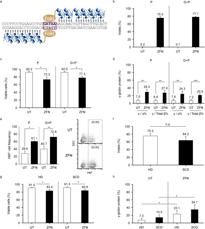
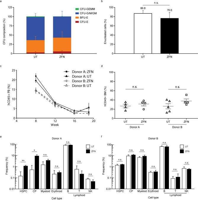
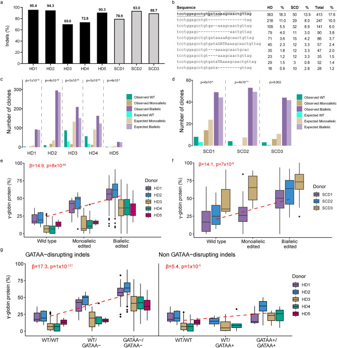
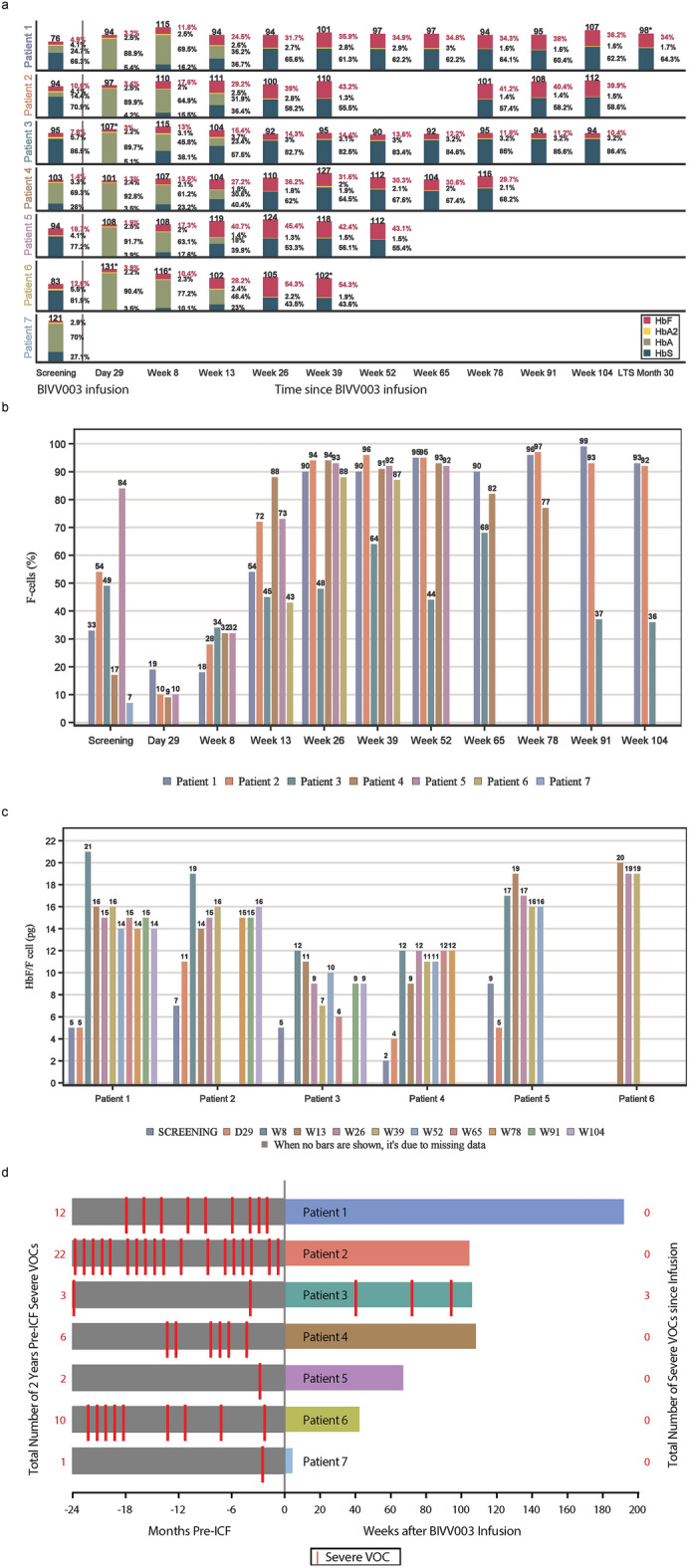
BIVV003 is a gene-edited autologous cell therapy in clinical development for the potential treatment of sickle cell disease (SCD). Hematopoietic stem cells (HSC) are genetically modified with mRNA encoding zinc finger nucleases (ZFN) that target and disrupt a specific regulatory GATAA motif in the BCL11A erythroid enhancer to reactivate fetal hemoglobin (HbF). We characterized ZFN-edited HSC from healthy donors and donors with SCD. Results of preclinical studies show that ZFN-mediated editing is highly efficient, with enriched biallelic editing and high frequency of on-target indels, producing HSC capable of long-term multilineage engraftment in vivo, and express HbF in erythroid progeny. Interim results from the Phase 1/2 PRECIZN-1 study demonstrated that BIVV003 was well-tolerated in seven participants with SCD, of whom five of the six with more than 3 months of follow-up displayed increased total hemoglobin and HbF, and no severe vaso-occlusive crises. Our data suggest BIVV003 represents a compelling and novel cell therapy for the potential treatment of SCD.


Conflict of interest statement

AH and SL are employees of Sanofi and may hold shares and/or stock options in the company. HL, KM, BV, DL, DR, PR, RP, TH, YL, GMR, IG and VH were employees of Sanofi at the time of the study. AR, MC, and BH are employees of Sangamo and may hold shares and/or stock options in the company. RB and BC were employees of Sangamo at the time of study. NU and JT have no conflict of interest. MA receives honorarium from Bristol Myers Squibb, hold stock from Cytodine Inc, and participates in speaker bureaus for Bristol Myers Squibb, Gilead, and Abbvie. MCW has served as consultant for Ensoma, Inc., Vertex pharmaceuticals, AllCells, Inc, and BioLabs, Inc.

Figures

Fig. 1
Fig. 1
ZFN-mediated gene editing is highly efficient and leads to HbF induction independently of mobilization strategy or disease state. (a) Schematic of zinc-finger nucleases (ZFN) targeting the BCL11A erythroid-specific enhancer. The FokI domains dimerize over the core GATAA motif to induce double-strand breaks. Arrows represent the ZFN-nucleotide contacts. (b) Indel frequency and (c) cell viability using acridine orange/propidium iodide in single- (n = 5) and dual-mobilized (n = 4) CD34 + HSPC 2 days after ZFN editing of the BCL11A erythroid enhancer. (d) Gamma-globin (γ-globin) protein levels measured by reverse phase ultra-performance liquid chromatography (RP-UPLC) at day 21 of erythroid differentiation (single-mobilized n = 4, dual-mobilized n = 4). (e) Fraction of HbF+ cells in enucleated glycophorin A–positive erythroid cells derived from single- and dual-mobilized CD34 + HSPC (left; single-mobilized n = 3, dual-mobilized n = 2). A representative flow cytometry plot of HbF+ cell frequency is shown on the right. (f) Indel levels and (g) cell viability in HSPC derived from healthy (n = 4) and SCD (n = 3) donors 2 days post-transfection of ZFNs. (h) γ-globin protein levels measured by RP-UPLC at terminal erythroid differentiation (day 18). Data expressed as mean ± standard deviation (*P < 0.05; **P < 0.01; ***P < 0.001). G + P, granulocyte colony-stimulating factor plus plerixafor; HbF, fetal hemoglobin; HD, healthy donor; HSC, hematopoietic stem cell; P, plerixafor; RP-UPLC, reversed-phase ultra-performance liquid chromatography; SCD, sickle cell disease donor; SSC, side scatter; UT, untreated; ZFN, zinc finger nuclease.
Fig. 2
Fig. 2
ZFN-mediated gene editing does not adversely affect HSC progenies. (a) Number of CFU-derived colonies of HSPC 2 days post-ZFN editing from plerixafor cells. Shown are the contribution of CFU-E, BFU-E, CFU-G/M/GM, and CFU-GEMM (n = 3). (b) Frequency of enucleated cells at Day 20 of in vitro differentiation (n = 4). (c) Proportion of human CD45+ (hCD45) cells in NSG mouse PB. Data from 9–10 mice per donor and condition at Weeks 8 and 12 and from 4–5 mice at Weeks 16 and 19. Error bars correspond to the standard error of the mean. (d) Proportion of hCD45+ cells in mouse BM at Week 19. (e and f) Frequencies of HSPC (Lin-CD34highCD38-), committed progenitors (Lin-CD34highCD38 +), myeloid (CD33 +), erythroid (CD71 +), lymphoid B (CD19 +), lymphoid T (CD3 +), and natural killer (CD56 +) within the hCD45 + population within the total bone marrow cells are shown for two healthy donors at 19 weeks after engraftment. Data expressed as mean ± standard deviation unless specified (*P < 0.05; **P < 0.01; ***P < 0.001). BFU-E, burst-forming unit-erythroid; CFU, colony-forming unit; CFU-E, CFU-erythroid; CFU-G/M/GM, CFU-granulocyte–macrophage; CFU-GEMM, CFU-granulocyte-erythrocyte-monocyte-megakaryocyte; CP, committed progenitor; HD, healthy donor; HSC, hematopoietic stem cell; HSPC, hematopoietic stem and progenitor cell; NK, natural killer; UT, untreated; ZFN, zinc finger nuclease.
Fig. 3
Fig. 3
ZFN-mediated gene editing does not compromise the functionality or progenies of HSPC and results in stable editing levels after engraftment in vivo. (a) PB indel levels at input (Week 0, n = 2 donors) and after engraftment (For each donor, n = 9–10 mice for week 8 and 12 post engraftment, and n = 4–5 mice for week 16 and 19). (b) Indel levels in human bone marrow (BM) as well as cell subpopulations within the hCD45 + compartment sorted using antibodies recognizing the indicated lineage markers at input (Week 0) and after engraftment (Week 19 post engraftment). No significant difference was observed between lineages at week 19 using a multivariate ANOVA controlling for donor differences (P > 0.05, excluding input). BM refers to the total human CD45 + cell population in the bone marrow. (c) Frequency of enucleated cells in terminal erythroid differentiation in vitro from BM-derived HSPC at Week 19. (d) Globin protein levels were analyzed at Day 16 of in vitro erythroid differentiation by UPLC. Average γ-globin protein levels for each group are plotted as percentage of all β-like globin protein (A-γ-, G-γ-, δ-, and adult β-globin protein) on the left, and average γ-globin protein levels as percentage of α-globin levels are plotted on the right. (e) Read counts of the 10 most frequent sequences identified by next-generation sequencing at input (Week 0) in healthy donor 1 (HD 1) and healthy donor 2 (HD 2). The unedited (wild type) sequence is shown in bold, with a rectangle highlighting the core GATAA site and lines representing ZFN contacts. (f) Fraction of total indels that are likely derived from MMEJ. MMEJ patterns represent those identified by the RGEN Microhomology-Predictor tool (see Methods). (g) Frequency of indels by size of insertion (> 0) or deletions (< 0). Data expressed as mean ± standard deviation (*P < 0.05; **P < 0.01; ***P < 0.001). HD, healthy donor; HSC, hematopoietic stem cell; MMEJ, microhomology-mediated end-joining; n.s., not significant; PB, peripheral blood; BM, bone marrow; UPLC; ultra-performance liquid chromatography; UT, untreated; ZFN, zinc finger nuclease.
Fig. 4
Fig. 4
ZFN-mediated gene editing leads to enriched biallelic edited cells and allele-additive increases in HbF. (a) Total editing levels in erythroid colonies derived from single-cell CD34 + HSPC from healthy and SCD donors. (b) Frequency of the most common indel patterns in single-cell derived colonies. The unedited (wild type) sequence is shown in bold, with a rectangle highlighting the core GATAA site and lines representing ZFN contacts. (c and d) Frequency of wild-type, monoallelic, and biallelic-edited genotypes derived from single-cell clonal analysis of plerixafor-mobilized HSC from (c) 5 healthy and (d) 3 SCD donors. The distribution of genotype was significantly skewed towards biallelic clones. The expected distribution of clones was calculated using an equation analogous to the Hardy–Weinberg equilibrium (Online Methods). P-values from χ tests comparing the observed and expected number of wild-type, monoallelic, and biallelic clones for each sample are reported on the figure. Significant P-values reflect a skew of the distributions towards wild-type and biallelic clones. (e and f) Association of γ-globin levels measured by reversed-phase ultra-performance liquid chromatography with genotypes in (e) healthy donors (linear regression P = 8 × 10−84; beta = 14.9; standard error = 0.7) and (f) SCD donors (linear regression P = 7 × 10−8; beta = 14.1; standard error = 2.5). Mean differences between biallelic-edited and unedited clones were 32% (HD 1), 38% (HD 2), 28% (HD 3), 30% (HD 4), 21% (HD 5), 28% (SCD 1), 32% (SCD 2), and 20% (SCD 3). (g) Effect of indels on gamma-globin expression in healthy donor cells, stratified by whether they disrupt the core GATAA site (GATAA-, left) or not (GATAA + , right). For (e, f, and g), beta and P-values were calculated by fitting linear regression models of HbF (gamma-globin) against genotype in all clones, adjusted for the different donors (HbF ~ genotype + donor). The beta coefficient for the genotype effect is reported. HbF, fetal hemoglobin; HD, healthy donor; HSC, hematopoietic stem cell; SCD, sickle cell disease; WT, wild type.
Fig. 5
Fig. 5
ZFN-mediated gene editing is highly specific and characterized by high frequency of on-target small indels. (a) Experimental strategy to confirm homozygote clones and identified large deletions. Left: With the original 140 bp sequencing assay, clones with only 1 indel pattern may represent homozygote clones or clones with an uncaptured deletion spanning a primer site. Center: 12 homozygous clones could be confirmed by a sequencing assay that captures an informative SNP in 2 healthy donors. Right: For samples with no informative SNPs or uncaptured events, a 12 kb region around the target site was sequenced. Example shows a heterozygous sample with a large deletion that was not captured by the small 140 bp amplicon and therefore ruled out as a homozygote by the subsequent analysis. (b) Frequency of identified allele lengths in clones (N = 1175) derived from healthy donors (n = 4). A 12 kb region was sequenced around the BCL11A enhancer in clones with alleles potentially missed by short amplicon sequencing. Indels > 1 kb represent less than 1% of events. Unknown events include 3 insertions for which the length could not be determined. (c) Depiction of the largest deletion observed (5.9 kb). The deletion is contained within the BCL11A erythroid super-enhancer, itself located within the 77 kb intron 2 of BCL11A. (d) Total fraction of indels that disrupt the core GATAA site. (e) Enrichment of MMEJ indels with large deletions. The figure shows the frequency of small indels (MMEJ or NHEJ) as the second allele of clones carrying a large deletion (> 50 bp), a large insertion (> 50 bp), a complex indel, or a NHEJ indel. MMEJ indels are over-represented as the second allele of clones with deletions > 50 bp (Fisher exact test P = 0.008). Complex indels and insertions show over- and under-representation of large deletions (P = 0.02 and 0.03, respectively). Data from healthy donor cells (n = 4). MMEJ patterns represent those identified by the RGEN Microhomology-Predictor tool (methods). bp, base pairs; HD, healthy donor; MMEJ, microhomology-mediated end joining; NHEJ, non-homologous end joining; SNP, single nucleotide polymorphism.
Fig. 6
Fig. 6
Preliminary safety and efficacy results from PRECIZN-1: an ongoing Phase 1/2 study of BIVV003, a ZFN-modified autologous CD34 + HSC therapy for SCD. (a) Total Hb and Hb fractionation in all participants (n = 7) following BIVV003 infusion. *Indicates the Hb value from local lab, since the central lab value was not collected. (b) F cells over time for treated participants. (c) HbF/F cell levels above the threshold for preventing HbS polymerization. (d) Number of VOCs (severe pain crises, acute chest syndrome) reported pre- and post-BIVV003 infusion. Note, some participants had back-to-back VOC events. The total number of events is reported in Table S2. Hb, hemoglobin; HbA, adult hemoglobin; HbA2, variant adult hemoglobin; HbF, fetal hemoglobin; HbS, sickle hemoglobin; VOC, vaso-occlusive crisis

References

    1. Frenette, P. S. & Atweh, G. F. Sickle cell disease: old discoveries, new concepts, and future promise. J Clin Invest117, 850–858 (2007). - PMC - PubMed
    1. Hebbel, R. P., Osarogiagbon, R. & Kaul, D. The endothelial biology of sickle cell disease: inflammation and a chronic vasculopathy. Microcirculation11, 129–151 (2004). - PubMed
    1. Kato, G. J. et al. Sickle cell disease. Nat Rev Dis Primers4, 18010 (2018). - PubMed
    1. Khemani, K., Katoch, D. & Krishnamurti, L. Curative Therapies for Sickle Cell Disease. Ochsner J19, 131–137 (2019). - PMC - PubMed
    1. Lettre, G. & Bauer, D. E. Fetal haemoglobin in sickle-cell disease: from genetic epidemiology to new therapeutic strategies. Lancet387, 2554–2564 (2016). - PubMed

Publication types