. 2024 Oct 16;7(1):1340.
doi: 10.1038/s42003-024-07043-2.

snPATHO-seq, a versatile FFPE single-nucleus RNA sequencing method to unlock pathology archives

Affiliations

snPATHO-seq, a versatile FFPE single-nucleus RNA sequencing method to unlock pathology archives

Taopeng Wang et al. Commun Biol. .

Abstract

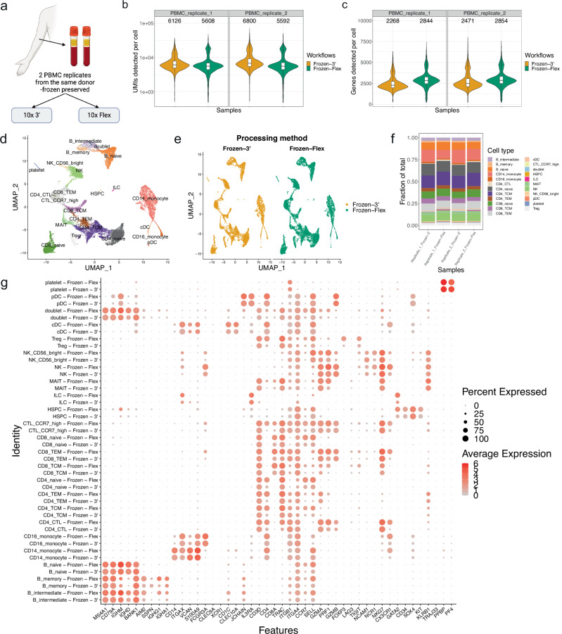
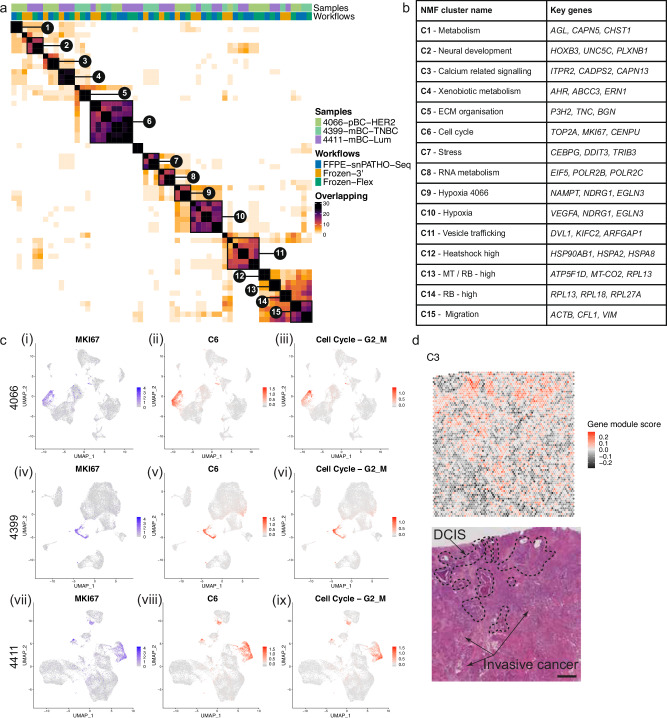
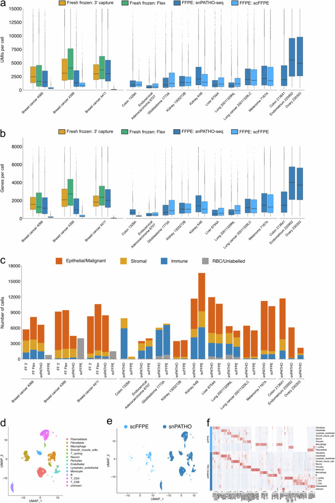
Formalin-fixed paraffin-embedded (FFPE) samples are valuable but underutilized in single-cell omics research due to their low RNA quality. In this study, leveraging a recent advance in single-cell genomic technology, we introduce snPATHO-seq, a versatile method to derive high-quality single-nucleus transcriptomic data from FFPE samples. We benchmarked the performance of the snPATHO-seq workflow against existing 10x 3' and Flex assays designed for frozen or fresh samples and highlighted the consistency in snRNA-seq data produced by all workflows. The snPATHO-seq workflow also demonstrated high robustness when tested across a wide range of healthy and diseased FFPE tissue samples. When combined with FFPE spatial transcriptomic technologies such as FFPE Visium, the snPATHO-seq provides a multi-modal sampling approach for FFPE samples, allowing more comprehensive transcriptomic characterization.


Conflict of interest statement

The authors declare no competing interests.

Figures

Fig. 1
Fig. 1. The 10x single-cell Flex chemistry produces results comparable to those of 10x 3’ chemistry when applied to PBMC samples.
a Illustration of experiment design for 10x 3’ and Flex assay comparison. Created in BioRender. Wang, T. (2024) BioRender.com/l86j691. b, c Violin plots of the number of UMIs (b) and Genes (c) detected per cell. The boxes in the violin plots show the UMIs (b) and Genes (c) median and interquartile range. d UMAP embedding of PBMC cells integrated using the Seurat CCA method and annotated by cell type. e UMAP embedding split by processing method. f Barplots showing the fraction of cell types detected in each technical replicate processed using 10x 3’ or Flex chemistry. g Dotplot of the expression of canonical PBMC cell type markers. N = 2 sample per protocol. The two replicates were conducted using samples from the same donor. Replicates are labeled in (b, c, f).
Fig. 2
Fig. 2. The snPATHO-seq workflow enables nuclei isolation and single-nucleus gene expression detection from human FFPE tissue samples.
a Illustration of the snPATHO-seq workflow. Created in BioRender. Wang, T. (2024) BioRender.com/u53s150. b, c Boxplots of the number of UMIs (b) and genes (c) detected per nucleus. The boxes show the UMIs (b) and Genes (c) median and interquartile range. Outliers were shown as dots. d UMAP embedding of unintegrated snRNA-seq data annotated by sample IDs. e UMAP embedding of unintegrated snRNA-seq data split by processing methods. f UMAP embedding of Seurat CCA integrated snRNA-seq data from patient 4411 annotated by cell type. g UMAP embedding of Seruat CCA integrated 4411 snRNA-seq data split by processing methods. h Barplot showing the fraction of cell types detected by different snRNA-seq methods in sample 4411. i Heatmap of the scaled expression of selected cell type markers detected by differential gene expression analyses in 4411 data. The top 200 significantly differentially expressed genes identified in each cell population (if available) were selected by fold change and used for plotting. A gene was considered significantly differentially expressed if the BH-adjusted P value was lower than 0.05. Genes were arranged by hierarchical clustering based on the expression in the FFPE-snPATHO-seq data on the x-axis. Cell types identified by different snRNA-seq workflows were manually arranged on the y-axis. N = 1 sample per protocol.
Fig. 3
Fig. 3. snPATHO-seq detected comparable transcriptomic signatures from FFPE samples as the conventional Flex chemistry using matching snap-frozen samples.
a Heatmap showing similarities between robust NMF programs derived from snPATHO-seq, 10x 3’, and 10x Flex data generated using 4066, 4399, and 4411. Robust NMF programs with similar gene compositions were clustered and highlighted in brackets. b Table of robust NMF program clusters annotated based on the shared gene compositions. c UMAP embedding of Seurat CCA integrated data from patients 4066 (i–iii), 4399 (iv–vi), and 4411 (vii–ix) overlaid with the expression of MKI67 (i, iv, vii), the module scores of robust NMF program cluster C6 (ii, v, viii) and the module scores of a published cell cycle gene program (iii, vi, ix). d Spatial enrichment pattern of the module score of robust NMF program cluster C3 in the Visium data from patient 4066 and the H&E image of this sample. Scale bar = 1 mm.
Fig. 4
Fig. 4. Comparison of the snPATHO-seq workflow to scFFPE workflow.
a, b Boxplots of the number of UMIs (a) and genes (b) detected per nuclei using different workflows. The boxes show the median and interquartile range of the UMIs (a) and Genes (b). Outliers were shown as dots. c Barplots of the number of nuclei/cells detected in each dataset colored by major cell lineages. d UMAP embedding of Seurat CCA integrated snPATHO-seq and scFFPE data from sample Colon_1328A colored by cell type annotations. e UMAP embedding split by processing methods. f Heatmap of top differentially expressed genes detected between cell types by snPATHO-seq and scFFPE workflows. The top 200 significantly differentially expressed genes identified in each cell population (if available) were selected by fold change and used for plotting. A gene was considered significantly differentially expressed if the BH-adjusted P value was lower than 0.05. N = 1 sample per protocol.

References

    1. Yuan, Y. et al. Pathology laboratory policies and procedures for releasing diagnostic tissue for cancer research. Arch. Pathol. Lab. Med.145, 222–226 (2021). - PMC - PubMed
    1. Greytak, S. R., Engel, K. B., Bass, B. P. & Moore, H. M. Accuracy of molecular data generated with FFPE biospecimens: lessons from the literature. Cancer Res.75, 1541–1547 (2015). - PMC - PubMed
    1. Friedrich, C. et al. Comprehensive micro-scaled proteome and phosphoproteome characterization of archived retrospective cancer repositories. Nat. Commun.12, 3576 (2021). - PMC - PubMed
    1. Munchel, S. et al. Targeted or whole genome sequencing of formalin fixed tissue samples: potential applications in cancer genomics. Oncotarget6, 25943–25961 (2015). - PMC - PubMed
    1. Turnbull, A. K. et al. Unlocking the transcriptomic potential of formalin-fixed paraffin embedded clinical tissues: comparison of gene expression profiling approaches. BMC Bioinforma.21, 30 (2020). - PMC - PubMed