. 2024 Oct 16;14(1):24228.
doi: 10.1038/s41598-024-76163-w.

A self-adjuvanted VLPs-based Covid-19 vaccine proven versatile, safe, and highly protective

Affiliations

A self-adjuvanted VLPs-based Covid-19 vaccine proven versatile, safe, and highly protective

Larissa Vuitika et al. Sci Rep. .

Abstract

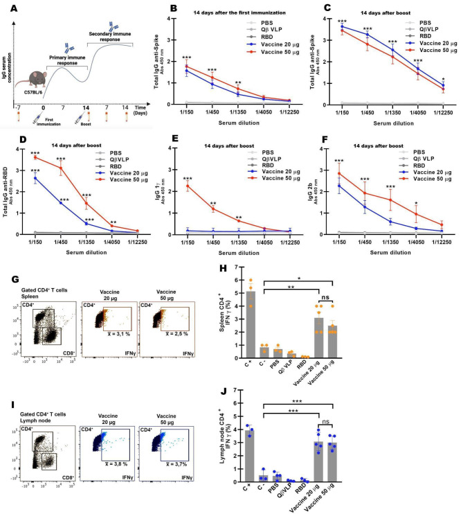
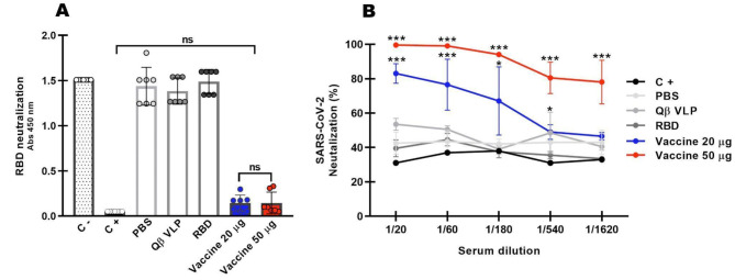
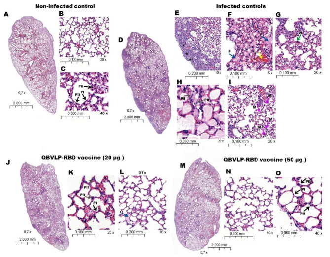
Vaccination has played a critical role in mitigating COVID-19. Despite the availability of licensed vaccines, there remains a pressing need for improved vaccine platforms that provide high protection, safety, and versatility, while also reducing vaccine costs. In response to these challenges, our aim is to create a self-adjuvanted vaccine against SARS-CoV-2, utilizing Virus-Like Particles (VLPs) as the foundation. To achieve this, we produced bacteriophage (Qβ) VLPs in a prokaryotic system and purified them using a rapid and cost-effective strategy involving organic solvents. This method aims to solubilize lipids and components of the cell membrane to eliminate endotoxins present in bacterial samples. For vaccine formulation, Receptor Binding Domain (RBD) antigens were conjugated using chemical crosslinkers, a process compatible with Good Manufacturing Practice (GMP) standards. Transmission Electron Microscopy (TEM) confirmed the expected folding and spatial configuration of the QβVLPs vaccine. Additionally, vaccine formulation assessment involved SDS-PAGE stained with Coomassie Brilliant Blue, Western blotting, and stereomicroscopic experiments. In vitro and in vivo evaluations of the vaccine formulation were conducted to assess its capacity to induce a protective immune response without causing side effects. Vaccine doses of 20 µg and 50 µg stimulated the production of neutralizing antibodies. In in vivo testing, the group of animals vaccinated with 50 µg of vaccine formulation provided complete protection against virus infection, maintaining stable body weight without showing signs of disease. In conclusion, the QβVLPs-RBD vaccine has proven to be effective and safe, eliminating the necessity for supplementary adjuvants and offering a financially feasible approach. Moreover, this vaccine platform demonstrates flexibility in targeting Variants of Concern (VOCs) via established conjugation protocols with VLPs.

Keywords: COVID-19; SARS-CoV-2; VLPs platform; Vaccine.


Conflict of interest statement

The authors declare no competing interests.

Figures

Fig. 1
Fig. 1
Flowchart of the experimental design.
Fig. 2
Fig. 2
VLPs-based vaccine production and characterization. (A) The QβVLPs (PDB 1QBE) tridimensional structures, highlighting the high exposure number of linker conjugation points, as well as the internal toll-like agonist and the core with ~ 27 nm, measured after electron microscopy analysis (E). (B) The coat protein gene of Qβ bacteriophage (Genbank M99039.1 and Proteinbank AAA1662.1) was cloned in pET28a and BL21 (DE3) E. coli competent cells were transformed with the construction. After IPTG induction QβVLPs are self-assembled in the bacteria cytoplasm. (C) The expression and purification of recombinant QβVLPs was analyzed using 15% SDS–PAGE under reducing conditions and Coomassie blue dye staining. Line 1 depicts E. coli cells before induction with 1 mM IPTG. Lane 2 shows the proteins of cells after induction IPTG. Lanes 3 depict the supernatant (soluble form) obtained through freeze, thawing and sonication lysis in the extraction buffer before purification. Lane 4 show purified recombinant protein CP Qβs. (D) Western blot of the bacterial extracts and purified CP QβVLPs using hyperimmune serum of the mouse immunized with QβVLPs. Line 1 depicts E. coli cells before induction with 1 mM IPTG. Lane 2 shows the proteins of cells after induction IPTG. Lanes 3 depict the supernatant (soluble form) after lysis. Lane 4 show purified recombinant protein CP QβVLPs. Molecular weight marker (Thermo Fisher Scientific Ref. 26619). (E) Transmission electron microscopy (EM) of QβVLPs nanoparticle at 129.300 magnitude. (F) Chemical modifications performed on the QβVLPs (SMPH) and RBD antigen (SATA and Hydroxylamine.HCl), and subsequent conjugation for the vaccine formulation. Bacteriophage Q beta capsid protein in T3 symmetry PDB Entry − 7TJM; SARS-CoV-2 E406W mutant RBD PDB Entry − 7TPK (G) SDS-PAGE 15% stained with Coomassie brilliant blue for characterization of the vaccine formulation. Line 1 QβVLPs, line 2 RBD antigen and line 3 the vaccine profile with different bands representing different vaccine oligomers. (H) Western blot performed with hyperimmune serum anti QβVLP-RBD vaccine and total anti-mouse IgG antibody conjugated with HRP. Line 1 QβVLPs, line 2 RBD antigen, line 3 shows the vaccine profile with different bands representing different vaccine oligomers. (I) Transmission EM of the VLPs-based vaccine and its tridimensional structure. (J) Stereomicroscopic images of popliteal draining LN in live mice illustrate the accumulation of QβVLPs labeled with AF488 QβVLPs-RBD vaccine labeled with AF594 (A-bright field image of LN (identified by arrowhead); B-C QβVLPs/AF488 in 5 and 10 min post injection taken with appropriate fluorescent filter sets; D-E QβVLPs-RBD vaccine/AF594 in 5 and 10 min and post injection taken with appropriate fluorescent filter sets. Magnitude 7x. The QβVLPs (PDB 1QBE) (10.1016/s0969-2126(96)00060-3), RBD (PDB 6M0J) (10.1038/s41586-020-2180-5), and the RNA (PDB 4GXY) (10.1038/nsmb.2405) tridimensional structures were rendered on 3D protein imager (10.1093/bioinformatics/btaa009).
Fig. 3
Fig. 3
Immunological assessment of the vaccine formulation. (A) - Schedule of mouse (C57BL/6) immunization in two doses (day 0 and boost) and serum collection after immunizations. (B-C) Profile of total IgG anti-Spike production of serum collected 14 days after first and second immunization with 20 µg, 50 µg, and controls (PBS, QβVLPs, and RBD), respectively. (D) Total IgG anti-RBD production of serum collected 14 days after boost with 20 µg, 50 µg, and controls. (E-F) Production of IgG2b and IgG1𝛾 anti-Spike present in the serum collected 14 days after the second immunization with 20 µg, 50 µg, and controls. ELISA results show an average of three experiments ± SEM. *** P ≤ 0.001 compared with PBS, RBD, and QβVLPs controls. The results show an average of three experiments ± SEM. *** P ≤ 0.001 compared with PBS, RBD, and QβVLPs controls. (G-J) Profile of intracellular IFNγ production by lymphocytes of the mice immunized with QβVLPs-RBD vaccine analyzed by flux cytometry. (G-H) Detection of IFNγ production in CD4+ T lymphocytes from the spleen extracted from mice immunized with the QβVLPs-RBD vaccine (20 µg and 50 µg) RBD, QβVLPs or PBS and subsequently stimulated with the Spike recombinant protein. (I-J) Detection of IFNγ production in T lymphocytes CD4+ from cervical lymph nodes extracted from mice immunized with the QβVLPs-RBD vaccine (20 µg and 50 µg), RBD, QβVLPs or PBS and subsequently stimulated with the Spike recombinant protein. Negative control: PBS. Positive control: PMA + Ionomycin. The results show an average of three experiments ± SEM. *** P ≤ 0.001 compared with negative control (C-).
Fig. 4
Fig. 4
Neutralizing antibodies assay. (A) The samples collected from vaccinated and control group of mice were diluted 1:10 and incubated with RBD conjugated with HRP and after were added in the 96 wells plate previously coating with ACE2, as described on the Kit cPass SARS-CoV-2 Neutralization Antibody Detection Kit (GenScript, EUA Ref. L00847). Negative (without antibody) and positive controls (positive neutralizing antibody) are purchased from the kit. The average ± standard errors are shown, with significance levels ***P ≤ 0.001 compared with positive control and PBS, RBD, and QβVLPs controls. (B) In vitro microneutralization assay. P1. SARS-CoV-2 were incubated with hyperimmune serum samples of 14 days after boost of the QβVLPs-RBD vaccine (20 µg and 50 µg), and controls (RBD, QβVLPs or PBS in a 1/3 dilution ratio. After, Vero E6 monolayer sub cultivated in 96 well plates were incubated with P.1 SARS-CoV-2 + hyperimmune sera samples for 72 h at 37 oC in 5% CO2. After this, it was added and the absorbance was measured in a plate reader in the range of 620 nm. The average ± standard errors are shown, with significance levels ***P ≤ 0.001 compared with positive control and PBS, RBD, and QβVLPs controls.
Fig. 5
Fig. 5
Challenge with SARS-CoV-2 and assessment of clinical signs and viral. Two-dose immunization (20 µg and 50 µg) was able to protect K18-hACE-2 animals (model for severe COVID-19 from mortality, weight loss and other clinical signs of the disease such as motility, bristling hair and hunchback position. (A) Survival (%) after 7 days post infection (DPI); (in purple and highlighted with a star the survival rate of animals vaccinated with 20 µg and 50 µg of QβVLPs-RBD). (B) Loss weight (%) after 7 DPI. Mice immunized with 50 µg QβVLPs-RBD showed no significant weight loss while non immunized had more than 20% of body weight loss, in addition to showing the clinical symptoms of the disease. The average ± standard errors are shown, with significance levels ***P ≤ 0.001 compared with positive control (C+). (C) Viral quantitative measurements by qRT-PCR showed a significantly lower viral load of SARS-CoV-2 in the lungs of mice vaccinated with two-dose immunization (20 µg and 50 µg) in comparison to non-immunized animals The average ± standard errors are shown, with significance levels ***P ≤ 0.001 compared with positive control (C+). (D) Cytokine profile expression measurements by qRT-PCR. Negative Control (C-): Samples from animals that were neither vaccinated nr exposed to the virus (naive animals). Positive Control (C+): Samples from animals that, while not vaccinated, were exposed to the virus.
Fig. 6
Fig. 6
Lung histopathological analysis of immunized B6.Cg-Tg(K18-ACE2)2Prlmn animals challenged with SARS-CoV-2. (A-C) Lung of non-infected animals. (A) Panoramic lung vision. (B) Lung parenchyma with normal appearance. (C) Type I and II pneumocytes with normal morphology (arrow black PI and arrow black PII). (D-I) Lung of the infected controls animals previously immunized with PBS, RBD, and QβVLPs. (D) Panoramic lung vision. (E) Perivascular inflammation (asterisk). (F) Alveolar fibrin and hemorrhage (blue F and yellow H arrows, respectively). (G) Syncytial and single cell necrosis (green N arrow). (H) Type II pneumocyte hyperplasia (gray PII arrows). (I) Macrophages (pink M arrows). (J-L) Lung os the infected controls animals previously immunized with QβVLPs-RBD vaccine (20 µg). (J) Panoramic lung vision. (K) Type II pneumocyte hyperplasia (gray PII arrows). (L) Alveolar fibrin (blue F arrow). (M-O) Lung os the infected controls animals previously immunized with QβVLPs-RBD vaccine (50 µg). (M) Panoramic lung vision. (N) Lung parenchyma with normal appearance. (O) Type I and II pneumocytes with normal morphology (arrow black PI and arrow black PII). The 4 μm thick sections were stained with hematoxylin and eosin and images were captured with Slide Digitizer − 3D Histech scanner and analyzed in Download CaseViewer 2.4, 64-bit version (3D- Histotech, Hungary - https://www.3dhistech.com/solutions/caseviewer/).

References

    1. Kumar, A. et al. SARS-CoV-2-specific virulence factors in COVID-19. J. Med. Virol. 93 (3), 1343–1350. 10.1002/jmv.26615 (2021). Epub 2020 Nov 1. PMID: 33085084. - PubMed
    1. World Health Organization. WHO Coronavirus (COVID-19) Dashboard. https://covid19.who.int/
    1. DeGrace, M. M. et al. Defining the risk of SARS-CoV-2 variants on immune protection. Nature. 605, 640–652 (2022). - PMC - PubMed
    1. Prates-Syed, W. A. et al. VLP-Based COVID-19 vaccines: an adaptable technology against the threat of New variants. Vaccines (Basel). 9 (12), 1409. 10.3390/vaccines9121409 (2021). PMID: 34960155; PMCID: PMC8708688. - PMC - PubMed
    1. Chirico, F., da Silva, J. A. T., Tsigaris, P. & Sharun, K. Safety & effectiveness of COVID-19 vaccines: a narrative review. Indian J. Med. Res. 155, 91–104 (2022). - PMC - PubMed

MeSH terms

LinkOut - more resources