. 2024 Nov;43(49):3586-3597.
doi: 10.1038/s41388-024-03182-2. Epub 2024 Oct 16.

Obesity-induced extracellular vesicles proteins drive the endometrial cancer pathogenesis: therapeutic potential of HO-3867 and Metformin

Affiliations

Obesity-induced extracellular vesicles proteins drive the endometrial cancer pathogenesis: therapeutic potential of HO-3867 and Metformin

Takahiko Sakaue et al. Oncogene. 2024 Nov.

Abstract

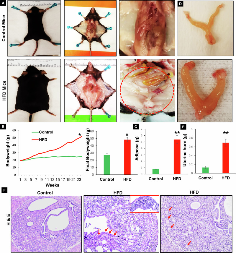
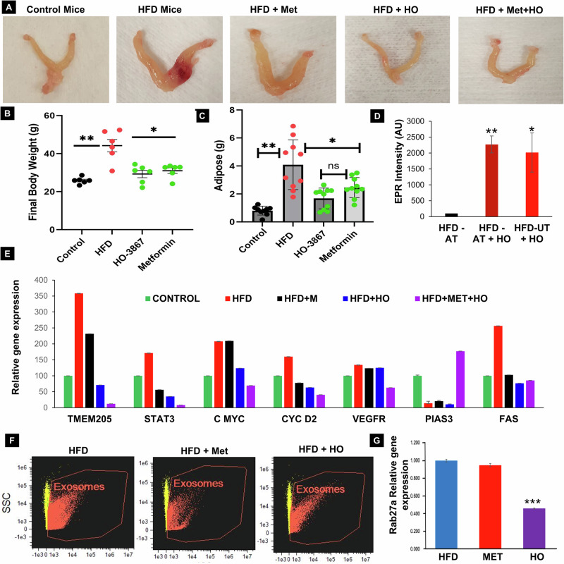
Endometrial cancer (EC) is the leading gynecologic malignancy in the United States with obesity implicated in 57% of cases. This research investigates the molecular complexities of extracellular vesicles (EV) secretion as carriers of oncogenic protein and their involvement in obesity-mediated EC. An understanding of these mechanisms is pivotal for unraveling pathways relevant to obesity-associated EC, thereby guiding the development of innovative prevention and treatment strategies. Our exploration revealed a significant increase in EV secretion carrying oncogenic proteins (TMEM205, STAT5, and FAS) in adipose and uterine tissues/serum samples from obese EC patients compared to control (without cancer). We identified alterations in EV-regulating proteins (Rab7, Rab11, and Rab27a) in obesity-mediated EC patients, adipose/uterine tissues, and serum samples. Through a 24-week analysis of the effects of a 45% kcal high-fat diet (HFD) on mice, we observed increased body weight, increased adipose tissue, enlarged uterine horns, and increased inflammation in the HFD group. This correlated with elevated levels of EV secretion and increased expression of oncogenic proteins TMEM205, FAS, and STAT5 and downregulation of the tumor suppressor gene PIAS3 in adipose and uterine tissues. Furthermore, our study confirmed that adipocyte derived EV increased EC cell proliferation, migration and xenograft tumor growth. Additionally, we identified that the small molecule inhibitors (HO-3867) or Metformin inhibited EV secretion in vitro and in vivo, demonstrating significant inhibition of high glucose or adipocyte-mediated EC cell proliferation and a reduction in body weight and adipose tissue accumulation when administered to HFD mice. Moreover, HO-3867 or Metformin treatment inhibited HFD induced hyperplasia (precursor of EC) by altering the expression of EV-regulated proteins and decreasing oncogenic protein expression levels. This study provides critical insights into the mechanisms underpinning obesity-mediated EV secretion with oncogenic protein expression, shedding light on their role in EC pathogenesis. Additionally, it offers pre-clinical evidence supporting the initiation of novel studies for EV-targeted therapies aimed at preventing obesity-mediated EC.


Conflict of interest statement

Competing interests: The authors declare no competing interests. Ethics approval and consent to participate: All procedures used in this study were authorized and conducted according to the guidelines of the Ohio State University Research Institute Ethics Committee. All animal experiments were following the Animal Experimentation Ethics of the Ohio State University Animal Experimentation Research Lab, and the ethics approval number for animal experimentation was 2012A00000008-R3. The use of stored human tissues in this study was approved by the Institutional Review Board of the Ohio State University Wexner Medical Center under Study Number: 2004C0124 and the Ohio State University’s OHRP Federal wide Assurance #00006378. No human subjects were directly consented for this study as the tissues were obtained from a biorepository.

Figures

Fig. 1
Fig. 1. Enhanced extracellular vesicle (EV) secretion associated with oncogenic proteins and EV regulatory proteins in endometrial cancer (EC) patient samples.
A, B Serum samples from control (without cancer), obese EC and non-obese EC patients were thawed on ice and diluted (1:4 to 1:10) in 1xPBS. EV were isolated from 100  µL of patient serum using the q-izon column. EV concentration was assessed by Image Stream Flow Cytometry (ISF), revealing a significant increase in EV concentration and fold change in obese EC patients compared to non-obese EC (n = 5, p < 0.01). C EV regulatory proteins, including Rab7, Rab11, and Rab27a, were analyzed by RT-PCR in control (obese benign) obese EC early-stage (EC-ES) and obese EC late-stage (EC-LS) tissue samples (n = 3, p < 0.001 or 0.005). D Oncogenic proteins (TMEM205, STAT5, FAS, and PIAS3) and their relative gene expression levels were examined in control (obese benign) and EC-ES patient tissues samples by RT-PCR (n = 3, p < 0.001 or 0.005). E Upregulated TMEM205, STAT5 and FAS protein expression levels in obese EC compare with control (without cancer) patient serum EV samples by ELISA assay (n = 12, p < 0.005).
Fig. 2
Fig. 2. High-Fat Diet (HFD) supplementation promotes adipose tissue accumulation and induces endometrial hyperplasia.
A, B Immunocompetent mice were treated with a high-fat diet (HFD, 45% protein calorie) for 24 weeks. Representative images depict accumulated levels of adipose tissue (circled) in mice fed with a HFD for 24 weeks. Changes in body weight were assessed at the beginning (8 weeks) and end (24 weeks) of the HFD treatment (n = 10/group, p < 0.01). C Increased accumulation of adipose tissue was observed in mice fed with a HFD compared to controls (n = 5, p < 0.005). D, E Increased uterine enlargement and weight were observed in mice fed with a HFD compared to controls (n = 5, p < 0.005). F Enhanced cell proliferation in the endometrium of mice fed a HFD compared to controls (normal diet) is illustrated by hematoxylin-Eosin (H&E) staining. The inset shows a higher magnification of the increased cell proliferation.
Fig. 3
Fig. 3. Increased extracellular vesicle (EV) secretion associated with EV regulatory proteins in HFD treated mice samples.
A Transmission Electron Microscopy (TEM) reveals increased formation of EV in uterine tissues of high-fat diet (HFD) treated as compared to control diet mice. B, C Quantification of EV in serum samples from control and HFD-treated obese mice using Image Stream analysis (n = 3, p*0.01). D, E Analysis of relative protein and gene expression of EV regulating proteins (Rab11 and Rab27a) levels in adipose and uterine tissues from HFD and control diet-treated mice assessed by ELISA and RT-PCR (n = 3, p*0.001 or 0.005).
Fig. 4
Fig. 4. Alterations in oncogenic and tumor suppressor proteins in HFD treated mice tissues.
A, B Analysis of targeted proteins (TMEM205, STAT5, FAS, and PIAS3) and their relative gene and proteins expression levels in adipose and uterine tissues of HFD and control diet-treated mice. The assessment was conducted using a combination of RT-PCR, ELISA, and Immunohistochemistry (IHC) techniques (n = 3, p < 0.01 or 0.005).
Fig. 5
Fig. 5. Adipocytes mediated EV secretion increased EC cells proliferation/migration and EC progression.
A Adipose-derived mesenchymal stem cells underwent adipocyte differentiation initiated 72 h post-confluency on Day 3, 5, and 7, followed by maintenance in adipocyte medium until day 15. Microscopic images (10x and 40x) at different time points show increasing lipid accumulation. B To confirm adipocyte formation, cells were cultured in a 12-well plate, fixed, and stained with 0.2% oil red O in 2-propanol for 10 min at room temperature. Adipogenesis was measured at various time points. C Adipocyte-mediated extracellular vesicle (EV) secretion and size confirmation by ISF. D Adipocyte-derived EV (ADEV) were co-cultured with IK cells for 48 h, and the expression of candidate proteins TMEM205, FAS, and STAT5 was analyzed by ELISA (n = 4, p < 0.01 or p < 0.005). E, F Cell proliferation assessed by 5-ethynyl 2´-deoxyuridine (EdU) incorporation using flow cytometry in IK cells co-cultured with ADEV, demonstrating an increased percentage of EdU+ve cells compared to IK control cells (untreated ADEV) (n = 3, p < 0.05 or 0.01). G Ishikawa (IK) cells (2 ×10^6 cells) were injected subcutaneously into the right flank of athymic nude mice. One week later, the mice were randomly divided into three groups: Cancer Control (IK cells only, n = 6), EV25µg group (IK cells + EV, n = 7), and EV100µG group (IK cells + EV, n = 7). Adipocyte-derived EV (25 µg and 100 µg per 100 µL) were injected subcutaneously near the tumors in the EV groups, while physiological saline was injected into the Cancer Control group. Injections were administered twice a week for 4 weeks. H, I Tumor growth was monitored weekly, and tumor size was measured using a linear digital caliper. Tumor volume was calculated and represented in the graph. J At the end of the ADEV treatment, the tumor-bearing mice were sacrificed. Tumor tissues were collected and subjected to ELISA to measure the expression of candidate proteins (TMEM205, STAT5, FAS, and Rab27a) in both the control and AEV-treated groups (n = 6; p < 0.001).
Fig. 6
Fig. 6. Effects of EV inhibitors on EV secretion in EC cells.
A, B EV secretion levels analyzed and quantified by ISF in EV inhibitors Amiloride (10uM), GW4869 (5uM), Metformin (10uM), or HO-3867 (5uM) for 24 h in EC cells (n = 3, p < 0.001). C Cell proliferation assay measured in IK cells treated with small molecule inhibitors HO-3867 or Metformin for 24 h (n = 5, p < 0.01).
Fig. 7
Fig. 7. Effects of Metformin and HO-3867 on EV secretion in HFD-Fed Mice: prevention of endometrial cancer.
A Representative images of uterine tissues in mice fed a high-fat diet (HFD) for 24 weeks, relative to those fed a control diet or treated with HO-3867 (2 mg/kg) or Metformin (5 mg/kg in drinking water or food). B, C Reduction in body weight and adipose tissue’s accumulation were observed in HFD-fed mice following treatment with HO-3867 or Metformin (n = 7 or 10, p*0.01 or 0.001). D Electron paramagnetic resonance (EPR) spectra obtained from adipose and uterine tissue biopsies of HFD-treated mice, illustrating the presence of HO-3867 in its oxidized (nitroxide) form. The levels of HO-3867 in the tissue samples were quantified using EPR (n = 4; p < 0.005). E Relative expression levels of target genes determined by RT-PCR in Metformin- or small molecule inhibitor (HO-3867)-treated uterine tissues of HFD-fed mice (n = 5, p < 0.05). F, G Isolation of EV from HFD and DAP-HO treated mice serum samples, with quantification of EV secretion levels by Image stream flow cytometry (ISF). EV regulating protein Rab27a was analyzed in Metformin or HO treated HFD mice uterine tissues by RT-PCR (n = 3; p < 0.001).

References

    1. Siegel RL, Miller KD, Wagle NS, Jemal A. Cancer statistics, 2023. CA Cancer J Clin. 2023;73:17–48. - PubMed
    1. Zhang S, Gong TT, Liu FH, Jiang YT, Sun H, Ma XX, et al. Global, Regional, and National Burden of Endometrial Cancer, 1990-2017: Results From the Global Burden of Disease Study, 2017. Front Oncol. 2019;9:1440. - PMC - PubMed
    1. Renehan AG, Tyson M, Egger M, Heller RF, Zwahlen M. Body-mass index and incidence of cancer: a systematic review and meta-analysis of prospective observational studies. Lancet. 2008;371:569–78. - PubMed
    1. Renehan AG, MacKintosh ML, Crosbie EJ. Obesity and endometrial cancer: unanswered epidemiological questions. BJOG. 2016;123:175–8. - PubMed
    1. Hetemaki N, Mikkola TS, Tikkanen MJ, Wang F, Hamalainen E, Turpeinen U, et al. Adipose tissue estrogen production and metabolism in premenopausal women. J Steroid Biochem Mol Biol. 2021;209:105849. - PubMed