. 2024 Oct 16;23(1):230.
doi: 10.1186/s12943-024-02150-4.

Tumor-intrinsic role of ICAM-1 in driving metastatic progression of triple-negative breast cancer through direct interaction with EGFR

Affiliations

Tumor-intrinsic role of ICAM-1 in driving metastatic progression of triple-negative breast cancer through direct interaction with EGFR

Jae-Hyeok Kang et al. Mol Cancer. .

Abstract

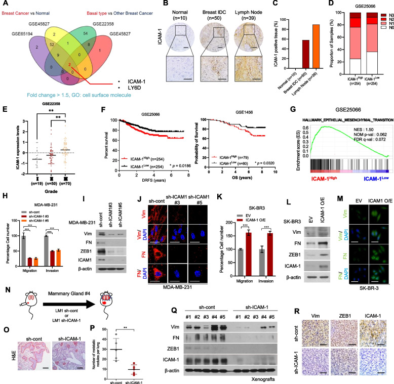
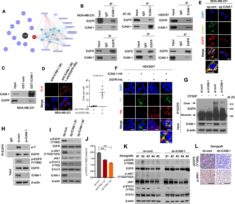
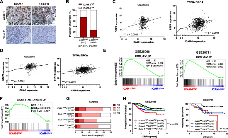
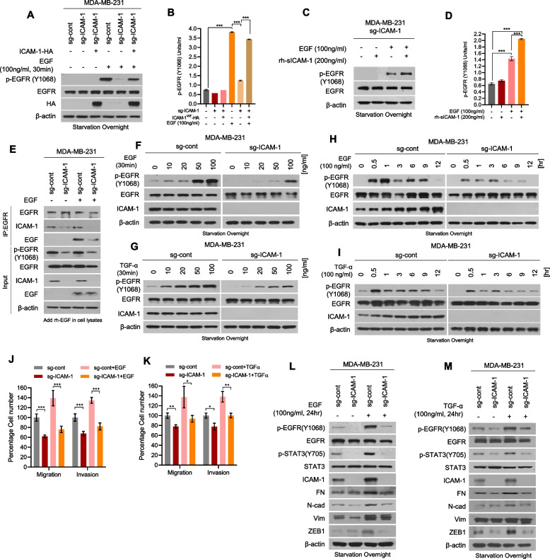
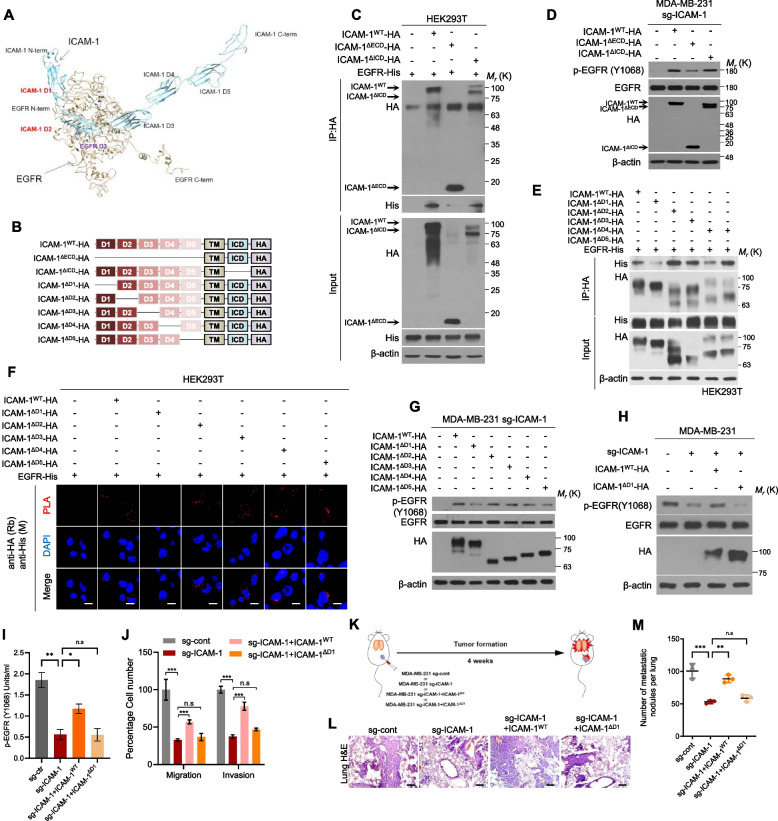
Triple-negative breast cancer (TNBC), the most aggressive subtype, presents a critical challenge due to the absence of approved targeted therapies. Hence, there is an urgent need to identify effective therapeutic targets for this condition. While epidermal growth factor receptor (EGFR) is prominently expressed in TNBC and recognized as a therapeutic target, anti-EGFR therapies have yet to gain approval for breast cancer treatment due to their associated side effects and limited efficacy. Here, we discovered that intercellular adhesion molecule-1 (ICAM-1) exhibits elevated expression levels in metastatic breast cancer and serves as a pivotal binding adaptor for EGFR activation, playing a crucial role in malignant progression. The activation of EGFR by tumor-expressed ICAM-1 initiates biased signaling within the JAK1/STAT3 pathway, consequently driving epithelial-to-mesenchymal transition and facilitating heightened metastasis without influencing tumor growth. Remarkably, ICAM-1-neutralizing antibody treatment significantly suppressed cancer metastasis in a breast cancer orthotopic xenograft mouse model. In conclusion, our identification of ICAM-1 as a novel tumor intrinsic regulator of EGFR activation offers valuable insights for the development of TNBC-specific anti-EGFR therapies.

Keywords: EGFR; ICAM-1; JAK1/STAT3 signaling; Targeted therapy; Triple-negative breast cancer.


Conflict of interest statement

The authors declare no competing interests.

Figures

Fig. 1
Fig. 1
Upregulation of ICAM-1 Correlates with Poor Prognosis and Metastasis of Breast Cancer. Expression of cell surface molecules and comparison of breast cancer tissue versus normal tissue and basal-type breast cancer patients versus other subtype breast cancer patients (GSE65194, GSE45827, GSE22358). Fold change > 1.5 (B) IHC of ICAM-1 in human breast cancer tissue array (BR1008). C IHC scoring of human breast cancer tissue array using IHC profiler. Scale bar = 200 μm. D Lymph node stage analysis using GEO dataset (GSE25066); ICAM-1High vs. ICAM-1Low. E ICAM-1 expression levels in three different grades of breast cancer patients (GSE22358). F Kaplan–Meier survival analysis of breast cancer patients (GSE25066, GSE1456); ICAM-1High vs. ICAM-1Low. G GSEA of hallmark epithelial-mesenchymal transition signature in human breast cancer patients (GSE25066); ICAM-1High vs. ICAM-1Low. NES, normalized enrichment score; Nom p-val, normalized p-value; FDR q-val, false discovery rate q-value. H Transwell migration and invasion assay of MDA-MB-231 cells transduced with ICAM-1 shRNA. I Western blots of EMT markers and regulators in MDA-MB-231 cells transduced with ICAM-1 shRNA. J ICC of vimentin (Vim) and fibronectin (FN) in MDA-MB-231 cells transduced with ICAM-1 shRNA. Scale bar = 20 μm. K Transwell migration and invasion assay of SK-BR3 transfected with ICAM-1 or control empty vector. L Western blots of EMT markers and regulators in SK-BR3 transfected with ICAM-1 or control empty vector. M ICC of vimentin (Vim) and fibronectin (FN) in SK-BR3 transfected with ICAM-1 or control empty vector. Scale bar = 20 μm. N Schematic illustration of animal experiments. O, P H&E staining (O) and the number of metastatic nodules (P) of lung in LM-1sh−cont or LM-1sh−ICAM−1 orthotopic xenograft mice. Scale bar = 200 μm. Q, R Western blots (Q) and IHC (R) of EMT markers, regulators, and ICAM-1 in LM-1sh−cont or LM-1.sh−ICAM−1 orthotopic xenograft tumors. Scale bar = 100 μm. Data are presented as mean ± SD and analyzed using one way ANOVA or Student’s t-tests. n.s, non-significant; *, p < 0.05; **, p < 0.01; ***, p < 0.001
Fig. 2
Fig. 2
ICAM-1 directly interacts with EGFR and promotes its activation in breast cancer. A Analysis of identified ICAM-1 interactors using Cytoscape. ICAM-1 interactors were detected using MS and cell surface molecules were selected. B Co-IP with ICAM-1 or EGFR antibody and western blot to evaluate the interaction between ICAM-1 and EGFR in basal-type breast cancer cell lines. C GST pull-down assay in MDA-MB-231 cells. D Representative images and quantification of in situ PLA showing the interaction between ICAM-1 and EGFR in MDA-MB-231 cells. Scale bar = 20 μm. E ICC of ICAM-1 and EGFR in MDA-MB-231 cells. Scale bar = 20 μm. F ICC of HA tag and His-tag in HEK293T cells transfected with either ICAM-1-HA or EGFR-His, or co-transfected with both. Scale bar = 20 μm. G Dimerization of EGFR in MDA-MB-231 cells transfected with siRNA targeting ICAM-1 or control siRNA. H Co-IP with EGFR antibody and western blotting to evaluate the total phospho tyrosine in MDA-MB-231 cells transfected with siRNA targeting ICAM-1 or control siRNA. I, J Western blots (I) and phospho EGFR ELISA (J) of MDA-MB-231 cells transfected with sgRNA targeting ICAM-1 or control as indicated. K, L Western blots (K) and IHC (L) of phospho EGFR, phospho JAK1, and phospho STAT3 in LM-1sh−cont or LM-1.sh−ICAM−1 orthotopic xenograft tumors. Scale bar = 100 μm. Data are presented as mean ± SD and analyzed using one-way ANOVA. n.s, non-significant; *, p < 0.05; **, p < 0.01; ***, p < 0.001
Fig. 3
Fig. 3
High levels of ICAM-1 expression correlate with EGFR expression and poor prognosis in patients with breast cancer. A IHC of ICAM-1 and p-EGFR in human breast cancer tissue array (BR2082b). Scale bar = 200 μm. B Contingency table for statistical analysis of IHC showing the relationship between ICAM-1 and p-EGFR in human breast cancer tissue array using IHC profiler. C Pearson correlation analysis of ICAM-1 and EGFR in patient cohorts from GEO dataset (GSE25066) and TCGA. D Pearson correlation analysis of ICAM-1 and STAT3 in patient cohorts from GEO dataset (GSE25066) and TCGA. E GSEA plots for a significant correlation between high expression of ICAM-1 and EGFR signaling (GSE25066, GSE20711). F GSEA plots for a significant correlation between high expression of ICAM-1 and STAT3 signaling (GSE25066). G Lymph node stage analysis using GEO dataset (GSE25066); ICAM-1High EGFRHigh vs. ICAM-1High EGFRLow vs. ICAM-1Low EGFRHigh vs. ICAM-1Low EGFRLow. H Kaplan–Meier survival analysis of patients with breast cancer (GSE25066, GSE20711); ICAM-1High EGFRHigh vs. ICAM-1High EGFRLow vs. ICAM-1Low EGFRHigh vs. ICAM-1Low EGFR.Low. Data are presented as mean ± SD and analyzed using one-way ANOVA. n.s, non-significant; *, p < 0.05; **, p < 0.01; ***, p < 0.001
Fig. 4
Fig. 4
ICAM-1 increases ligand-binding affinity for EGFR and promotes metastasis of breast cancer. A, B Western blots (A), and phospho EGFR ELISA (B) of MDA-MB-231 sg-cont and MDA-MB-231 sg-ICAM-1 cells transfected with either ICAM-1 or control empty vector. Cells were starved overnight and treated with EGF or PBS. C, D Western blots (C), and phospho EGFR ELISA (D) of MDA-MB-231 sg-ICAM-1 cells. Cells were starved overnight and treated with either rh-sICAM-1 or EGF, or co-treated. E Co-IP with EGFR antibody and western blotting to evaluate the interaction between EGF and EGFR in MDA-MB-231 sg-cont and MDA-MB-231 sg-ICAM-1 cells. rh-EGF was added in cell lysates. F, G Western blots of EGFR phosphorylation in MDA-MB-231 sg-cont and MDA-MB-231 sg-ICAM-1 cells. Cells were starved overnight and treated with increasing doses of EGF (F) or TGF-α (G). H, I Western blots of EGFR phosphorylation in MDA-MB-231 sg-cont and MDA-MB-231 sg-ICAM-1 cells. Cells were starved overnight and treated with EGF (H) or TGF-α (I) for various time intervalsin a time-dependent manner. J, K Migration and invasion by MDA-MB-231 sg-cont and MDA-MB-231 sg-ICAM-1 cells. Cells were starved overnight and treated with EGF (J) or TGF-α (K) for 24 h. L, M Western blots of EMT markers and regulators in MDA-MB-231 sg-cont and MDA-MB-231 sg-ICAM-1 cells. Cells were starved overnight and treated with EGF (L) or TGF-α (M) for 24 h. Data are presented as mean ± SD and analyzed using one-way ANOVA or Student’s t-tests. n.s, non-significant; *, p < 0.05; **, p < 0.01; ***, p < 0.001
Fig. 5
Fig. 5
The ICAM-1 D1 domain is necessary for EGFR activation and its downstream signaling. A Protein–protein docking simulation to evaluate the interaction domain between ICAM-1 and EGFR. B Schematic diagram of expression constructs encoding ICAM-1WT, ICAM-1ΔECD, ICAM-1ΔICD, ICAM-1ΔD1, ICAM-1ΔD2, ICAM-1ΔD3, ICAM-1ΔD4, and ICAM-1ΔD5C Co-IP with HA antibody and western blot to evaluate the interaction domain between ICAM-1 and EGFR in HEK293T cells co-transfected with EGFR and ICAM-1WT, ICAM-1ΔECD, or ICAM-1ΔICD vector. D Western blots of EGFR phosphorylation in MDA-MB-231 sg-ICAM-1 cells transfected with ICAM-1WT, or ICAM-1ΔECD, or ICAM-1ΔICD, or control empty vector. E, F Co-IP (E) and in situ PLA (F) to evaluate the interaction domain between ICAM-1 and EGFR in HEK293T cells co-transfected with EGFR and either ICAM-1WT, ICAM-1ΔD1, ICAM-1ΔD2, ICAM-1ΔD3, ICAM-1ΔD4, or ICAM-1ΔD5. Scale bar = 20 μm. G Western blots of EGFR phosphorylation in MDA-MB-231 sg-ICAM-1 cells transfected with either control empty vector or ICAM-1WT, ICAM-1ΔD1, ICAM-1ΔD2, ICAM-1ΔD3, ICAM-1ΔD4, or ICAM-1ΔD5. H-J Western blotting (H), phospho EGFR ELISA (i), migration and invasion assay (J) in MDA-MB-231 sg-ICAM-1 cells transfected with either control empty vector, ICAM-1WT, or ICAM-1ΔD1. K Schematic illustration of animal experiments. L, M H&E staining (L) and the number of metastatic nodules (M) of lung in MDA-MB-231 sg-cont or MDA-MB-231 sg-ICAM-1 transfected with control empty vector, ICAM-1WT, or ICAM-1.ΔD1 orthotopic xenograft mice. Scale bar = 200 μm. Data are presented as mean ± SD and analyzed using one-way ANOVA. n.s, non-significant; *, p < 0.05; **, p < 0.01; ***, p < 0.001
Fig. 6
Fig. 6
Blockade of ICAM-1 using a neutralizing antibody can suppress breast cancer metastasis. A, B Western blotting (A) and phospho EGFR ELISA (B) of MDA-MB-231 cells treated with various doses of ICAM-1 neutralizing antibody for 24 h. C, D Transwell migration, invasion assay (C) and (D) western blotting of MDA-MB-231 cells treated with ICAM-1 neutralizing antibody (500 ng/ml, 24 h). E Co-IP with EGFR antibody and western blotting to evaluate the interaction between ICAM-1 and EGFR in MDA-MB-231 cells treated with ICAM-1 neutralizing antibody. F Co-IP with EGFR antibody and western blotting to evaluate the interaction between EGFR and EGF in MDA-MB-231 cells treated with ICAM-1 neutralizing antibody. rh-EGF was added in cell lysates. G Schematic illustration of animal experiments. H, I H&E staining (H) and the number of metastatic nodules (I) of lung in MDA-MB-231 orthotopic xenograft mice treated with ICAM-1 neutralizing antibody. Scale bar = 200 μm. J-M Phospho EGFR ELISA (J), IHC (K), western blotting (L), and in situ PLA (M) of in MDA-MB-231 orthotopic xenograft tumors mice treated with ICAM-1 neutralizing antibody. Scale bar = 100 μm. N Schematic illustration of the mechanism underlying ICAM-1-mediated EGFR activation, leading to cancer metastasis in breast cancer. Highlighted arrows indicate the pathways suggested by our study. Data are presented as mean ± SD and analyzed using one-way ANOVA or Student’s t-tests. n.s, non-significant; *, p < 0.05; **, p < 0.01; ***, p < 0.001

References

    1. Siegel RL, Miller KD, Fuchs HE, Jemal A. Cancer Statistics, 2021. CA Cancer J Clin. 2021;71:7–33. - PubMed
    1. Fahad Ullah M. Breast Cancer: Current Perspectives on the Disease Status. Adv Exp Med Biol. 2019;1152:51–64. - PubMed
    1. Scully OJ, Bay BH, Yip G, Yu Y. Breast cancer metastasis. Cancer Genomics Proteomics. 2012;9:311–20. - PubMed
    1. Kozłowski J, Kozłowska A, Kocki J. Breast cancer metastasis - insight into selected molecular mechanisms of the phenomenon. Postepy Hig Med Dosw (Online). 2015;69:447–51. - PubMed
    1. Fares J, Fares MY, Khachfe HH, Salhab HA, Fares Y. Molecular principles of metastasis: a hallmark of cancer revisited. Signal Transduct Target Ther. 2020;5:28. - PMC - PubMed

MeSH terms

LinkOut - more resources