Review
. 2024 Oct 16;17(1):31.
doi: 10.1186/s13072-024-00554-6.

Epigenetic frontiers: miRNAs, long non-coding RNAs and nanomaterials are pioneering to cancer therapy

Affiliations
Review

Epigenetic frontiers: miRNAs, long non-coding RNAs and nanomaterials are pioneering to cancer therapy

Rajkumar Prabhakaran et al. Epigenetics Chromatin. .

Abstract

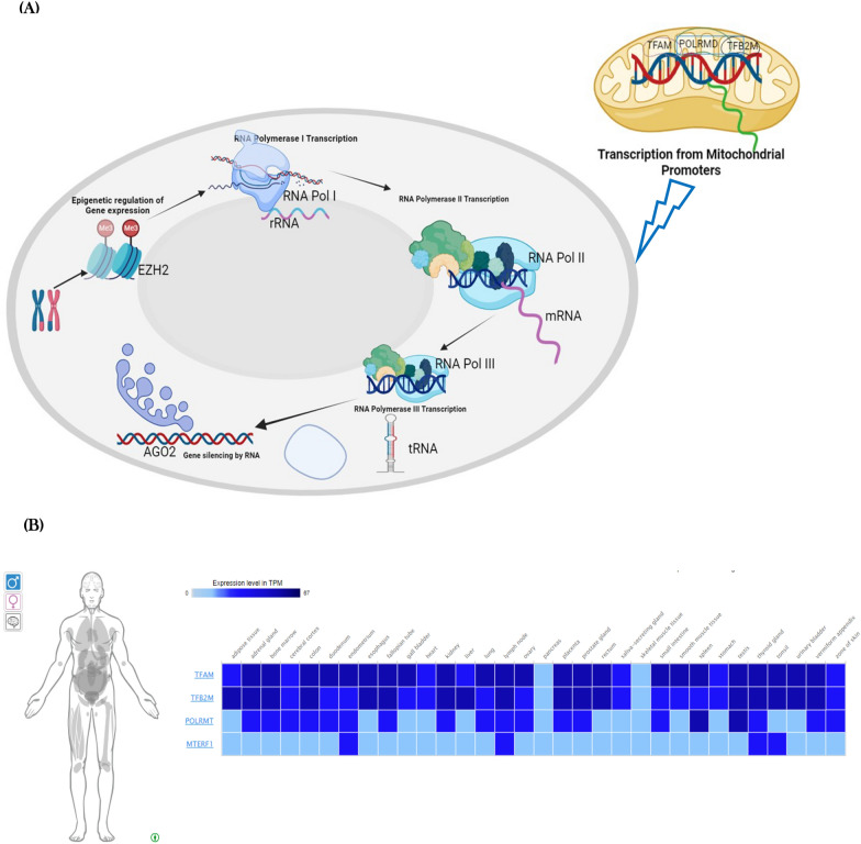
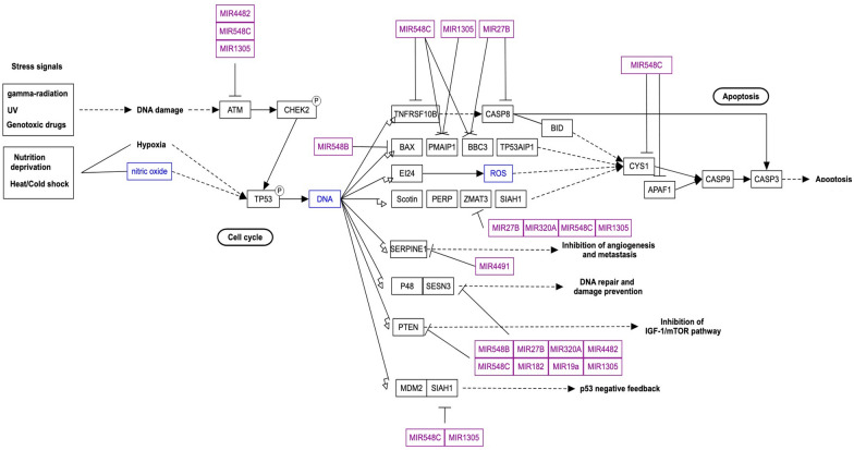
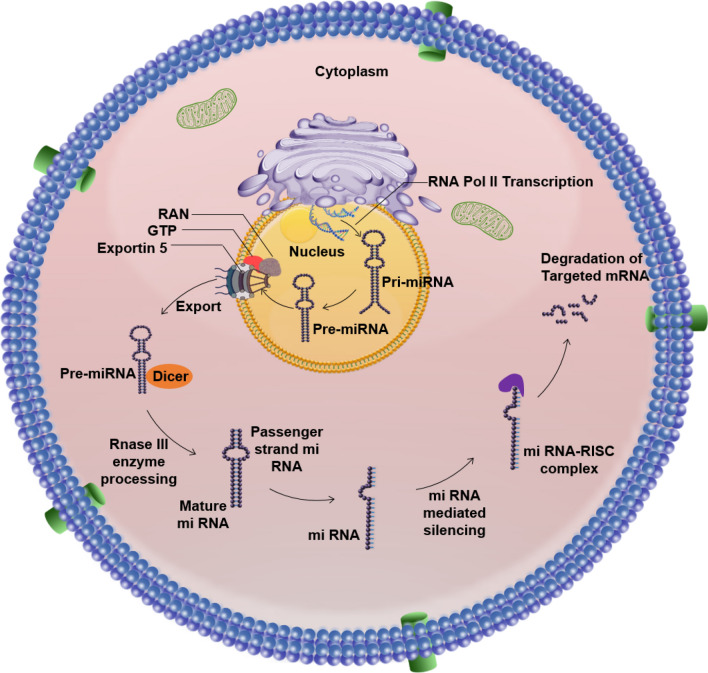
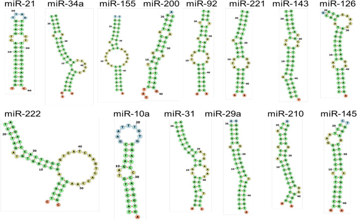
Cancer has arisen from both genetic mutations and epigenetic changes, making epigenetics a crucial area of research for innovative cancer prevention and treatment strategies. This dual perspective has propelled epigenetics into the forefront of cancer research. This review highlights the important roles of DNA methylation, histone modifications and non-coding RNAs (ncRNAs), particularly microRNAs (miRNAs) and long non-coding RNAs, which are key regulators of cancer-related gene expression. It explores the potential of epigenetic-based therapies to revolutionize patient outcomes by selectively modulating specific epigenetic markers involved in tumorigenesis. The review examines promising epigenetic biomarkers for early cancer detection and prognosis. It also highlights recent progress in oligonucleotide-based therapies, including antisense oligonucleotides (ASOs) and antimiRs, to precisely modulate epigenetic processes. Furthermore, the concept of epigenetic editing is discussed, providing insight into the future role of precision medicine for cancer patients. The integration of nanomedicine into cancer therapy has been explored and offers innovative approaches to improve therapeutic efficacy. This comprehensive review of recent advances in epigenetic-based cancer therapy seeks to advance the field of precision oncology, ultimately culminating in improved patient outcomes in the fight against cancer.

Keywords: Cancer; DNA methylation; Histone modifications; Nanomedicine; Non-coding RNAs epigenetic therapy.


Conflict of interest statement

The authors declare no competing interests.

Figures

Fig. 1
Fig. 1
A Schematic diagram illustrating the pathway and expression of miRNA21. This figure shows the interaction of miRNA21 within nucleosomes and its role in the regulation of transcription from mitochondrial promoters. Key components include miRNA21, nucleosome complexes, and mitochondrial transcription factors. The pathways and interactions are detailed to illustrate how miRNA21 influences gene expression at the chromatin level. B Schematic diagram showing the expression of miRNA21 across different tissues. The figure integrates RNA-seq data from 122 human individuals representing 32 different tissues, highlighting the differential expression of miRNA21. The diagram includes details on how miRNA21 expression is mapped across various tissue types, with annotations indicating specific tissues and expression levels. For further details, refer to the RNA-seq dataset available at (https://reactome.org/PathwayBrowser/#/R-HSA-74160&SEL=R-HSA-75944&DTAB=EX) [49]
Fig. 2
Fig. 2
Diagram illustrating the regulation of the p53 pathway by miRNAs in prostate cancer (WP3982). The figure depicts how elevated levels of specific miRNAs affect the p53 signaling pathway in prostate cancer cells. Key elements shown include the targeted genes within the p53 pathway, the interactions between miRNAs and these genes, and the overall impact on pathway activity. For a detailed view of the pathway, refer to (https://www.wikipathways.org/pathways/WP3982.html)
Fig. 3
Fig. 3
The schematic representation reveals the biogenesis of microRNAs (miRNAs). The biogenesis of microRNAs (miRNAs) unfolds through five key stages: Transcription: miRNA precursors originate from autonomously transcribed genes, co-transcripts with other genes, or introns of host genes. RNA polymerase II transcribes most miRNAs, though some come from RNA polymerase III co-transcripts with adjacent repetitive elements. The initial transcript, known as primary microRNA (pri-miRNA), includes an imperfectly double-stranded region within a hairpin loop, with longer sequences extending from both the 5′ and 3′ ends. Cleavage by DROSHA: The DROSHA nuclease, in association with the RNA-binding protein DGCR8 (forming the Microprocessor complex), endoribonucleolytically cleaves the 5′ and 3′ ends of the pri-miRNA. This cleavage produces a short hairpin structure, about 60 to 70 nucleotides long, called pre-microRNA (pre-miRNA). Nuclear Export by Exportin-5: The pre-miRNA associates with Exportin-5, Ran, and GTP to be transported through the nuclear pore into the cytoplasm. Cleavage by DICER1: In the cytoplasm, the pre-miRNA is processed by the RISC loading complex, which includes DICER1, an Argonaute protein, and either TARBP2 or PRKRA. DICER1 cleaves the pre-miRNA, resulting in a double-stranded miRNA approximately 21 to 23 nucleotides in length, with protruding single-stranded 3′ ends of 2–3 nucleotides. Incorporation into RNA-Induced Silencing Complex (RISC) and Strand Selection: The double-stranded miRNA is incorporated into an Argonaute protein within the RISC loading complex. The passenger strand is removed and degraded, while the guide strand is retained, directing the Argonaute complex (RISC) to target mRNAs
Fig. 4
Fig. 4
The secondary structure of microRNAs is visualized using a Force Directed Graph Layout. This figure presents the secondary structure of specific miRNAs (miR-21, miR-34a, miR-155, miR-200, miR-92, miR-221, miR-143, miR-126, miR-222, miR-10a, miR-31, miR-29a, miR-210, and miR145) as visualized through interactive graph layout software. The diagram displays the folding pattern of the miRNA molecule, highlighting key structural features such as hairpins, loops, and stems. These structural elements are crucial for miRNA function and its interaction with target mRNAs (http://mirwalk.umm.uni-heidelberg.de/human/mirna/MIMAT0000437/)
Fig. 5
Fig. 5
Provides a schematic representation of epigenetic biomarkers used for cancer diagnosis and prognosis. Key examples include DNA methylation, where hypermethylation of tumor suppressor genes like p16INK4a and hypomethylation of oncogenes such as MYC can signal various cancers, including melanoma and colon cancer. Histone modifications also play a crucial role; for instance, increased acetylation of histone H3 lysine 27 (H3K27ac) is linked to active gene expression in prostate cancer, while specific methylation marks like trimethylation of histone H3 lysine 4 (H3K4me3) are associated with active promoters in leukemias. Non-coding RNAs, including miR-21, are associated with poor prognosis in breast cancer and lung cancer, and HOTAIR lncRNA overexpression is linked to metastasis in breast cancer and colorectal cancer. Chromatin remodeling factors such as mutations in BRG1 (part of the SWI/SNF complex) are found in small-cell lung cancer and endometrial cancer, affecting gene expression. Additionally, genomic imprinting anomalies, such as overexpression of the IGF2 gene, are seen in Wilms’ tumor and can influence cancer progression. These biomarkers are pivotal in advancing cancer diagnosis, predicting disease outcomes, and guiding personalized treatments
Fig. 6
Fig. 6
Schematic representation of various types of cancer therapies and their mechanisms of action. This figure categorizes and illustrates different cancer treatment modalities including chemotherapy, radiotherapy, targeted therapy, immunotherapy, and hormone therapy. Each therapy type is depicted with its specific mechanism of action, such as direct cytotoxic effects, DNA damage induction, targeted inhibition of cancer-specific pathways, immune system activation, and hormone receptor modulation. The diagram aims to provide a comprehensive overview of how each therapy targets cancer cells and the underlying biological mechanisms involved

References

    1. Cusenza VY, et al. The lncRNA epigenetics: the significance of m6A and m5C lncRNA modifications in cancer. Front Oncol. 2023;13:1063636. 10.3389/fonc.2023.1063636. - PMC - PubMed
    1. Nencioni A, et al. Fasting and cancer: molecular mechanisms and clinical application. Nat Rev Cancer. 2018;18(11):707–19. 10.1038/s41568-018-0061-0. - PMC - PubMed
    1. Rupaimoole R, Slack FJ. MicroRNA therapeutics: towards a new era for the management of cancer and other diseases. Nat Rev Drug Discov. 2017;16(3):203–22. 10.1038/nrd.2016.246. - PubMed
    1. Zhang J, et al. Emerging epigenetic-based nanotechnology for cancer therapy: modulating the tumor microenvironment. Adv Sci. 2023;10(7): e2206169. 10.1002/advs.202206169. - PMC - PubMed
    1. Janin M, Davalos V, Esteller M. Cancer metastasis under the magnifying glass of epigenetics and epitranscriptomics. Cancer Metastasis Rev. 2023. 10.1007/s10555-023-10120-3. - PMC - PubMed

LinkOut - more resources