. 2024 Oct 15;5(11):e686.
doi: 10.1002/mco2.686. eCollection 2024 Nov.

Vitamin B12 protects necrosis of acinar cells in pancreatic tissues with acute pancreatitis

Affiliations

Vitamin B12 protects necrosis of acinar cells in pancreatic tissues with acute pancreatitis

Yulin Chen et al. MedComm (2020). .

Abstract

Pharmacological agents regarding the most optimal treatments of acute pancreatitis remain. One-carbon metabolism nutrients as therapeutic agents in many diseases might be involved in acute pancreatitis. The roles are acquired exploration in acute pancreatitis. We utilized Mendelian randomization to assess the causal impact of folate, homocysteine, and vitamin B12 (VB12) on acute pancreatitis. Wild-type and corresponding genetically modified mouse models were used to verify the genetic correlating findings. A negative association between genetically predicted serum VB12 levels and risks of acute pancreatitis was identified in human population. The transcobalamin receptor (TCblR)/CD320 gene ablation that decreased cellular VB12 uptake and ATP production in pancreatic tissues promoted necrosis, resulting in much severe pathological changes of induced acute pancreatitis in mice. VB12 pretreatment and posttreatment dramatically increased ATP levels in pancreatic tissues and reduced the necrosis, then the elevated levels of amylase in serum, the levels of CK-19, the activity of trypsin, and T lymphocyte infiltration in pancreatic tissues, prevented the pancreatic gross loss and ameliorated histopathological changes of mouse pancreases with induced acute pancreatitis. The results reveal that VB12 is potential as a therapeutic agent to inhibit tissue injuries and adaptive inflammatory responses in the pancreas in patients with acute pancreatitis.

Keywords: CD320‐ablation mouse; Mendelian randomization (MR); acute pancreatitis; vitamin B12.


Conflict of interest statement

The authors declare they have no conflicts of interest.

Figures

FIGURE 1
FIGURE 1
Mendelian randomization (MR) estimates for the association between folate, homocysteine, and vitamin B12 (VB12) with acute pancreatitis. Odds ratio (OR) was estimated using the random‐effects inverse variance weighted method. CI, confidence interval; SNPs, single‐nucleotide polymorphisms.
FIGURE 2
FIGURE 2
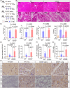
Acute pancreatitis induced by L‐Arg was aggravated in CD320‐ablation mice. (A) The flow charts of experiment. (B) The representative histological images of the pancreas were obtained 72 h after the final administration of L‐Arg (scale bar, 50 µm). (C) The histological evaluation of pancreatic edema, inflammatory infiltration, necrosis, and the overall sum of these features. (D) Serum amylase and lipase levels were measured at 72 h after the final administration of L‐Arg. (E) The representative immunohistochemical images for CK‐19 and CD3+ T lymphocyte cell of mouse pancreatic tissues at 72 h after the final administration of L‐Arg (scale bar, 50 µm). (F and G). Quantification of immunohistochemical analysis for CK‐19 and CD3+ T lymphocyte cell of mouse pancreatic tissues at 72 h after the final administration of L‐Arg, respectively. The data were obtained from 5−10 mice per group and presented as mean ± standard deviation (SD) from individual mice with one‐way analysis of variance (ANOVA) and Tukey's multiple‐comparison posttest.
FIGURE 3
FIGURE 3
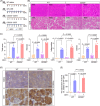
Acute pancreatitis induced by cerulein (CER) was aggravated in CD320‐ablation mice. (A) The flow charts of experiment. (B) The representative histological images of the pancreas were obtained 12 h after the first administration of CER (scale bar, 50 µm). (C) The histological evaluation of pancreatic edema, inflammatory infiltration, necrosis, and the overall sum of these features. (D) The representative immunohistochemical images for CK‐19 of mouse pancreatic tissues at 12 h after the first administration of CER (scale bar, 50 µm). (E) Quantification of immunohistochemical analysis for CK‐19 of mouse pancreatic tissues at 12 h after the first administration of CER. The data were obtained from five mice per group and presented as mean ± standard deviation (SD) from individual mice with one‐way analysis of variance (ANOVA) and Tukey's multiple‐comparison posttest.
FIGURE 4
FIGURE 4
Vitamin B12 (VB12) exhibits therapeutic effects on experimental acute pancreatitis induced by cerulein (CER). (A) The flow charts of experiment. (B) The representative histological and immunohistochemical images of the pancreas for CK‐19 were obtained 12 h after the first administration of CER (scale bar, 50 µm). (C) The histological evaluation of pancreatic edema, inflammatory infiltration, necrosis, and the overall sum of these features. (D) Serum amylase levels were measured at 12 h after the first administration of CER. (E). Quantification of immunohistochemical analysis for CK‐19 of mouse pancreatic tissues at 12 h after the first administration of CER. (F) Representative of trypsin activity in pancreatic tissues at 12 h after the first administration of CER. The data were obtained from five to six mice per group and presented as mean ± standard deviation (SD) from individual mice with one‐way analysis of variance (ANOVA) and Tukey's multiple‐comparison posttest.
FIGURE 5
FIGURE 5
Vitamin B12 (VB12) exhibits therapeutic effects on experimental acute pancreatitis induced by L‐Arg. (A) The flow charts of experiment. (B) The representative histological images of pancreas were obtained 72 h after the final administration of L‐Arg (scale bar, 50 µm). (C) The histological evaluation of pancreatic edema, inflammatory infiltration, necrosis, and the overall sum of these features. (D) Serum amylase and lipase levels were measured at 72 h after the final administration of L‐Arg. (E) Representative of trypsin activity in pancreatic tissues at 72 h after the final administration of L‐Arg. (F) The representative immunohistochemical images for CK‐19 and CD3+ T lymphocyte cell of mouse pancreatic tissues at 72 h after the final administration of L‐Arg (scale bar, 50 µm). (G and H) Quantification of immunohistochemical analysis for CK‐19 and CD3+ T lymphocyte cell of mouse pancreatic tissues at 72 h after the final administration of L‐Arg, respectively. The data were obtained from 5−10 mice per group and presented as mean ± standard deviation (SD) from individual mice with one‐way analysis of variance (ANOVA) and Tukey's multiple‐comparison posttest.
FIGURE 6
FIGURE 6
Vitamin B12 (VB12) displays preventive effects on acute pancreatitis induced by L‐Arg in mouse models. (A) The flow charts of experiment. (B) The representative histological images of pancreas were obtained 72 h after the final administration of L‐Arg (scale bar, 50 µm). (C) The histological evaluation of pancreatic edema, inflammatory infiltration, necrosis, and the overall sum of these features. (D) The levels of serum amylase and lipase were measured at 72 h after the final administration of L‐Arg. (E) The representative immunohistochemical images for CK‐19 and CD3+ T lymphocyte cell of mouse pancreatic tissues at 72 h after the final administration of L‐Arg (scale bar, 50 µm). (F and G) Quantification of immunohistochemical analysis for CK‐19 and CD3+ T lymphocyte cell of mouse pancreatic tissues at 72 h after the final administration of L‐Arg, respectively. The data were obtained from 5−10 mice per group and presented as mean ± standard deviation (SD) from individual mice with one‐way analysis of variance (ANOVA) and Tukey's multiple‐comparison posttest.
FIGURE 7
FIGURE 7
Vitamin B12 (VB12) protects the acinar cells via ATP production in pancreatic tissues in CD320‐ablation mice. (A) The flow charts of experiment. (B) The representative histological and immunohistochemical images of the pancreas for CK‐19 were obtained 12 h after the first administration of cerulein (CER) (scale bar, 50 µm). (C) The histological evaluation of pancreatic edema, inflammatory infiltration, necrosis, and the overall sum of these features. (D) The levels of serum amylase and lipase were measured at 12 h after the first administration of CER. (E) Representative of trypsin activity in pancreatic tissues at 12 h after the first administration of CER. (F) Quantification of immunohistochemical analysis for CK‐19 of mouse pancreatic tissues at 12 h after the first administration of CER. (G) Representative of activity of MPO in pancreatic tissue at 12 h after the first administration of CER. The data were obtained from 5−10 mice per group and presented as mean ± standard deviation (SD) from individual mice with one‐way analysis of variance (ANOVA) and Tukey's multiple‐comparison posttest.

References

    1. Garg PK, Singh VP. Organ failure due to systemic injury in acute pancreatitis. Gastroenterology. 2019;156(7):2008‐2023. - PMC - PubMed
    1. Boxhoorn L, Voermans RP, Bouwense SA, et al. Acute pancreatitis. Lancet. 2020;396(10252):726‐734. - PubMed
    1. Chua TY, Walsh RM, Baker ME, Stevens T. Necrotizing pancreatitis: diagnose. Clin J Med. 2017;84(8):639‐648. - PubMed
    1. Hines OJ, Pandol SJ. Management of severe acute pancreatitis. BMJ. 2019;367:l6227. - PubMed
    1. Vaccaro JA, Qasem A, Naser SA. Folate and vitamin B12 deficiency exacerbate inflammation during Mycobacterium avium paratuberculosis (MAP) infection. Nutrients. 2023;15(2):261. - PMC - PubMed