. 2024 Oct 4;5(5):1014-1020.
doi: 10.1002/jha2.1018. eCollection 2024 Oct.

The genomic landscape of transformed splenic diffuse red pulp small B-cell lymphoma

Affiliations

The genomic landscape of transformed splenic diffuse red pulp small B-cell lymphoma

Marta Grau et al. EJHaem. .

Abstract

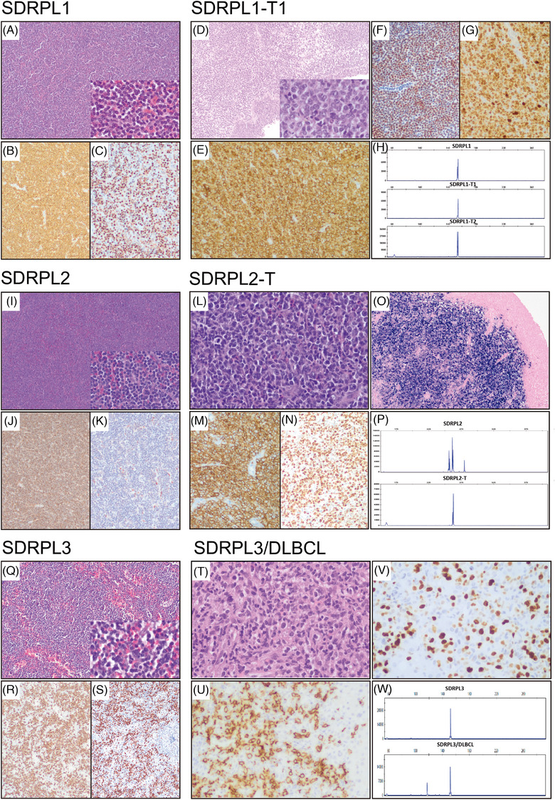
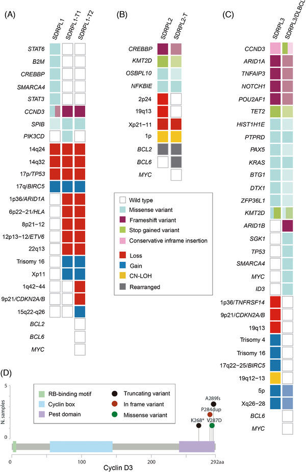
The genetic landscape underlying the transformation of splenic diffuse red pulp small B-cell lymphoma (SDRPL) is not well understood. The present study aimed to unravel the genomic alterations involved in the progression and transformation of SDRPL. We performed genetic studies on both SDRPL and subsequent or synchronous diffuse large B cell lymphoma (DLBCL) samples in three SDRPL patients who eventually developed DLBCL. Our findings revealed that SDRPL cases progressing to DLBCL acquired genomic alterations in genes related to the cell cycle (CDKN2A/B, TP53, MYC and CCND3) and B cell development (BCL6).

Keywords: B‐cell developement; cell cycle; clonal relationship; genetic alterations; histologic transformation; splenic diffuse red pulp small B‐cell lymphoma.


Conflict of interest statement

Elias Campo has been a consultant for GenMab, and Takeda; has received research support from AstraZeneca; received honoraria from Janssen, EUSPharma, Takeda and Roche for speaking at educational activities; and is an inventor on a Lymphoma and Leukemia Molecular Profiling Project patent ‘Method for subtyping lymphoma subtypes by means of expression profiling’ (PCT/US2014/64161) and a bioinformatic tool (IgCaller) licenced to Diagnostic Longwood. Armando López‐Guillermo. served on the advisory board of Roche, Celgene, Novartis and Gilead/Kite, and received grants from Celgene and Gilead/Kite. Ferran Nadeu has received honoraria from Janssen, AbbVie, AstraZeneca and SOPHiA GENETICS for speaking at educational activities; has received research support from Gilead; and has licensed the use of the protected IgCaller algorithm to Diagnóstica Longwood. The remaining authors declare no competing financial interests.

Figures

FIGURE 1
FIGURE 1
Morphologic and immunohistochemical features of splenic diffuse red pulp small B‐cell lymphoma (SDRPL) and transformed splenic diffuse red pulp lymphoma (SDRPL‐T). Histology of SDRPL1 (A–C): The panels show a proliferation of small lymphocytes diffusely infiltrating the red pulp of the spleen. They have a monomorphic appearance with round nuclei, clumped chromatin, and scant cytoplasm (A, hematoxylin and eosin (H&E) 10x; inset, 40x). On immunohistochemical analysis, they show positivity for CD20 (B, 10x), cyclin D3 (C, 20x), IgG and aberrant coexpression of CD2, and negativity for CD5, CD23, CD43, IgD, IgM, cyclin D1 and annexin A1. By flow cytometry, the cells are CD11c, FMC7 and kappa positive and negative for CD10, CD25 and CD103. Histology of SDRPL1‐T1 (D–G): There is a neoplastic proliferation of large cells invading the bladder muscular wall. The large cells have scant cytoplasm, irregular nuclei with coarse chromatin and nucleoli on higher power (D, H&E, 10x; inset, 40x). The cells express CD20 (E, 20x), BCL6, MUM1, MYC, cyclin D3 (F, 20x), IgG and aberrant CD2 and CD5. The Ki67 proliferation index is 95% (G, 20x). The cells are negative for CD10 and CD23. (H) IGH gene rearrangement studies in SDRPL1 (upper part) and SDRPL1‐T1 (lower part) showed same clonal peak in FR2. Histology of SDRPL2 (I–K): Histologic findings are similar to case 1 (I, H&E, 10x; inset, 40x). The cells are CD20 positive (J, 10x) with coexpression of IgG and DBA44, being negative for cyclin D1, annexin A1, cyclin D3 (K, 20x), CD25, LMO2, and BCL6. On flow cytometry the cells are positive for CD103, FMC7 and kappa, and negative for CD43, CD23, IgM, CD10 and CD5. Histology of SDRPL2‐T (L–O): Ileum with extensive ulceration and transmural infiltration by large, atypical cells (L, H&E, 40x). Mitotic figures are present. The large cells are positive for CD20 (M, 20x), BCL6 and MUM1, and negative for cyclin D1, IgD, CD10, CD5 and CD23. The Ki67 proliferation index is 90% (N, 20x). in situ hybridization for Epstein–Barr virus is positive (O, 10x). (P) IGH gene rearrangement studies in SDRPL2 (upper part) and SDRPL2‐T (lower part) showed the same clonal peak in FR2 displaying IGHJ4*02–IGHD1‐14*01–IGHV1‐18*04 rearrangement in both samples. Histology of SDRPL3 (Q–S): The splenic histology is similar to cases 1 and 2 (Q, H&E, 10x; inset, 40x). On immunohistochemistry the cells are CD20 (R, 10x), BCL2, IgD, IgM and cyclin D3 (S, 10x) positive and are negative for cyclin D1, CD23, CD10 and CD5. On flow cytometry, cells are FMC7 and Lambda positive and CD5, CD23, CD43 and CD103 negative. Histology of SDRPL3/DLBCL (T–V): Liver biopsy with a patchy lymphoid infiltration composed predominantly of large, atypical cells and some small lymphocytes (T, H&E, 40x). The neoplastic cells are CD20 (U, 40x), BCL2 and IgD positive, being negative for CD5 and CD10. Ki67 immunostaining shows that most of the large cells are proliferating in the context of an inflammatory background (V, 40x). (W) IGH gene rearrangement studies in SDRPL3 (upper part) and SDRPL3/DLBCL (lower part) showing the same clonal peak in FR3 shared between both samples, corresponding to IGHJ4*02‐IGHD3‐3*01 ‐ IGHV3‐21*01 rearrangements and an additional peak in SDRPL3/DLBCL showing IGHJ4*02 ‐ IGHD4‐23*01 ‐ IGHV3‐11*04 rearrangement.
FIGURE 2
FIGURE 2
Genetic landscape of splenic diffuse red pulp small B‐cell lymphoma (SDRPL) and transformed splenic diffuse red pulp lymphoma (SDRPL‐T). Oncoprint displaying the genomic alterations: copy number alterations (CNA), copy neutral loss of heterozygosity (CN‐LOH), single nucleotide variants (SNVs) and small insertions and deletions (indels) found in paired SDRPL and SDRPL‐T samples of case 1(A), case 2(B) and case 3(C). SNVs and indels are depicted in the upper part, and CNA and CN‐LOH in the lower part. Each column represents an individual sample. Rows correspond to individual genetic alterations. The decreasing intensity colour squares represent lower variant allele frequency in comparison to other samples of the same patient. Some genetic data of SDRPL2 and SDRPL3 have been previously published [3]. (D) Distribution of CCND3 SNVs and indels across the cyclin D3 protein sequence found in cases 1 and 3. Different colours indicate different types of variants scattered within the functional domains of cyclin D3 protein.aa, amino acids.

References

    1. Campo E, Jaffe ES, Cook JR, Quintanilla‐Martinez L, Swerdlow SH, Anderson KC, et al. The international consensus classification of mature lymphoid neoplasms: a report from the Clinical Advisory Committee. Blood. 2022;140(11):1229–1253. - PMC - PubMed
    1. Alaggio R, Amador C, Anagnostopoulos I, Attygalle AD, Araujo IBDeO, Berti E, et al. The 5th edition of the World Health Organization classification of haematolymphoid tumours: lymphoid neoplasms. Leukemia. 2022;36(7):1720–1748. - PMC - PubMed
    1. Martinez D, Navarro A, Martinez‐Trillos A, Molina‐Urra R, Gonzalez‐Farre B, Salaverria I, et al. NOTCH1, TP53, and MAP2K1 mutations in splenic diffuse red pulp small B‐cell lymphoma are associated with progressive disease. Am J Surg Pathol. 2016;40(2):192–201. - PubMed
    1. Jallades L, Baseggio L, Sujobert P, Huet S, Chabane K, Callet‐Bauchu E, et al. Exome sequencing identifies recurrent BCOR alterations and the absence of KLF2, TNFAIP3 and MYD88 mutations in splenic diffuse red pulp small B‐cell lymphoma. Haematologica. 2017;102(10):1758–1766. - PMC - PubMed
    1. Curiel‐Olmo S, Mondéjar R, Almaraz C, Mollejo M, Cereceda L, Marès R, et al. Splenic diffuse red pulp small B‐cell lymphoma displays increased expression of cyclin D3 and recurrent CCND3 mutations. Blood. 2017;129(8):1042–1045. - PubMed

LinkOut - more resources