[Preprint]. 2024 Oct 31:2024.10.09.617269.
doi: 10.1101/2024.10.09.617269.

Danicamtiv reduces myosin's working stroke but enhances contraction by activating the thin filament

Affiliations

Danicamtiv reduces myosin's working stroke but enhances contraction by activating the thin filament

Brent Scott et al. bioRxiv. .

Abstract

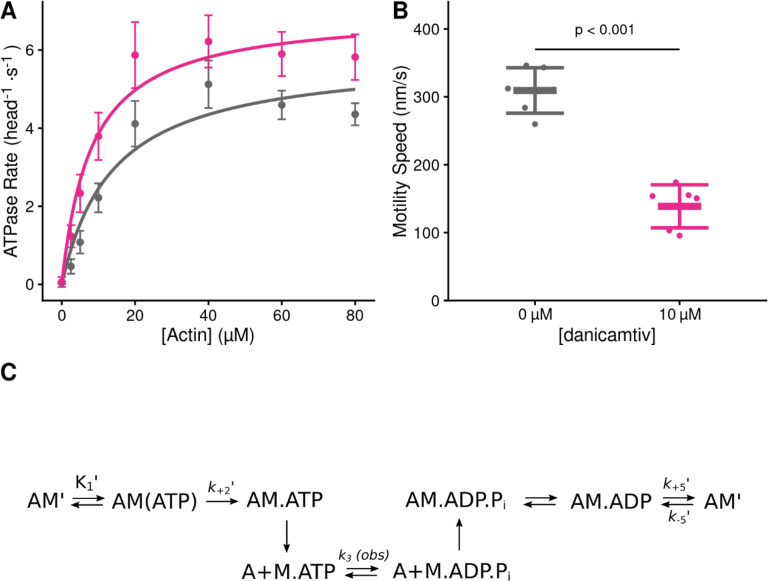
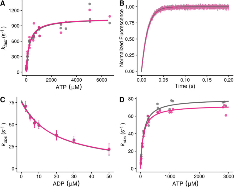
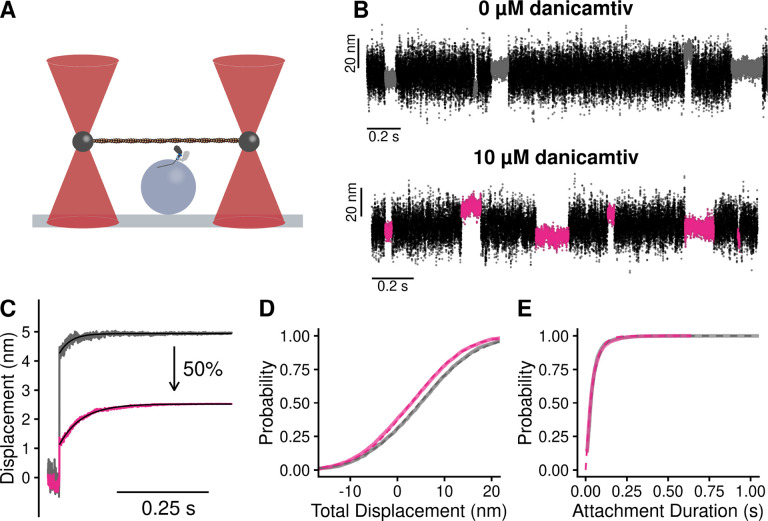
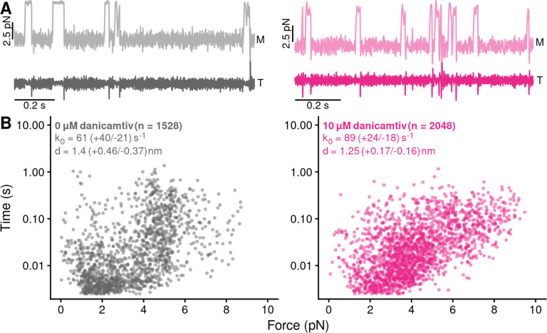
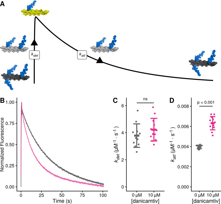
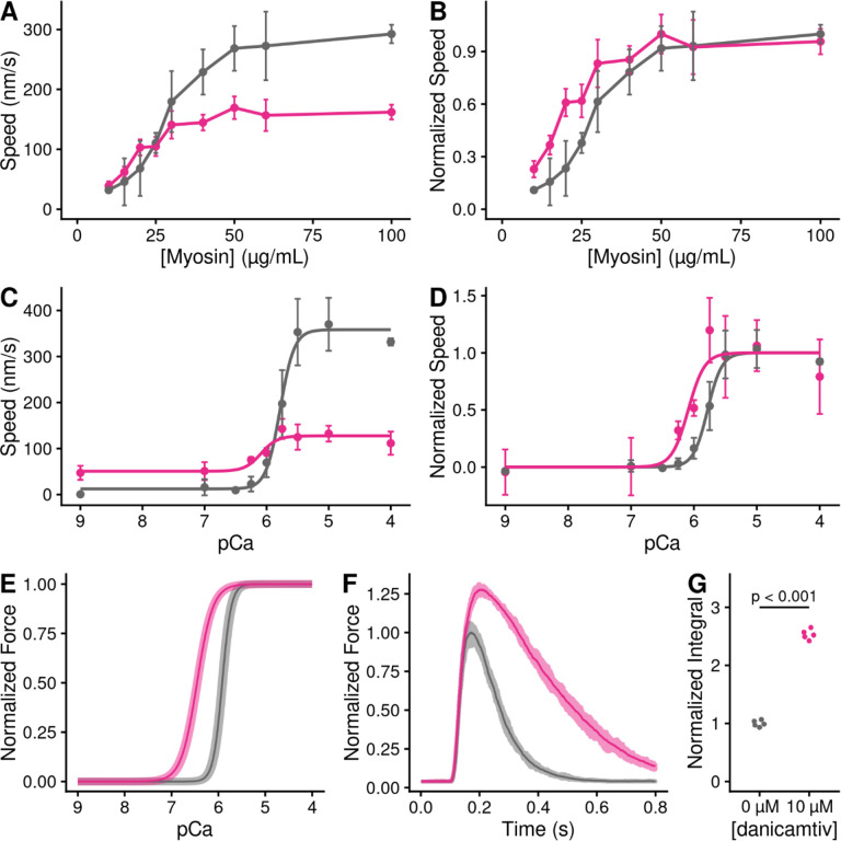
Heart failure is a leading cause of death worldwide, and even with current treatments, the 5-year transplant-free survival rate is only ~50-70%. As such, there is a need to develop new treatments for patients that improve survival and quality of life. Recently, there have been efforts to develop small molecules for heart failure that directly target components of the sarcomere, including cardiac myosin. One such molecule, danicamtiv, recently entered phase II clinical trials; however, its mechanism of action and direct effects on myosin's mechanics and kinetics are not well understood. Using optical trapping techniques, stopped flow transient kinetics, and in vitro reconstitution assays, we found that danicamtiv reduces the size of cardiac myosin's working stroke, and in contrast to studies in muscle fibers, we found that it does not affect actomyosin detachment kinetics at the level of individual crossbridges. We demonstrate that danicamtiv accelerates actomyosin association kinetics, leading to increased recruitment of myosin crossbridges and subsequent thin filament activation at physiologically-relevant calcium concentrations. Finally, we computationally model how the observed changes in mechanics and kinetics at the level of single crossbridges contribute to increased cardiac contraction and improved diastolic function compared to the related myotrope, omecamtiv mecarbil. Taken together, our results have important implications for the design of new sarcomeric-targeting compounds for heart failure.

Keywords: cardiac myosin; contractility; single molecule.


Conflict of interest statement

All experiments were conducted in the absence of any commercial or financial relationships that could be construed as potential conflicts of interest. M.J.G. discloses research funding from Edgewise Therapeutics on an unrelated project.

Figures

Figure 1.
Figure 1.. Steady state properties of β-cardiac myosin treated with danicamtiv.
A) The steady-state myosin ATPase rate was measured using the NADH coupled assay. The steady state myosin ATPase rate is plotted verses a function of actin concentration. Data were fitted by a hyperbolic function to calculate the maximal cycling rate (Vmax) and actin affinity (Km) with the Michaelis Menten equation. Treatment with 10 μM danicamtiv increased the maximal rate by ~1.2 fold from 5.9 to 7.0 s−1 (P = 0.028) and decreased the Km from 14.0 to 8.1 μM (P = 0.01). Each point represents the average rate from four independent trials with error bars showing the standard deviation. Statistical testing done using a 2-tailed T-test. Black = DMSO control. Pink = 10 μM danicamtiv. B) Speed of actin translocation in the unregulated in vitro motility assay. The addition of 10 μM danicamtiv decreased motility speed ~55% (P = 4×10⁻⁶). Thick horizontal lines show the average speed with standard deviation shown by the thin horizontal lines. Points represent the average speed of all filaments in a field of view for a single technical replicate measured across N = 3 independent experiments. Statistical testing done using a 2-tailed T-test. C) Scheme of myosin’s mechanochemical cross-bridge cycle. Myosin’s rate limiting step is actin attachment, so the predominant population of motors reside in the pre-working stroke M.ADP.Pi state during steady state cycling. The steady-state ATPase is thus limited by actin attachment (katt) which is rapidly followed by the mechanical working stroke and phosphate release. In vitro motility speed is limited by the ADP release rate (k+5’).
Figure 2.
Figure 2.. Stopped-flow kinetics measured with and without 10 μM danicamtiv.
Black = DMSO control. Pink = 10 μM danicamtiv. A) The rates of ATP-induced actomyosin dissociation were measured in the stopped flow. Transients were well fitted by the sum of two exponential functions, where the observed rate of the fast phase (kfast) is plotted as a function of ATP concentration. Data were fitted with a hyperbolic function to obtain K1’ and k+2’. There are no differences in either K1’ or k+2’ with or without danicamtiv (P = 0.64 and P = 0.99, respectively). Each point represents an independent measurement over 3 experimental days. B) The rate of ADP release from actomyosin was measured using stopped flow techniques, and the fluorescence transients were fitted with single exponential functions. Note, both 0 and 10 μM danicamtiv are plotted and overlay. There is no statistically significant difference in the rate of ADP release the absence or presence of danicamtiv (P = 0.96). C) The overall ADP binding affinity to actomyosin was measured by mixing an actomyosin solution containing increasing concentrations of ADP with 50 μM ATP (concentrations after mixing) measured using a competition experiment. The observed rate as a function of ADP concentration was fitted with a hyperbolic function to determine the ADP affinity (see methods). Each point shows the average of 3 separate trials and error bars show the standard deviations. There is no difference in the ADP binding affinity (k-5; P = 0.40). D) The rate of ATP hydrolysis by myosin was measured using stopped flow techniques. The rate of hydrolysis is reported by the change in tryptophan fluorescence at saturating ATP concentrations. Fluorescence transients are well fitted with single exponential functions. The observed rates of ATP hydrolysis were plotted against their respective ATP concentration and fitted with a hyperbolic function. The plateau represents the sum of the forwards and backwards rate of ATP hydrolysis (k3 (obs)). While there was a slight decrease in the observed hydrolysis rate with danicamtiv (P = 0.03), this slight decrease is not biologically meaningful. For all stopped-flow values, see Table 1.
Figure 3.
Figure 3.. Single molecule optical trapping reveals that danicamtiv reduces the size of myosin’s working stroke without altering detachment kinetics.
Black = DMSO control. Pink = 10 μM danicamtiv. A) Cartoon schematic of the optical trapping assay. An actin filament is strung between two optically-trapped beads and lowered onto a pedestal bead sparsely bound with myosin. B) Optical trapping data traces showing the stochastic binding of myosin to actin. Binding interactions are shown in grey or pink and detached states are shown in black. C) Time forward ensemble averages of myosin’s working stroke reveal a ~50% reduction in the size of myosin’s total working stroke in the presence of danicamtiv. D) The cumulative distribution of the total working stroke displacements at 10 μM ATP is well fit by a single cumulative Gaussian function (dotted lines) with average values of 4.9 ± 9.7 nm versus 3.0 ± 9.0 nm for DMSO control and 10 μM danicamtiv, respectively (P < 0.001 using a two-tailed T-test). N = 2076 binding interactions for control and 4776 binding events for 10 μM danicamtiv. E) The cumulative distributions of attachment durations at 10 μM ATP. Single exponential functions were fit to the distributions using maximum likelihood estimation. 95% confidence intervals were calculated using bootstrapping methods. There is no statistical difference between control and 10 μM danicamtiv, 23 (−2.5/+2.5) s−1 vs. 24 (−0.8/+0.9) s−1 (P = 0.48). For all trapping values, see Table 2.
Figure 4.
Figure 4.. Danicamtiv does not alter myosin’s load-dependent detachment kinetics at 1 mM ATP.
Black = DMSO control. Pink = 10 μM danicamtiv. A) An isometric force clamp was used to maintain actin at an isometric position during myosin binding interactions. To do this, the motor bead (M) was moved to hold the transducer bead (T) at an isometric position. Data traces are shown. B) Plots of actomyosin attachment duration versus the average resistive force applied during the binding event. Data are exponentially distributed at each force. Each point represents an independent actomyosin binding interaction. The data were fitted with the Bell equation using maximum likelihood estimation and 95% confidence intervals were calculated for each parameter by bootstrapping. The detachment rate in the absence of load, k0, was not different between control and 10 μM danicamtiv, 61 (−21/+40) vs 89 (−18/+24) s−1 (P = 0.17). These values are consistent with our measurements of the rates of ADP release from stopped-flow experiments. The distance to the transition state, d, which measures the load-sensitivity of the detachment rate, was not different between control and 10 μM danicamtiv, 1.40(−0.37/+0.46) vs 1.25 (−0.16/+0.17) nm (P = 0.43).
Figure 5.
Figure 5.. Danicamtiv increases myosin’s attachment rate in a single turnover stopped flow assay.
Black = DMSO control. Pink = 10 μM danicamtiv. A) Schematic conceptually describing the single turnover assay used to measure myosin’s attachment and detachment rates. In this assay, myosin and pyrene labeled actin are pre-incubated and then mixed with a sub-saturating concentration of ATP. The pyrene fluorescence increases as myosins detach from actin, and the increase in fluorescence reports the rate of detachment of myosin from actin (kdet). Myosin then reattaches to actin, quenching the fluorescence and reporting the attachment rate (katt). B) Fluorescence transients from the single turnover assay. Data were fitted as described in the Supplemental Methods. C) The average second-order rate of detachment (kdet) was similar with and without 10 μM danicamtiv (3.8 ± 0.9 vs. 4.2 ± 0.8 s−1; P = 0.23). D) The second-order rate of attachment (katt) increased with the addition of 10 μM danicamtiv (0.0040 ± 0.0002 vs 0.0063 ± 0.0007 μM−1·s−1; P = < 0.001). For C and D, the thick lines show the average values, and the error bars show the standard deviation. The individual points are the fitted second-order rates to individual transients collected across three experimental replicates. Statistical testing was done using a two-tailed T-test after passing a normality test.
Figure 6.
Figure 6.. Danicamtiv increases motility speed in the presence of regulatory proteins and these effects on muscle contraction can be recapitulated computationally.
Black = DMSO control. Pink = 10 μM danicamtiv. A and B) Unregulated in vitro motility speed as a function of the myosin concentration on the flowcell surface. The speed decreases if there is not at least one active myosin head bound to actin at any given time. Thus, if the duty ratio increases with drug, less myosin will be required to reach saturation. 10 μM danicamtiv decreases motility speed at higher myosin concentrations but increases speed at low myosin concentrations, indicative of a higher duty ratio, despite having a smaller working stroke. A) shows the measured speed and B) shows normalized data. ~40 filaments were tracked across four fields of view from two different experimental preparations. C and D) Regulated in vitro motility speed using thin filaments decorated with troponin and tropomyosin as a function of calcium. The data were fitted with the Hill equation and the fitted values ± standard error are: Vmax values are 386 ± 8 vs. 135 ± 7 nm/s (P < 0.001), pCa50 values are 5.76 ± 0.02 vs. 6.1 ± 0.09 (P = 0.01), and the Hill coefficients are 3.4 ± 0.4 vs 3.1 ± 1.5 (P = 0.85) for the control vs. 10 μM danicamtiv, respectively C) Shows the measured speed and D) shows the data normalized to the fitted Vmax and Vmin. Each point represents average speed with error bars showing the standard deviation of ~40–60 filaments imaged from 4–6 fields of view from 2–3 experimental replicates. E) Simulated force-calcium relationship from FiberSim. To simulate danicamtiv, we incorporated increased actin attachment, reduced myosin working stroke, and an increase in the population of active myosin heads. The simulations recapitulate the shift seen in the motility experiments. 5 replicates were conducted, the shaded region shows the range of values, and the solid line shows the mean. F) Simulated twitch in response to a calcium transient using the same simulation parameters. Danicamtiv increases the maximal force, slows kinetics, and G) increases the force-time integral.μ

Similar articles

References

    1. Heidenreich P. A. et al., 2022 AHA/ACC/HFSA Guideline for the Management of Heart Failure: Executive Summary: A Report of the American College of Cardiology/American Heart Association Joint Committee on Clinical Practice Guidelines. J Am Coll Cardiol 79, 1757–1780 (2022). - PubMed
    1. Hsich E. M. et al., Heart Transplantation: An In-Depth Survival Analysis. JACC Heart Fail 8, 557–568 (2020). - PMC - PubMed
    1. Rogers J. G. et al., Chronic mechanical circulatory support for inotrope-dependent heart failure patients who are not transplant candidates: results of the INTrEPID Trial. J Am Coll Cardiol 50, 741–747 (2007). - PubMed
    1. Green E. M. et al., A small-molecule inhibitor of sarcomere contractility suppresses hypertrophic cardiomyopathy in mice. Science 351, 617–621 (2016). - PMC - PubMed
    1. Malik F. I. et al., Cardiac myosin activation: a potential therapeutic approach for systolic heart failure. Science 331, 1439–1443 (2011). - PMC - PubMed

Publication types