[Preprint]. 2024 Oct 13:2024.10.10.617667.
doi: 10.1101/2024.10.10.617667.

Catabolism of extracellular glutathione supplies amino acids to support tumor growth

Affiliations

Catabolism of extracellular glutathione supplies amino acids to support tumor growth

Fabio Hecht et al. bioRxiv. .

Abstract

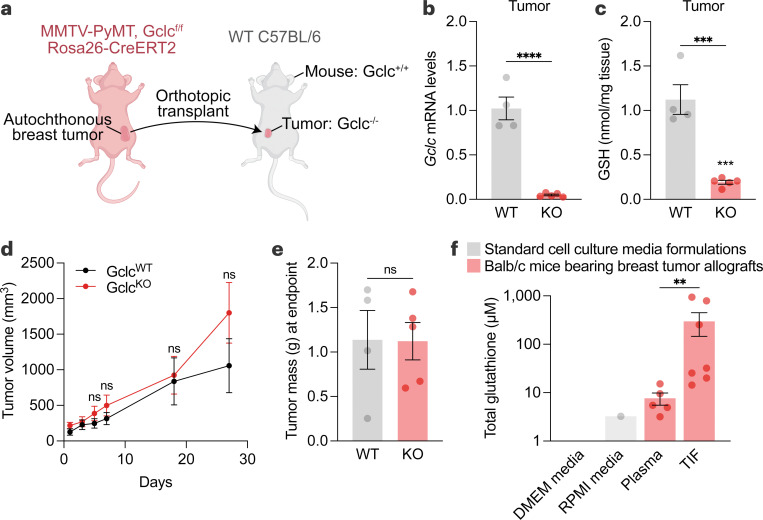
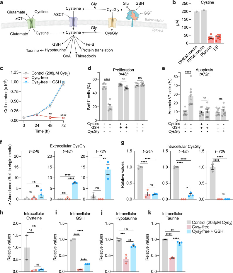
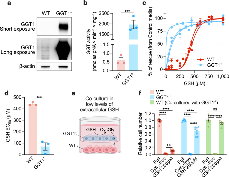
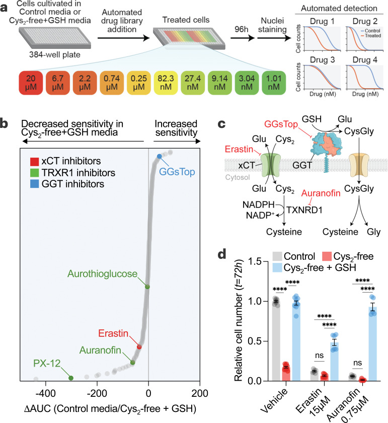
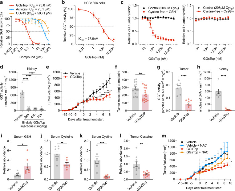
Restricting amino acids from tumors is an emerging therapeutic strategy with significant promise. While typically considered an intracellular antioxidant with tumor-promoting capabilities, glutathione (GSH) is a tripeptide of cysteine, glutamate, and glycine that can be catabolized, yielding amino acids. The extent to which GSH-derived amino acids are essential to cancers is unclear. Here, we find that GSH catabolism promotes tumor growth. We show that depletion of intracellular GSH does not perturb tumor growth, and extracellular GSH is highly abundant in the tumor microenvironment, highlighting the potential importance of GSH outside of tumors. We find supplementation with GSH can rescue cancer cell survival and growth in cystine-deficient conditions, and this rescue is dependent on the catabolic activity of γ-glutamyltransferases (GGTs). Finally, pharmacologic targeting of GGTs' activity prevents the breakdown of circulating GSH, lowers tumor cysteine levels, and slows tumor growth. Our findings indicate a non-canonical role for GSH in supporting tumors by acting as a reservoir of amino acids. Depriving tumors of extracellular GSH or inhibiting its breakdown is potentially a therapeutically tractable approach for patients with cancer. Further, these findings change our view of GSH and how amino acids, including cysteine, are supplied to cells.


Conflict of interest statement

Competing Interests Statement All authors declare no competing interests.

Figures

Figure 1.
Figure 1.. Intracellular production of GSH is dispensable for tumor growth.
a, Schematic of the tumor-specific Gclc knockout mouse model. Autochthonous tumors from MMTV-PyMT Gclcf/f Rosa26-CreERT2 mice were excised and orthotopically transplanted into mammary fat pads of wild-type C57BL/6 mice. C57BL/6 mice were treated with vehicle (corn oil; WT) or 160 mg/kg tamoxifen for 5 days (KO). b-c, Relative Gclc mRNA levels (b) and GSH levels (c) of WT tumors (n=4) and KO tumors (n=5). Statistical significance assessed by unpaired two-tailed t test. d-e, Tumor volume (d) and mass (e) of mice described in (a-c). Statistical significance was assessed by two-way ANOVA (p=0.38) followed by Šídák’s multiple comparisons test for tumor volume and by unpaired t test (p=0.97) for tumor mass. f, Concentration of total glutathione in cell culture media formulations (grey bars), and in tumor interstitial fluid (n=7) and matched plasma (n=5) from Balb/c mice bearing 4T1 orthotopic allografts (red bars). Statistical significance was assessed by Mann-Whitney test. Data represented as mean ± s.e.m., ns, not significant; *p-value<0.05; **p-value<0.01; ***p-value<0.001; ****p-value<0.0001.
Figure 2.
Figure 2.. Extracellular GSH supplies amino acids to promote cancer cell growth and survival in cystine-free environments.
a, Schematic of the different mechanisms of cysteine acquisition and utilization. b, Concentration of cystine in cell culture media formulations (grey bars), and in tumor interstitial fluid (n=7) and matched plasma (n=5) from Balb/c mice bearing 4T1 orthotopic allografts (red bars). c-e, HCC-1806 breast cancer cells were grown in control (208 µM cystine; Cys2), cystine-free (Cys2-free), cystine-free/GSH-supplemented (750 µM) or cystine-free/CysGly-supplemented (750 µM) medium and cell numbers (c), percentages of proliferative (BrdU+) (d) and apoptotic (Annexin V+) (e) cells was determined at indicated timepoints. Statistical significance was analyzed by two-way ANOVA followed by Tukey’s multiple comparisons test (n=4 independent experiments) for (c), two-way ANOVA followed by Šídák’s multiple comparisons test (n=3 independent experiments) for (d-e). f-g, Levels of extracellular CysGly in media (f) and intracellular CysGly in HCC-1806 breast cancer cells (g) grown in indicated media and at indicated time points. h-k, Levels of intracellular cysteine (h), GSH (i), hypotaurine (j), and taurine (k) in indicated media and at indicated time points. Statistical significance was assessed by one-way ANOVA followed by Tukey’s multiple comparisons test. Data is representative of an experiment with 3 biological replicates and is represented as mean ± s.e.m., *p-value<0.05; **p-value<0.01; ***p-value<0.001; ****p-value<0.0001; ns, not significant.
Figure 3.
Figure 3.. GGT1 is sufficient to provide an environment permissive of cell survival under cystine-free conditions.
a-b, Immunoblot analysis of human GGT1 (a) and GGT activity (b) in wild-type (WT) and GGT1-overexpressing (GGT1+) PC3 prostate cancer cells. Statistical significance was assessed by unpaired two-tailed t-test (n=3 with two independent experiments). c, WT and GGT1+ PC3 cells were grown in control media (208 µM cystine) or cystine-free media supplemented with indicated GSH concentrations. After 72 hours, cell numbers were quantified, and the percentage of rescue ((cell numbers in indicated media – cell numbers in cystine-free media)/(cell numbers in control media) was determined (n=3 independent experiments). d, Half-maximal effective concentration (EC50) of GSH for WT and GGT1+ cells calculated from (c). Statistical significance was assessed by an unpaired two-tailed t-test. e, Schematic of non-contact co-culture experiments using 0.4 μm PET membrane transwell inserts in media containing low concentrations of GSH (250 µM), which were insufficient to rescue the growth of WT cells in cystine-depleted conditions. f, Relative cell numbers of WT, GGT1+, or WT cells co-cultured with GGT1+ cells in control, cystine-depleted, or cystine-depleted/GSH-supplemented (250 µM) conditions. Statistical significance was evaluated by two-way ANOVA followed by Tukey’s multiple comparisons test (n=3 independent experiments). Data represented as mean ± s.e.m., ***p-value<0.001; ****p-value<0.0001; ns, not significant.
Figure 4.
Figure 4.. Utilization of GSH as a cysteine source drives drug resistance in cancer cells.
a, Schematic of multifunctional approach to pharmacologic screening (MAPS). HCC1806 cells in control (208 µM cystine) or cysteine-free/GSH-supplemented (500 µM) media were treated with libraries of drugs, each arrayed at 10 dose points (20 µM – 1 nM). After 96 hours, cell numbers were determined, and dose-response curves were generated for each drug. b, MAPS results showing each drug ranked by the difference in the area under the curve (ΔAUC) obtained for each drug curve in the conditions. c, Schematic showing the targets of selected hits from (b). d, HCC1806 cells grown in 6-well plates and treated with erastin and auranofin for 72 hours in different media conditions and relative cell numbers were determined. Statistical significance was evaluated by two-way ANOVA followed by Tukey’s multiple comparisons test (n=3 independent experiments). Data represented as mean ± s.e.m., ***p-value<0.001; ****p-value<0.0001; ns, not significant. Cys2, cystine; CysGly, cysteinylglycine; GGT, γ-glutamyl-transpeptidase; Glu, glutamate; GSH, glutathione; Gly, glycine; NADPH, reduced nicotinamide adenine dinucleotide phosphate; NADP+, oxidized nicotinamide adenine dinucleotide phosphate.
Figure 5.
Figure 5.. GSH catabolism is necessary to support cysteine supply and tumor growth.
a, Mouse kidney extracts were assayed for GGT activity in the presence of GGT inhibitors. b, HCC-1806 cells were grown in a control medium with indicated doses of GGsTop for 4 hours, and GGT activity was determined. c, Relative cell number of HCC-1806 cells treated with GGsTop for 96 hours in control (208 µM cystine) or cystine-free/GSH- (left) or CysGly-supplemented (right). d, GGT activity in the kidney extracts of C57BL/6 mice treated intraperitoneally with vehicle (n=6 mice) or with 5 mg/kg of GGsTop every 12 hours for 1 (n=9), 2 (n=9), or 3 days (n=8). Statistical significance was evaluated by one-way ANOVA followed by Dunnett’s multiple comparisons test. e, Volume of orthotopically implanted HCC-1806 cell xenografts in mice treated intraperitoneally with vehicle (sterile saline) (n=15) or 5 mg/kg GGsTop (n=18) every 12 hours for 8 days. Statistical significance was assessed by two-way ANOVA followed by Tukey’s multiple comparisons test. f-h, Tumor mass (f), and GGT activity in tumor (g) and kidney (h) of mice from (e) at the endpoint. i-k, Serum levels of GSH (NEM-GSH) (i), cysteine (NEM-cysteine) (j) and cystine (k) measured by LCMS. l, Tumor levels of cysteine (NEM-cysteine) measured by LCMS. Statistical significance in (f-l) was assessed by unpaired two-tailed t test. m, Volume of orthotopically implanted HCC-1806 cell xenografts in mice treated 5 mg/kg GGsTop every 12 hours while being supplemented with n-acetyl-cysteine (NAC, 30 mM) in their drinking water (Vehicle, n=5 mice; GGsTop, n=8; Vehicle with NAC, n=7, GGsTop with NAC, n=7). Statistical significance was assessed by two-way ANOVA. Data represented as mean ± s.e.m., *p-value<0.05; **p-value<0.01; ***p-value<0.001; ****p-value<0.0001; ns, not significant.

References

    1. Sivanand S. & Vander Heiden M. G. Emerging Roles for Branched-Chain Amino Acid Metabolism in Cancer. Cancer Cell 37, 147–156 (2020). 10.1016/j.ccell.2019.12.011 - DOI - PMC - PubMed
    1. DeBerardinis R. J. & Chandel N. S. Fundamentals of cancer metabolism. Sci Adv 2, e1600200 (2016). 10.1126/sciadv.1600200 - DOI - PMC - PubMed
    1. Martinez-Reyes I. & Chandel N. S. Cancer metabolism: looking forward. Nat Rev Cancer 21, 669–680 (2021). 10.1038/s41568-021-00378-6 - DOI - PubMed
    1. Muir A. et al. Environmental cystine drives glutamine anaplerosis and sensitizes cancer cells to glutaminase inhibition. Elife 6 (2017). 10.7554/eLife.27713 - DOI - PMC - PubMed
    1. Sullivan M. R. et al. Quantification of microenvironmental metabolites in murine cancers reveals determinants of tumor nutrient availability. Elife 8 (2019). 10.7554/eLife.44235 - DOI - PMC - PubMed

Publication types

LinkOut - more resources