[Preprint]. 2024 Oct 9:2024.10.09.617473.
doi: 10.1101/2024.10.09.617473.

Structural insights into inhibitor mechanisms on immature HIV-1 Gag lattice revealed by high-resolution in situ single-particle cryo-EM

Affiliations

Structural insights into inhibitor mechanisms on immature HIV-1 Gag lattice revealed by high-resolution in situ single-particle cryo-EM

Chunxiang Wu et al. bioRxiv. .

Abstract

HIV-1 inhibitors, such as Bevirimat (BVM) and Lenacapavir (LEN), block the production and maturation of infectious virions. However, their mechanisms remain unclear due to the absence of high-resolution structures for BVM complexes and LEN's structural data being limited to the mature capsid. Utilizing perforated virus-like particles (VLPs) produced from mammalian cells, we developed an approach to determine in situ cryo-electron microscopy (cryo-EM) structures of HIV-1 with inhibitors. This allowed for the first structural determination of the native immature HIV-1 particle with BVM and LEN bound inside the VLPs at high resolutions. Our findings offer a more accurate model of BVM engaging the Gag lattice and, importantly, demonstrate that LEN not only binds the mature capsid but also targets the immature lattice in a distinct manner. The binding of LEN induces a conformational change in the capsid protein (CA) region and alters the architecture of the Gag lattice, which may affect the maturation process. These insights expand our understanding of the inhibitory mechanisms of BVM and LEN on HIV-1 and provide valuable clues for the design of future inhibitors.


Figures
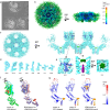
Fig. 1 |
Fig. 1 |. High-resolution in situ structural analysis of the immature GagCA-SP1 lattice assembly from VLPs.
a. An example micrograph of VLPs with picked single particles (white circles). The scale bar is 100 nm. b. Local resolution map of the immature GagCA-SP1 lattice assembly with both LEN and BVM bound at 2.10 Å global resolution. The GagCA-SP1 lattice assembly and ligands including LEN, BVM, and IP6 are well-resolved with high local resolution. c. The cryo-EM map for the immature GagCA-SP1 lattice assembly (cyan) with both LEN and BVM bound from the top (left) and clipped side (right) views. The map regions for LEN, BVM, and IP6 are shown in royal blue, purple, and lime respectively. d. Examples of cryo-EM density of selected residues. e. Zoom-in view of the LEN, IP6, and BVM binding pockets with densities of LEN (royal blue), BVM (purple), and IP6 (green). f. LEN induces a conformational change that alters the relative orientation of the NTD to the CTD of CA by about 8°, compared to the forms without LEN bound in both intact/apo or BVM-bound VLPs. Left, alignment of immature GagCA-SP1 protomers by superposition of the CTDs from VLPs without LEN (green, T8I VLP) and LEN-bound (cyan, NL-MA/NC VLP). Right, alignment of BVM-bound (slate, NL-MA/NC VLP) and LEN/BVM-bound (pink, T8I VLP) structures. g. Left, a comparison of models of intact T8I and PFO-treated NL-MA/NC VLPs shows that PFO treatment and the T8I mutation do not change the structure of GagCA-SP1. Center and Right, comparisons of models of intact and BVM-bound (center) VLPs and models of LEN-bound and LEN/BVM-bound (right) VLPs show that BVM does not change the overall structure of GagCA-SP1, with or without LEN bound.
Fig. 2 |
Fig. 2 |. Comparison of LEN binding pocket in the immature and the mature CA lattices.
a. LEN binding site in the mature capsid (PDB 6V2F). The binding site is mainly composed of the NTD of the major binder CA chain (orange) and the CTD of another CA chain (blue) within the same hexamer. LEN is shown in grey color. b. LEN binding site in the immature GagCA-SP1 lattice. The binding site is mediated by NTDs only from three GagCA-SP1 chains, including a major binder (green, oriented the same way as the major binder in a), a second chain within the same hexamer (yellow), and a third chain from an adjacent hexamer (cyan). LEN is shown in purple color. c. Alignment of the LEN conformations in the mature capsid (grey) and immature GagCA-SP1 (purple) lattices. The regions engaged in unique interactions are boxed. d. Comparison of the LEN binding pocket at the major binding interface in the mature and the immature lattices. The mature CA is shown in orange, and the immature GagCA-SP1 is shown in green. The residues with conserved interactions are shown as sticks. e. Unique interfaces at the LEN binding site in the immature GagCA-SP1 lattice. The main binding interface is highlighted in green (top left), with additional unique interactions displayed in the insets. The methylsulfonylbutyl group of LEN interacts with L20 and P17 (cyan) from a GagCA-SP1 chain of an adjacent immature hexamer (bottom left). The diazatricyclo-nonane group interacts with R18 (cyan) from the same adjacent immature hexamer (bottom right). Additionally, the trifluoroethyl group interacts with R82 (yellow) from the GagCA-SP1 chain within the same hexamer as the major binder chain (top right).
Fig. 3 |
Fig. 3 |. LEN binding induces conformational reorientation of the CANTD.
a. Alignment of the immature CANTD (left) and CACTD-SP1 (right) from VLPs with or without LEN, which shows that the overall folding of the individual CANTD or CACTD-SP1 is not changed upon LEN binding. The models are colored by the RMSD between the two GagCA-SP1 structures. b. Illustration of the conformational change of immature GagCA-SP1 hexamer before (lime green) and after (blue) LEN binding. The models of GagCA-SP1 hexamers are aligned and displayed from the side view (left) and the CANTD is displayed from the top view (right). LEN induces an ~8° rotation of the CANTD relative to the CACTD. This conformational change rearranges the CANTD organization in the lattice assembly. c. Key interfaces that mediate the CA NTD-NTD interaction in the native GagCA-SP1 lattice without LEN bound. Two CA-SP1 protomers in the same immature GagCA-SP1 hexamer are colored in green and yellow respectively, while a third CA-SP1 protomer from an adjacent hexamer is colored in cyan. d. New interfaces that are induced by LEN binding in between the CANTD protomers. Three CA-SP1 protomers are colored the same as in 3c. The LEN bound at the CA NTD-NTD interface is colored in slate. At the corresponding interface compared to the native GagCA-SP1 lattice, LEN reorganizes the hydrogen bond network and alters the orientation of the CANTD.
Fig. 4 |
Fig. 4 |. Binding poses of BVM and IP6 in the immature GagCA-SP1 lattice
a. BVM binding in the immature GagCA-SP1 lattice from PFO-treated NL-MA/NC VLPs. The cryo-EM density is shown in white and the atomic model is shown in green. IP6, BVM, and residues that form the binding pocket for BVM (K227, I231 of CA and M4, T8 of SP1) are shown in sticks. b. BVM binding in the immature GagCA-SP1 lattice from PFO-treated T8I VLPs treated with both BVM and LEN. Left: The cryo-EM density and the atomic model are shown in slate color. IP6, BVM, and residues that form the binding pocket for BVM are shown in sticks. Right: Local resolution of the cryo-EM map. c. Fit of BVM models in the cryo-EM density. Top: The observed cryo-EM map is fitted with six copies of BVM, arranged with C6 symmetry. Bottom: For comparison, a computationally generated surface contour is shown, based on the six-copy BVM model with C6 symmetry. The surface was generated using ChimeraX "molmap" utility at a resolution of 3 Å. d. Change of the IP6 pose upon BVM binding. Side (upper panels) and top (lower panels) views of the IP6 density from PFO-perforated VLPs treated with BVM (left), PFO-perforated VLPs treated with BVM and LEN (middle), or intact VLPs without any inhibitor treatment or PFO perforation (right). The CA-SP1 residues interacting with IP6 are labeled.
Fig. 5 |
Fig. 5 |. MD simulations of BVM binding
a. BVM binding characterized by potentials of mean force (PMF). Left: The binding site of BVM in the CACTD-SP1 hexamer is probed by pulling BVM along a reaction coordinate defined as the z-axis projection of the distance between the center of masses (c.o.m.) of the CACTD and BVM. Right: The PMF for the interaction of BVM with CACTD-SP1 is measured along the reaction coordinate for two possible orientations of BVM (dimethyl-succinate up or down) and in the presence or absence of myo-IP6. The PMF curves characterize the position and energy barrier for the binding of BVM in the CACTD-SP1 6-helix bundle. CACTD is colored in dark blue and SP1 in lavender. b. BVM binding mode from MD simulations in relation to cryo-EM density. Front, side, and bottom views of the BVM binding pose at the energy minimum from the PMF after MDFF refinement. The BVM binding position from the PMF matches the observed cryo-EM density with high cross-correlation coefficients. CACTD, BVM, and IP6 are colored by local cross-correlations according to the color bar. Cryo-EM density is shown in white.

References

    1. Freed E.O. HIV-1 assembly, release and maturation. Nature Reviews Microbiology 13, 484–496 (2015). - PMC - PubMed
    1. Wright E.R. et al. Electron cryotomography of immature HIV-1 virions reveals the structure of the CA and SP1 Gag shells. The EMBO Journal 26, 2218–2226 (2007). - PMC - PubMed
    1. Briggs J.A.G. et al. Structure and assembly of immature HIV. Proceedings of the National Academy of Sciences 106, 11090–11095 (2009). - PMC - PubMed
    1. Ganser-Pornillos B.K., Yeager M. & Pornillos O. Assembly and architecture of HIV. Adv Exp Med Biol 726, 441–65 (2012). - PMC - PubMed
    1. Qu K. et al. Maturation of the matrix and viral membrane of HIV-1. Science 373, 700–704 (2021). - PMC - PubMed

Publication types

LinkOut - more resources