[Preprint]. 2025 Jun 10:2024.09.30.615960.
doi: 10.1101/2024.09.30.615960.

Branched actin polymerization drives invasive protrusion formation to promote myoblast fusion during skeletal muscle regeneration

Affiliations

Branched actin polymerization drives invasive protrusion formation to promote myoblast fusion during skeletal muscle regeneration

Yue Lu et al. bioRxiv. .

Abstract

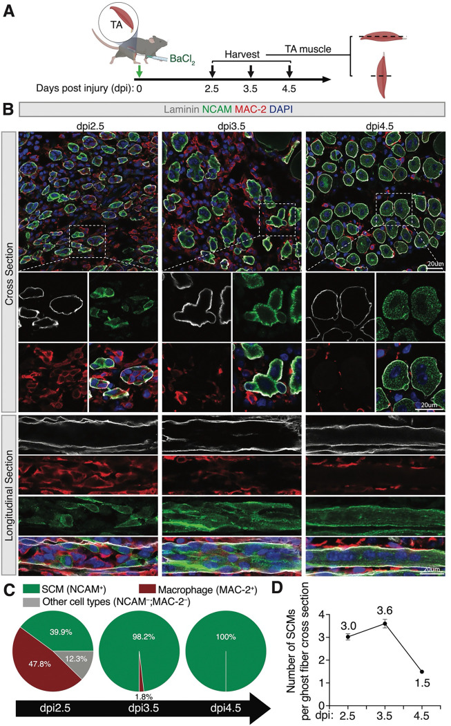
Skeletal muscle regeneration is a multistep process involving the activation, proliferation, differentiation, and fusion of muscle stem cells, known as satellite cells. The fusion of satellite cell-derived mononucleated muscle cells (SCMs) is indispensable for the generation of multinucleated, contractile myofibers during muscle repair. However, the molecular and cellular mechanisms underlying SCM fusion during muscle regeneration remain poorly understood. In this study, we uncovered an essential role for branched actin polymerization in SCM fusion. Using conditional knockouts of the Arp2/3 complex and its actin nucleation-promoting factors, N-WASP and WAVE, we demonstrated that branched actin polymerization is required for the SCM fusion, but not for satellite cell proliferation, differentiation, and migration. We showed that the N-WASP and WAVE complexes have partially redundant functions in regulating SCM fusion. Furthermore, we revealed that branched actin polymerization is essential for generating invasive protrusions at the fusogenic synapses in SCMs. Taken together, our study has identified new components of the myoblast fusion machinery in skeletal muscle regeneration and demonstrated a critical role for branched actin-propelled invasive protrusions in this process.


Conflict of interest statement

Declaration of interests The authors declare no competing financial interests. Correspondence and requests for materials should be addressed to E.H.C. (Elizabeth.Chen@UTSouthwestern.edu) and Y.L. (Yue.Lu@UTSouthwestern.edu).

Figures

Figure 1.
Figure 1.. Spatiotemporal coordination of macrophages and SCMs during skeletal muscle regeneration.
(A) Diagram of the TA muscle injury scheme. The TA muscles of the wild-type mice were injured by intramuscular injection of BaCI2. The injured TA muscles were collected at dpi 2.5, 3.5, and 4.5 for cross and longitudinal sectioning and immunostaining. (B) Immunostaining with anti-Laminin, anti-NACM, and anti-MAC-2 of the cross and longitudinal sections of TA muscles at the indicated time points. Note the decrease in the macrophage number within the ghost fiber at dpi 3.5 (compared to dpi 2.5), and the fusion of SCMs between dpi 3.5 and 4.5. Scale bars: 20 μm. (C) Quantification of the percentage of macrophages and differentiated SCMs within ghost fibers at the indicated time points. n = 3 mice were analyzed for each time point and > 98 ghost fibers in each mouse were examined. Mean ± s.d. values are shown. (D) Quantification of the number of differentiated SCMs in a single cross section of a ghost fiber at indicated time points. n = 3 mice were analyzed for each time point and > 98 ghost fibers in each mouse were examined. Mean ± s.e.m values are shown.
Figure 2.
Figure 2.. Branched actin polymerization is required for skeletal muscle regeneration.
(A) Schematic diagram of tamoxifen and BaCI2 treatment and subsequent CSA analysis at dpi 14. (B) Dystrophin and DAPI staining of the cross sections of TA muscles at dpi 14 from the control (Ctrl) and mutant mice. Note that the myofiber CSA is moderately decreased in N-WASPcKO and CYFIP1cKO mice, and severely reduced in dcKO, ArpC2cKO and MymXcKO mice. Scale bar: 100 μm. (C) The fold change of myofiber CSA in mutant mice vs. control mice. n = 3 mice were analyzed for each time point and > 200 fibers in each mouse were examined. Mean ± s.d. values are shown in the bar graph, and significance was determined by two-tailed student’s t-test. ****: p < 0.0001. (D) Frequency distribution of myofiber CSA of TA muscles in the control and mutant mice at dpi 14. n = 3 mice of each genotype were examined and > 200 ghost fibers in each mouse were examined.
Figure 3.
Figure 3.. Branched actin polymerization is required for SCM fusion.
(A) Schematic diagram of tamoxifen and BaCI2 treatment and subsequent SCM number analysis at dpi 4.5. (B) Immunostaining with anti-laminin, anti-NCAM, and anti-MAC-2 of the cross sections of TA muscles at dpi 4.5 from the control and mutant mice. Note that each ghost fiber in the control mice contained 1–2 centrally nucleated myofiber at dpi 4.5, indicating the near completion of SCM fusion. The ghost fibers in N-WASPcKO and CYFIP1cKO mice contained more SCMs, indicating impaired SCM fusion. Note that even more SCMs were seen in dcKO, ArpC2cKO and MymXcKO mice. Scale bar: 20 μm. (C) Quantification of the SCM number in a single cross section of a ghost fiber from TA muscles of the control and mutant mice at dpi 4.5. n = 3 mice were analyzed for each time point and > 80 ghost fibers in each mouse were examined. Mean ± s.d. values are shown in the bar graph, and significance was determined by two-tailed student’s t-test. ***: p < 0.001 and ****: p < 0.0001. (D) Frequency distribution of SCM number in a single cross section of a ghost fiber from TA muscles of the mutant mice and their littermate control. n = 3 mice of each genotype were analyzed and > 80 ghost fibers in each mouse were examined. (E) ArpC2 is required for SCM fusion in cultured cells. The satellite cells isolated from ArpC2cKO mice were maintained in GM without or with 2 μM 4OH-tamoxifen (4OHT) for 10 days. Subsequently, the cells were plated at 70% confluence in GM. After 24 hours, the cells were cultured in DM for 48 hours, followed by immunostaining with anti-MHC and DAPI. Note the robust fusion of the control (–4OHT) SCMs and the severe fusion defects in ArpC2 KO (+4OHT) SCMs. Scale bar: 100 μm. (F, G) Quantification of the differentiation index (% of nuclei in MHC+ cells vs. total nuclei) and fusion index (% of nuclei in MHC+ myotubes with ≥ 3 nuclei vs. total nuclei) of the two types of cells shown in (E). n = 3 independent experiments were performed. Mean ± s.d. values are shown in the bar graphs, and significance was determined by two-tailed student’s t-test. ****: p < 0.0001; n.s: not significant. (H) ArpC2 is required in both fusion partners. Fluorescence images from cell-mixing experiments using differentially labelled SCMs are shown. The satellite cells isolated from ArpC2cKO mice were infected with retroviruses encoding GFP or mScarleti (mScar). Next, the GFP+ cells were maintained in GM for 10 days (Ctrl GFP+ cells), and the mScar+ cells were maintained in GM without (Ctrl mScar+ cells) or with 2 μM 4OH-tamoxifen (ArpC2cKO mScar+ cells) for 10 days. Subsequently, the Ctrl GFP+ cells were mixed with Ctrl mScar+ cells or with ArpC2cKO mScar+ cells with a ratio of 1:1 and plated at 70% confluence in GM. After 24 hours, the cells were cultured in DM for 48 hours followed by direct fluorescent imaging. Arrowheads indicate syncytia derived from both GFP and mScar cells. Scale bar: 100 μm. (I) Percentage of GFP+mScar+ syncytia in total cells shown in (H). Mean ± s.d. values are shown in the bar graph, and significance was determined by two-tailed student’s t-test. **: p < 0.01.
Figure 4.
Figure 4.. Branched actin polymerization is required for invasive protrusion formation during SCM fusion.
(A) Still images of a fusion event between two LifeAct-mScar and Arp2-mNG co-expressing SCMs (see Supplemental Video 4). The boxed area is enlarged in (A’). Note the presence of two invasive protrusions (16 minute, arrowheads) enriched with LifeAct-mScar and Arp2-mNG at the fusogenic synapse. n = 8 fusion events were observed with similar results. Scale bar: 5 μm. (B) TEM of TA muscle cells in wild-type control, ArpC2cKO, and MymXcKO mice at dpi 3.5. The invading SCMs are pseudo-colored in light magenta. Note the finger-like protrusions projected by SCMs invading their neighboring cells in control and MymXcKO, but not in the ArpC2cKO, mice. Scale bars: 500 nm. (C) Quantification of the percentage of SCMs with invasive protrusions in a single cross section of a ghost fiber in the mice with genotypes shown in (B) at dpi 3.5. At least 83 SCMs from n = 20 ghost fibers in each genotype were quantified. Mean ± s.d. values are shown in the dot plots, and significance was determined by two-tailed student’s t-test. ***p < 0.001; n.s.: not significant. (D) A model depicting the function of Arp2/3-mediated branched actin polymerization in promoting invasive protrusion formation to promote SCM fusion during skeletal muscle regeneration. BM: basement membrane.

References

    1. Berger S, Schäfer G, Kesper DA, Holz A, Eriksson T, Palmer RH, Beck L, Klämbt C, Renkawitz-Pohl R, Onel SF. 2008. WASP and SCAR have distinct roles in activating the Arp2/3 complex during myoblast fusion. J Cell Sci 121: 1303–1313. - PubMed
    1. Bi P, McAnally JR, Shelton JM, Sanchez-Ortiz E, Bassel-Duby R, Olson EN. 2018. Fusogenic micropeptide Myomixer is essential for satellite cell fusion and muscle regeneration. Proc Natl Acad Sci U S A 115: 3864–3869. - PMC - PubMed
    1. Bi P, Ramirez-Martinez A, Li H, Cannavino J, McAnally JR, Shelton JM, Sanchez-Ortiz E, Bassel-Duby R, Olson EN. 2017. Control of muscle formation by the fusogenic micropeptide myomixer. Science 356: 323–327. - PMC - PubMed
    1. Capkovic KL, Stevenson S, Johnson MC, Thelen JJ, Cornelison DD. 2008. Neural cell adhesion molecule (NCAM) marks adult myogenic cells committed to differentiation. Exp Cell Res 314: 1553–1565. - PMC - PubMed
    1. Chen EH. 2011. Invasive podosomes and myoblast fusion. Curr Top Membr 68: 235–258. - PMC - PubMed

Publication types

LinkOut - more resources