[Preprint]. 2024 Oct 11:2024.10.09.617077.
doi: 10.1101/2024.10.09.617077.

Identification of DLK1, a Notch ligand, as an immunotherapeutic target and regulator of tumor cell plasticity and chemoresistance in adrenocortical carcinoma

Affiliations

Identification of DLK1, a Notch ligand, as an immunotherapeutic target and regulator of tumor cell plasticity and chemoresistance in adrenocortical carcinoma

Nai-Yun Sun et al. bioRxiv. .

Update in

Abstract

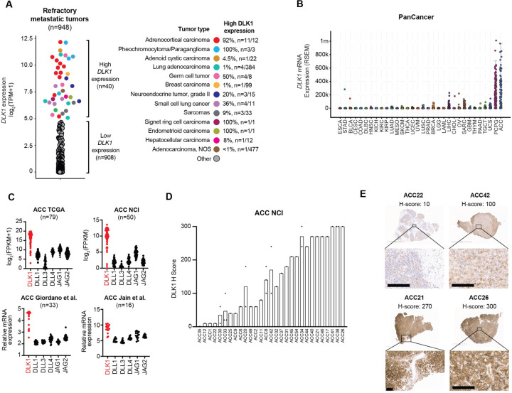
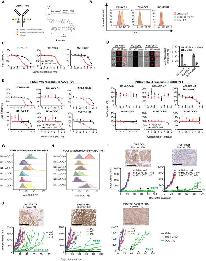
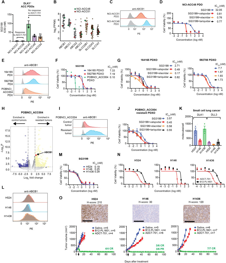
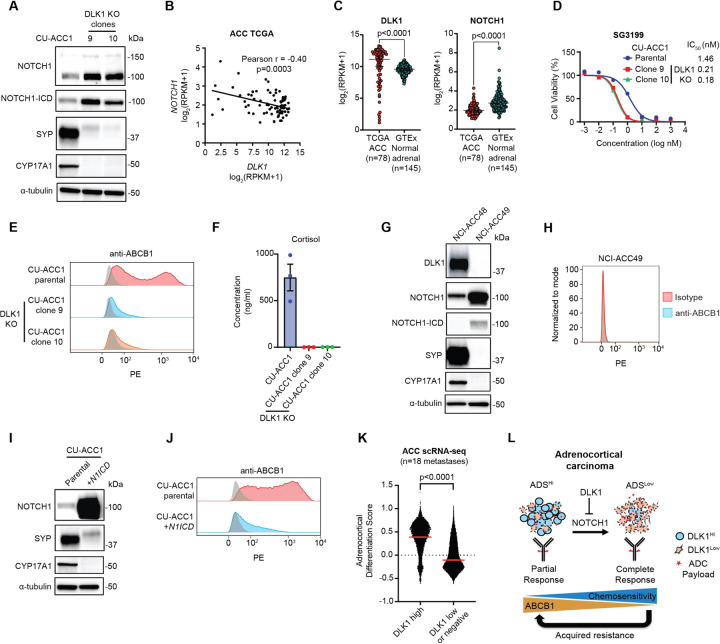
Immunotherapeutic targeting of cell surface proteins is an increasingly effective cancer therapy. However, given the limited number of current targets, the identification of new surface proteins, particularly those with biological importance, is critical. Here, we uncover delta-like non-canonical Notch ligand 1 (DLK1) as a cell surface protein with limited normal tissue expression and high expression in multiple refractory adult metastatic cancers including small cell lung cancer (SCLC) and adrenocortical carcinoma (ACC), a rare cancer with few effective therapies. In ACC, ADCT-701, a DLK1 targeting antibody-drug conjugate (ADC), shows potent in vitro activity among established cell lines and a new cohort of patient-derived organoids as well as robust in vivo anti-tumor responses in cell line-derived and patient-derived xenografts. However, ADCT-701 efficacy is overall limited in ACC due to high expression and activity of the drug efflux protein ABCB1 (MDR1, P-glycoprotein). In contrast, ADCT-701 is extremely potent and induces complete responses in DLK1+ ACC and SCLC in vivo models with low or no ABCB1 expression. Genetic deletion of DLK1 in ACC dramatically downregulates ABCB1 and increases ADC payload and chemotherapy sensitivity through NOTCH1-mediated adrenocortical de-differentiation. Single cell RNA-seq of ACC metastatic tumors reveals significantly decreased adrenocortical differentiation in DLK low or negative cells compared to DLK1 positive cells. This works identifies DLK1 as a novel immunotherapeutic target that regulates tumor cell plasticity and chemoresistance in ACC. Our data support targeting DLK1 with an ADC in ACC and neuroendocrine neoplasms in an active first-in-human phase I clinical trial (NCT06041516).


Conflict of interest statement

Nitin Roper and Jaydira Del Rivero have received research funding from ADC Therapeutics for this study. The other authors have no competing interests to report.

Figures

Figure 1.
Figure 1.. Identification of DLK1 as the most highly expressed Notch ligand in adrenocortical carcinoma.
(A) DLK1 mRNA expression across adult refractory metastatic cancers (n=948). Tumor types with high DLK1 expression are highlighted in the colors shown. The percentage of each tumor type with high DLK1 expression is shown on the right. (B) DLK1 mRNA expression in the TCGA PanCancer dataset. (C) Expression of Notch ligands from four independent bulk ACC RNA-seq datasets. (D) Quantification of DLK1 IHC staining in ACC tumors with IHC images of four representation tumors with varying levels of DLK1 expression. Scale bars represent 200 μM. IHC: immunohistochemistry.
Figure 2.
Figure 2.. ADCT-701, a DLK1 targeting antibody-drug conjugate, has potent in vitro activity and induces robust in vivo anti-tumor responses in adrenocortical carcinoma.
(A) Schematic structure of ADCT-701, a DLK1 targeting antibody drug conjugate. (B) Representative surface expression of DLK1 among ACC cell lines: CU-ACC1, CU-ACC2, and H295R. (C) ADCT-701 cytotoxicity among CU-ACC1, CU-ACC2, and H295R cells. Cells were treated with ADCT-701 and B12-PL1601 (non-targeted control ADC) for 7 days. Each point represents the mean±SEM. (D) Representative imaging flow cytometry images and signal intensity analysis (n=3 biological replicates) showing cellular internalization of DLK1 antibodies in CU-ACC1, CU-ACC2, and H295R. (E) Cytotoxic activity of ADCT-701 responsive (n=6) and (F) non-responsive (n=6) ACC patient-derived organoids (PDOs). Flow cytometry histograms assessing DLK1 among ADCT-701 (G) responsive and (H) non-responsive ACC PDOs. Shaded gray histograms represent unstained controls for each condition. (I) CU-ACC1 and H295R xenograft tumor growth curves after treatment with saline, B12-PL1601, or ADCT-701 (1 mg/kg). Additional doses of ADCT-701 indicated by arrows. (J) ACC PDXs 164165, 592788, and POBNCI_ACC004 tumor growth curves after treatment with saline, B12-PL1601 or ADCT-701 (1 mg/kg). X symbols indicate the administration of ADCT-701 re-dosing. Arrow indicates unexpected death of 1 POBNCI_ACC004 tumor-bearing mouse prior to endpoint. DLK1 immunohistochemistry with H-scores shown above each individual xenograft or PDX tumor. Scale bars represent 200 μM.
Figure 3.
Figure 3.. ABCB1, a drug efflux protein, mediates intrinsic and acquired resistance to ADCT-701.
(A) Cytotoxicity of SG3199 among ADCT-701 responsive and non-responsive DLK1+ ACC patient-derived organoids (PDOs). (B) Drug transporter mRNA expression in NCI-ACC patients as measured by RNA-seq. (C) Flow cytometry histograms assessing ABCB1 of DLK1+ ADCT-701 responsive (NCI-ACC51) and non-responsive (NCI-ACC48) ACC PDOs. (D) SG3199 cytotoxicity in the NCI-ACC48 PDO with and without treatment with ABCB1 inhibitors (1 μM valspodar, 10 μM elacridar and 1 μM tariquidar). (E) Flow cytometry histograms assessing ABCB1 among 164165, 592788 and POBNCI_ACC004 PDXs. (F) SG3199 cytotoxicity in 164165, 592788 and POBNCI_ACC004 PDX-derived organoids. (G) SG3199 cytotoxicity in the 164165 and 592788 PDX-derived organoids treated with or without ABCB1 inhibitors (1 μM valspodar, 10 μM elacridar and 1 μM tariquidar). (H) Volcano plot of differentially expressed genes in control tumors versus post-ADCT-701 acquired resistant tumors in POBNCI_ACC004 PDX. (I) Flow cytometry histograms assessing ABCB1 among ADCT-701 resistant and control POBNCI_ACC004 PDX tumors. (J) SG3199 cytotoxicity in the ADCT-701 resistant POBNCI_ACC004 PDX-derived organoid treated with or without ABCB1 inhibitors (1 μM valspodar, 10 μM elacridar and 1 μM tariquidar). (K) DLK1 molecules/cell relative to DLL3 among small cell lung cancer (SCLC) cell lines (H524, H146, and H1436). (L) Flow cytometry histograms assessing ABCB1 in SCLC cell lines. (M) SG3199 cytotoxicity in SCLC cell lines. Cells were treated with SG3199 for 3 days. (N) ADCT-701 cytotoxicity among SCLC cell lines. Cells were treated with ADCT-701 or B12-PL1601 for 7 days. Each point represents the mean±SEM. (O) Tumor growth curves of SCLC xenograft models after treatment with saline, B12-PL1601 or ADCT-701 (1 mg/kg) treatment. Arrows indicate re-treatment with ADCT-701. Scale bars represent 200 μM. For flow cytometry histograms, shaded gray histograms represent isotype controls for each condition.
Figure 4.
Figure 4.. DLK1 is a major regulator of ABCB1, adrenocortical differentiation, and chemoresistance in adrenocortical carcinoma.
(A) Immunoblot analysis of NOTCH1 signaling, total NOTCH1 and NOTCH1 intracellular domain (ICD), NE marker synaptophysin (SYP), and loading control (α-tubulin) proteins with and without DLK1 KO in CU-ACC1 cells. Two single-cell KO clones are shown. (B) Correlation between NOTCH1 and DLK1 expression among TCGA ACC tumors. (C) DLK1 and NOTCH1 expression in TCGA ACC tumors and normal adrenals. (D) SG3199 cytotoxicity in CU-ACC1 parental and DLK1 KO clones. (E) Flow cytometry histograms assessing ABCB1 in CU-ACC1 cells with and without DLK1 KO. (F) Concentration of cortisol in conditioned media from CU-ACC1 parental and DLK1 KO clones. (G) Immunoblot analysis of DLK1, total NOTCH1 and NOTCH1-ICD, SYP, the steroidogenic enzyme CYP17A1, and α-tubulin proteins in DLK1+ NCI-ACC48 and DLK1 ACC49 patient-derived organoids. (H) Flow cytometry histograms assessing ABCB1 in DLK1 negative NCI-ACC49 patient-derived organoids. (I) Immunoblot analysis of DLK1, total NOTCH1 (to detect the NOTCH1-ICD plasmid expression), SYP, CYP17A1, and α-tubulin proteins in CU-ACC1 cells with and without NOTCH1-ICD overexpression. (J) Flow cytometry histograms assessing ABCB1 in CU-ACC1 cells with and without N1ICD overexpression. (K) Single cell RNA-seq adrenocortical differentiation score comparing DLK1 high cells to DLK1 low or negative cells from 18 ACC metastatic tumors. (L) Model summarizing the findings of the current study. For flow cytometry histograms, shaded gray histograms represent isotype controls for each condition.

References

    1. Kunnimalaiyaan M. & Chen H. Tumor suppressor role of Notch-1 signaling in neuroendocrine tumors. The oncologist 12, 535–542 (2007). 10.1634/theoncologist.12-5-535 - DOI - PubMed
    1. Zhou B. et al. Notch signaling pathway: architecture, disease, and therapeutics. Signal Transduction and Targeted Therapy 7, 95 (2022). 10.1038/s41392-022-00934-y - DOI - PMC - PubMed
    1. Saunders L. R. et al. A DLL3-targeted antibody-drug conjugate eradicates high-grade pulmonary neuroendocrine tumor-initiating cells in vivo. Science translational medicine 7, 302ra136 (2015). 10.1126/scitranslmed.aac9459 - DOI - PMC - PubMed
    1. Puca L. et al. Delta-like protein 3 expression and therapeutic targeting in neuroendocrine prostate cancer. Science translational medicine 11 (2019). 10.1126/scitranslmed.aav0891 - DOI - PMC - PubMed
    1. Paz-Ares L. et al. Tarlatamab, a First-in-Class DLL3-Targeted Bispecific T-Cell Engager, in Recurrent Small-Cell Lung Cancer: An Open-Label, Phase I Study. Journal of clinical oncology : official journal of the American Society of Clinical Oncology 41, 2893–2903 (2023). 10.1200/JCO.22.02823 - DOI - PMC - PubMed

Publication types

Associated data

LinkOut - more resources