[Preprint]. 2024 Oct 13:2024.10.13.617647.
doi: 10.1101/2024.10.13.617647.

A noncanonical glycoprotein H complex enhances cytomegalovirus entry

Affiliations

A noncanonical glycoprotein H complex enhances cytomegalovirus entry

Michael J Norris et al. bioRxiv. .

Update in

Abstract

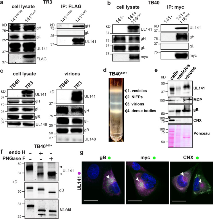
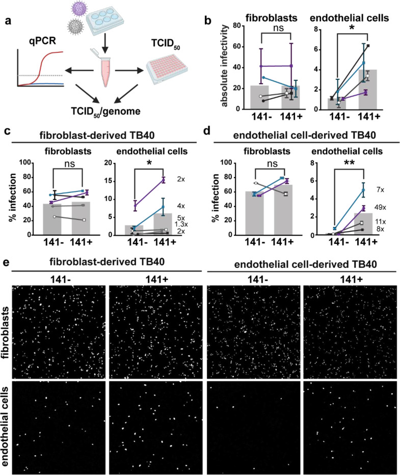
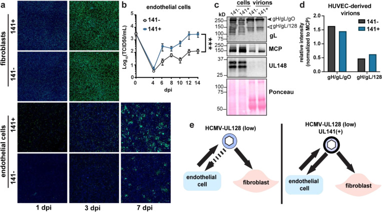
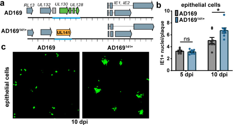
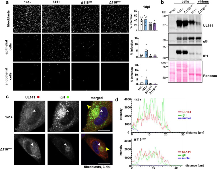
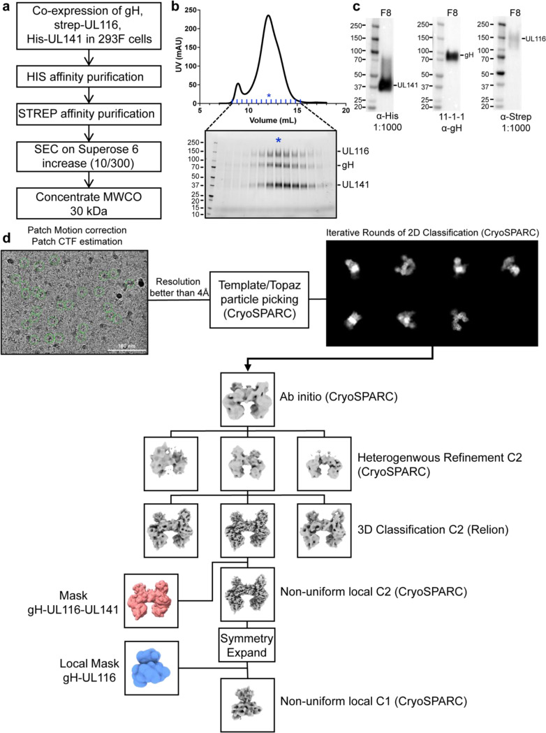
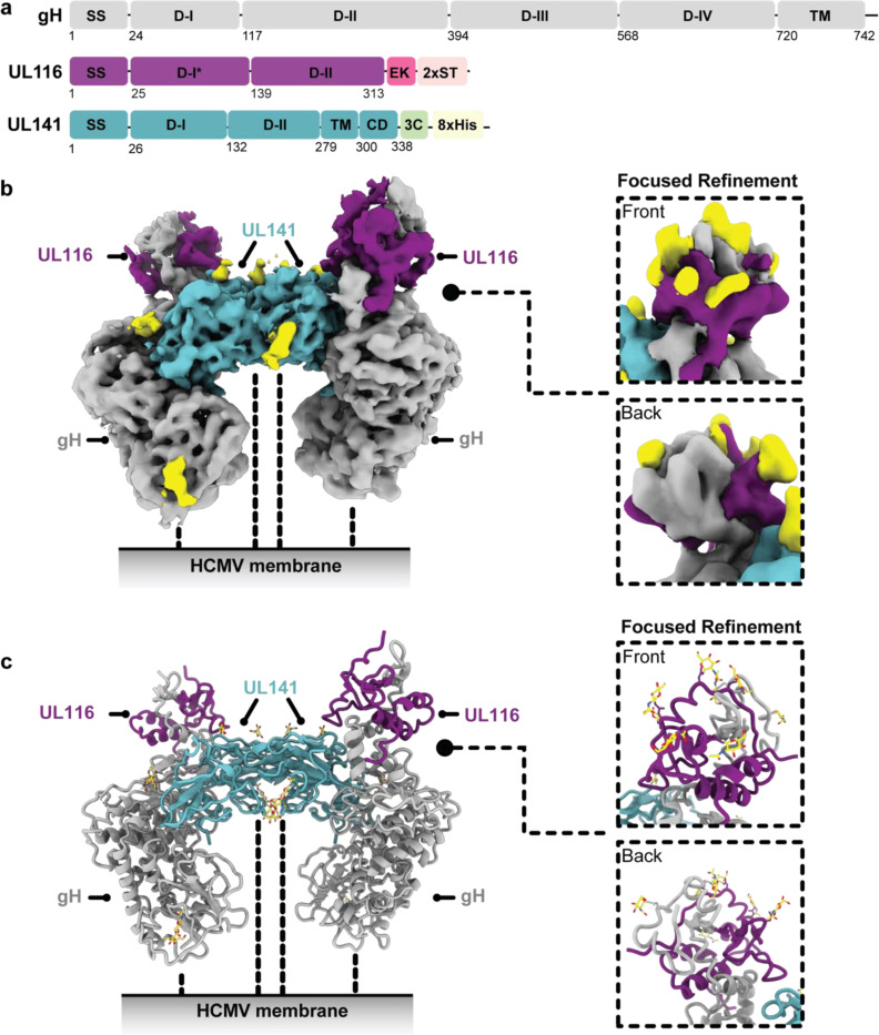
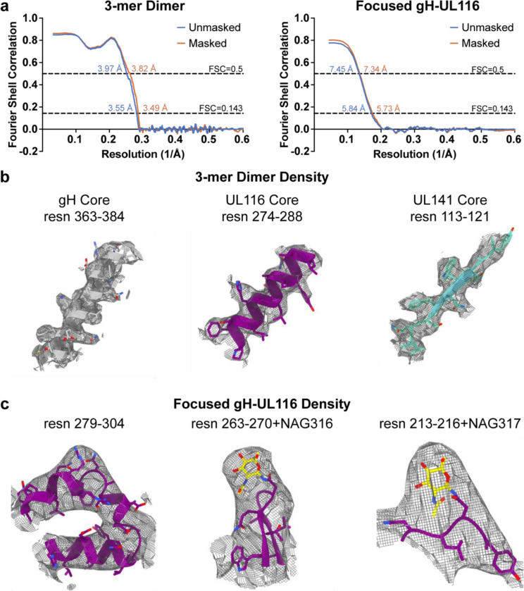
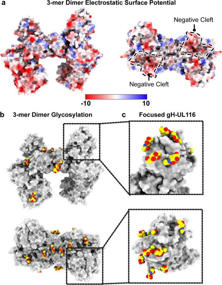
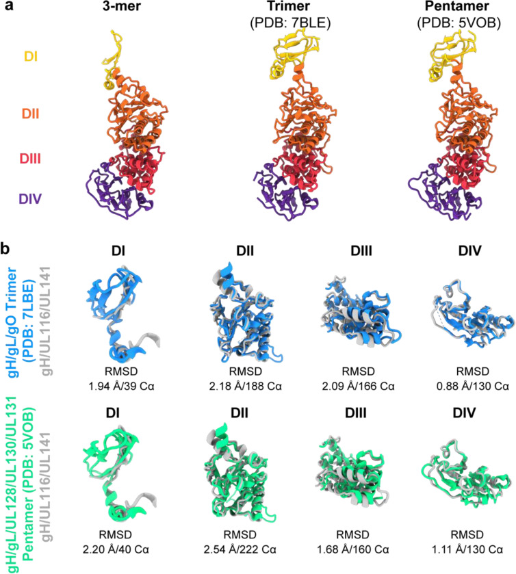
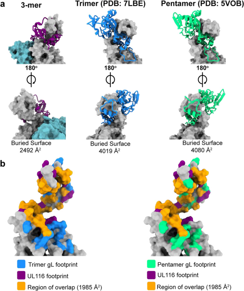
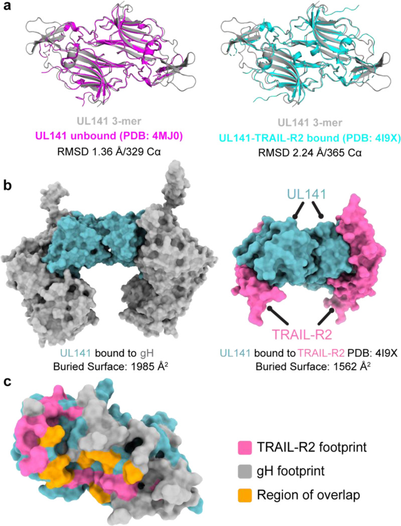
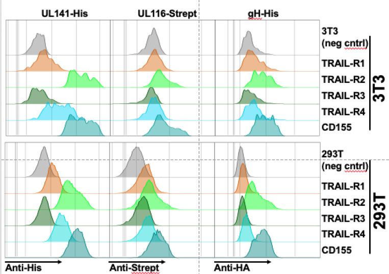
Human cytomegalovirus (HCMV) causes severe birth defects, lifelong health complications, and $4 billion in annual costs in the United States alone. A major challenge in vaccine design is the incomplete understanding of the diverse protein complexes the virus uses to infect cells. In Herpesviridae, the gH/gL glycoprotein heterodimer is expected to be a basal element of virion cell entry machinery. For HCMV, gH/gL forms a "trimer" with gO and a "pentamer" with UL128, UL130, and UL131A, with each complex binding distinct receptors to enter varied cell types. Here, we reveal a third glycoprotein complex, abundant in HCMV virions, which significantly enhances infection of endothelial cells. In this "3-mer" complex, gH, without gL, associates with UL116 and UL141, an immunoevasin previously known to function in an intracellular role. Cryo-EM reveals the virion-surface 3-mer is structurally unique among Herpesviridae gH complexes, with gH-only scaffolding, UL141-mediated dimerization and a heavily glycosylated UL116 cap. Given that antibodies directed at gH and UL141 each can restrict HCMV replication, our work highlights this virion surface complex as a new target for vaccines and antiviral therapies.


Conflict of interest statement

Competing interests. The authors declare no competing interests.

Figures

Fig. 1.
Fig. 1.. UL141 is incorporated into virions and assembles into a gH/UL116 complex.
a, The HCMV BAC-derived strain TR3 was modified to introduce a FLAG tag at the C-terminus of UL141 (TR3–141F). Infected fibroblasts were lysed 72 h post infection (hpi), and IP was performed with anti-FLAG. IP eluates were resolved by SDS-PAGE and analyzed by immunoblot with the indicated antibodies. b, Fibroblasts were infected with BAC-derived TB40/E (TB40-BAC4) repaired for UL141 expression and engineered to express a myc-tagged UL116 (141+,116myc). Anti-myc IP was carried out at 72 hpi and eluates were analyzed by Western blot. c, Infected cell lysates and purified virions from HCMV strains TB40 (141-) and TR3 (141+) were compared for the indicated viral glycoproteins. d, TB40–141+ viruses were grown in fibroblasts and concentrated through a 20% sorbitol cushion prior to glycerol-sodium tartrate gradient purification to separate infectious virions (band 3) from cell debris (bands 1 and 4) and non-infectious enveloped particles (NIEPs, band 2). e, Western blot analysis of fibroblasts’ whole cell lysates at 6 dpi, vesicles (c, band 1), and virions (c, band 3). Major capsid protein (MCP) identifies the fraction that is enriched for infectious virions. f, Lysates of membranes from TB40–141+ infected cells were treated with endoglycosidase H (endoH) or protein N-glycosidase F (PNGase F) and analyzed by Western blot. g, Fibroblasts were infected with TB40–141+ at MOI 1 for 3 days prior to staining for UL141 (magenta) and gB, UL116-myc and CNX (all green). The cytoplasmic viral assembly compartment is shown by the white arrowheads, and CNX identifies the ER. Scale bars are 25 μm.
Fig. 2.
Fig. 2.. The 3-mer improves endothelial cell infectivity.
a, Schematic of methods used to measure the absolute infectivity of TB40 virions. Viral genomes/mL were calculated from a standard curve with a 10-fold dilution series of TB40 BAC DNA. TCID50 assays were performed on fibroblasts and HUVEC in parallel by staining for IE1 at 3 days post infection, and together were used to calculate TCID50/genome (BioRender.com). b, Absolute infectivity shown as TCID50/103 genomes for fibroblasts and endothelial cells, and data represent 4 biological replicates. c, Infectivity of fibroblast-derived and d, endothelial cell-derived 141+ or 141− HCMV TB40 with fold differences shown as #x for each biological replicate. Fibroblasts and endothelial cells were infected for 24h in parallel with 50 or 100 genome equivalents/cell, respectively, stained for IE1 and counterstained with Hoechst 33342 to calculate the percentage of infected cells. e, Representative images of c and d. Statistical significance was determined by a paired t-test, with each point representing a biological replicate consisting of 2–3 technical replicates each. Error bars represent ± SEM for each biological replicate. Shaded bars are the mean % infection for all biological replicates.
Figure 3.
Figure 3.. UL141 promotes endothelial cell tropism independently of the pentamer complex.
a, Representative images of UL141-dependent spread in endothelial cells. Fibroblasts and endothelial cells were infected with 50 or 100 genome equivalents/cell, respectively, of 141− or 141+ TB40 viruses produced by fibroblasts. Cells were stained for IE1 (green) and Hoechst (blue) at the indicated days post-infection to monitor viral spread. b, Low MOI (0.01 TCID50) multicycle viral growth kinetics in HUVEC that were infected with 141− or 141+ viruses up to 14 dpi. This graph is a compilation of 3 biological replicates. Lognormal data were logarithmically transformed to fit a Gaussian distribution prior to calculating statistical significance via 2-way ANOVA for 12 dpi data points. c, Non-reducing SDS-PAGE of HUVEC cell lysates and HUVEC-derived virions. Cells were infected with 141− and 141+ viruses to measure virion incorporation of known HCMV entry complexes, trimer (gH/gL/gO) and pentamer (gH/gL/128). HUVEC-derived virions were concentrated through a 20% sorbitol cushion at 12 dpi and 14 dpi prior to lysis. Whole cell lysates were collected at 14 dpi. Lysates were immunoblotted for gL to identify covalently linked entry complexes, major capsid protein (MCP) to measure virion abundance, and UL148 to assess the purity of the virion preparations. d, Quantification of band intensities for gH/gL/gO and gH/gL/128 abundance in HUVEC-derived virions from c. Band intensities of 141− and 141+ virions were normalized to MCP. e, Graphical summary illustrating the role of UL141 as an endotheliotropic factor that restores the ability of TB40 virions to subsequently infect endothelial cells.
Fig. 4.
Fig. 4.. UL141 enhances the infectivity of the pentamer-null AD169 strain.
a, Schematic of the pentamer-null AD169 viruses used in the following experiments. b, ARPE-19 were infected with AD169 or UL141-restored AD169 (AD169141) at MOI 0.1. Cells were stained for IE1 at 5 and 10 dpi to measure the size of foci, or plaques in IE1+ nuclei per plaque. Each point in the bar graph represents a biological replicate (N=7–8). Data was logarithmically transformed to fit a Gaussian distribution prior to measuring statistical significance via Welch’s t test. c, Representative image of AD169 versus AD169141+ plaques in ARPE-19 cells at 10 dpi. Cells were stained for IE1 (green).
Fig. 5.
Fig. 5.. UL116 is required for UL141-dependent entry and virion incorporation of the 3-mer.
a, Representative images of fibroblasts, epithelial cells, and endothelial cells following infection with UL116-sufficient versus UL116-deficient (Δ116) TB40 that encode UL141 or not (141− and Δ116141−; 141+ or Δ116141+). Fibroblasts and epithelial cells (ARPE-19) were infected with 50 genome equivalents/cell, while endothelial cells (HUVEC) were infected with 100 genome equivalents/cell. The cells were stained for IE1 and counterstained with Hoechst at 1 dpi to measure the percentage of infected cells in each condition. Each point represents a biological replicate. b, Western blot analysis of whole cell and crude virion lysates from 141+ or Δ116141+ infected fibroblasts. c, Immunofluorescence of the cytoplasmic viral assembly compartment (cVAC, white arrowheads) in fibroblasts infected with 141+ or Δ116141+ at 3 dpi (MOI 1). Scale bars represent 20 μm. d, Intensity profiles of UL141 (red) and gH (green) throughout the cVAC. The regions of interest used to measure the intensity profiles for each condition are represented by the yellow dashed arrows in c.
Fig. 6.
Fig. 6.. Purification and cryo-electron microscopy processing of the HCMV gH/UL116/UL141 3-mer.
a, Schematic representation of the expression and purification process for the HCMV gH/UL116/UL141 complex. b, Size exclusion chromatography profile of the gH/UL116/UL141 complex. Fractions were analyzed by SDS-PAGE under non-reducing conditions, with the fraction indicated by blue asterisks used for cryo-EM studies. c, Western blot analysis of fraction 8, probed with anti-His, anti-gH, and anti-strep antibodies to detect UL141, gH, and UL116, respectively. d, Overview of the representative cryo-EM data processing workflow for the gH/UL116/UL141 complex.
Fig. 7.
Fig. 7.. Cryo-EM structure of HCMV gH/UL116/UL141 3-mer.
a, Schematic representation of the domain organization of HCMV gH, UL116, and UL141. b, Cryo-EM map of the HCMV gH/UL116/UL141 3-mer complex ectodomain, with gH shown in grey, UL116 in purple, and UL141 in teal. The dashed lines indicate the hypothetical locations of the protein stalks. The inset displays front and back views of a symmetry-expanded focused local refinement around the gH-UL116 interaction. Resolved locations of N-linked glycans from focused refinements are highlighted in yellow. c, Ribbon diagram of the gH/UL116/UL141 3-mer. The inset presents front and back views of the ribbon diagram focusing on the gH-UL116 interaction. Resolved N-linked glycans from focused refinements are also highlighted in yellow.
Fig. 8.
Fig. 8.. Cryo-EM structure validation.
a, Gold-standard Fourier shell correlation (FSC) curves for the refinements of the HCMV gH/UL116/UL141 dimer (left) and the symmetry-expanded focused local refinement of the gH-UL116 interface (right). b, Conical FSC (cFSC) analysis of the half maps. The blue cFSC summary plot displays the mean, minimum, maximum, and standard deviation of correlations at each spatial frequency. The green histogram shows the distribution of 0.143 threshold crossings, corresponding to the spread of resolution values across different directions. c, Euler angle distribution plot of the particles used in the final 3D reconstructions, demonstrating complete coverage of projections as generated in CryoSPARC. d, Final reconstructions filtered and colored by local resolution, as estimated in CryoSPARC.
Fig. 9.
Fig. 9.. Cryo-EM structure validation and model quality assessment.
a, Map versus model FSC curves calculated with and without masking, using the Phenix package. Curves are shown for the HCMV gH/UL116/UL141 dimer (left) and the symmetry-expanded focused local refinement of the gH-UL116 interface (right). b, Stereo views of cryo-EM density maps for fragments of gH (left), UL116 (middle), and UL141 (right) from the 3-mer dimer, demonstrating the quality of the density. The cryo-EM density is displayed as a mesh. c, Stereo views of cryo-EM density maps for a fragment of gH and UL116 from the symmetry-expanded focused refinement of the gH-UL116 interface, illustrating the quality of the cryo-EM density. The density is shown as a mesh.
Fig. 10.
Fig. 10.. Electrostatic surface potential and glycosylation of the HCMV gH/UL116/UL141 3-mer.
a, Electrostatic surface potential of the HCMV 3-mer displayed on a space-filling model, with positively charged regions in blue and negatively charged regions in red. The negatively charged cleft is outlined. Electrostatic potential maps were generated using the PDB2PQR and APBS software. b, Side and top views of the glycosylation site distribution on the HCMV gH/UL116/UL141 3-mer. c, Inset showing the glycosylation site distribution at the gH-UL116 interaction site, as resolved in the symmetry-expanded focused refinement of the gH-UL116 interface.
Fig. 11.
Fig. 11.. Structural comparison of gH from the HCMV 3-mer, trimer, and pentamer.
a, Structural representation and domain organization of gH in the HCMV 3-mer (left), trimer (middle), and pentamer (right). The gH domains I–IV are colored yellow, orange, red, and purple, respectively. In the 3-mer, the gH DI domain undergoes a significant rotational shift relative to the trimer and pentamer, transforming the gH subunit from a straight rod in the trimer and pentamer to a crescent shape in the 3-mer structure. b, Structural alignment of individual gH domains comparing the 3-mer with the trimer (top) and the pentamer (bottom). The structures were aligned using the indicated number of Cα atoms from the respective PDB files, and the alignment was quantified by the indicated r.m.s.d. values.
Fig. 12.
Fig. 12.. UL116 and gL share similar binding sites on gH.
a, Front (top) and back (bottom) views of UL116 (left), gL in the trimer (middle), and gL in the pentamer (right) bound to gH. UL116 and gL are depicted as ribbon diagrams, while gH is shown as a surface model. The calculated buried surface area of each interaction pair is indicated below each structure. b, Comparison of the UL116 binding footprint on gH with that of gL from the trimer (left) and the pentamer (right). The UL116 binding footprint is highlighted in purple, gL from the trimer in blue, and gL from the pentamer in green, with the overlapping region shown in orange. The buried surface area of the overlapping region is indicated.
Fig. 13.
Fig. 13.. gH and TRAIL-R2 share a similar binding site on UL141.
a, Structural comparison of UL141 from the 3-mer with unbound UL141 (left) and UL141 bound to TRAIL-R2 (right). The dimer structures were aligned as “dimers” using all Cα atoms in the respective PDB files, with the alignment quantified by the indicated r.m.s.d. values. b, Surface models of UL141 in the 3-mer (left) and UL141 bound to TRAIL-R2 (right) show that gH and TRAIL-R2 occupy similar binding sites on UL141. The calculated buried surface area for each interaction is indicated below. c, Surface model of a UL141 monomer with the TRAIL-R2 binding footprint highlighted in pink, the gH footprint in grey, and the region of overlap in orange. The calculated buried surface area of the overlapping region is indicated, accounting for approximately 25% of the TRAIL-R2 binding site.
Fig. 14.
Fig. 14.. The 3-mer binds to UL141 interacting proteins.
3T3 or 293T cells were transfected with the four known human TRAIL receptors or CD155, using expression constructs where the receptor ectodomains were fused to a gpi-addition signal to facilitate cell-surface expression and avoid apoptosis mediated by overexpression of full-length TRAIL death receptors. Recombinant 3-mer protein engineered to express the indicated epitope tags on the individual subunits was then incubated with transfected cells (5μg/ml), and binding was detected using the indicated antibodies followed by flowcytometry.

References

    1. Baker N.A., Sept D., Joseph S., Holst M.J., McCammon J.A., 2001. Electrostatics of nanosystems: application to microtubules and the ribosome. Proc. Natl. Acad. Sci. U. S. A. 98, 10037–10041. - PMC - PubMed
    1. Bepler T., Morin A., Rapp M., Brasch J., Shapiro L., Noble A.J., Berger B., 2019. Positive-unlabeled convolutional neural networks for particle picking in cryo-electron micrographs. Nat. Methods 16, 1153–1160. - PMC - PubMed
    1. Bossen C., Ingold K., Tardivel A., Bodmer J.-L., Gaide O., Hertig S., Ambrose C., Tschopp J., Schneider P., 2006. Interactions of tumor necrosis factor (TNF) and TNF receptor family members in the mouse and human. J. Biol. Chem. 281, 13964–13971. - PubMed
    1. Caló S., Cortese M., Ciferri C., Bruno L., Gerrein R., Benucci B., Monda G., Gentile M., Kessler T., Uematsu Y., Maione D., Lilja A.E., Carfí A., Merola M., 2016. The Human Cytomegalovirus UL116 Gene Encodes an Envelope Glycoprotein Forming a Complex with gH Independently from gL. J. Virol. 90, 4926–4938. - PMC - PubMed
    1. Caposio P., van den Worm S., Crawford L., Perez W., Kreklywich C., Gilbride R.M., Hughes C.M., Ventura A.B., Ratts R., Marshall E.E., Malouli D., Axthelm M.K., Streblow D., Nelson J.A., Picker L.J., Hansen S.G., Früh K., 2019. Characterization of a live-attenuated HCMV-based vaccine platform. Sci. Rep. 9, 19236. - PMC - PubMed

Publication types

LinkOut - more resources