Review
. 2024 Oct 2:12:1478283.
doi: 10.3389/fcell.2024.1478283. eCollection 2024.

Radial glia progenitor polarity in health and disease

Affiliations
Review

Radial glia progenitor polarity in health and disease

Valeria Viola et al. Front Cell Dev Biol. .

Abstract

Radial glia (RG) are the main progenitor cell type in the developing cortex. These cells are highly polarized, with a long basal process spanning the entire thickness of the cortex and acting as a support for neuronal migration. The RG cell terminates by an endfoot that contacts the pial (basal) surface. A shorter apical process also terminates with an endfoot that faces the ventricle, with a primary cilium protruding in the cerebrospinal fluid. These cell domains have particular subcellular compositions that are critical for the correct functioning of RG. When altered, this can affect proper development of the cortex, ultimately leading to cortical malformations, associated with different pathological outcomes. In this review, we focus on the current knowledge concerning the cell biology of these bipolar stem cells and discuss the role of their polarity in health and disease.

Keywords: cortical development; cortical malformations; local translation; neuronal migration; organelles; proliferation.


Conflict of interest statement

The authors declare that the research was conducted in the absence of any commercial or financial relationships that could be construed as a potential conflict of interest.

Figures

FIGURE 1
FIGURE 1
(A) Schematic of a section of the mouse developing cortex. Cortical zones are indicated on the left and separated by dashed lines. The different cell types are depicted on the right. (B) Illustration of a radial glia cell (RG), highlighting its different cell compartments and features. A migrating neuron (orange) is shown. Abbreviations: VZ, ventricular zone; SVZ, subventricular zone; IZ, intermediate zone; CP, cortical plate; MZ, marginal zone.
FIGURE 2
FIGURE 2
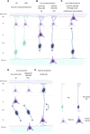
Different scenarios of changed polarity affecting RG and leading to cortical malformations. (A) RG and bRG (both light blue-green) are present during normal cortical development and neurons (light purple) are correctly positioned in the cortical plate following migration along the basal process. RG apical detachment giving rise to bRG from RG is regulated by factors such as Plekha7 (Tavano et al., 2018). (B) Perturbed RG (dark blue) with loss of apical processes can lead to cortical malformations in the mouse such as subcortical heterotopia (SH, left) e.g., due to mutations in Eml1, RhoA (Zaidi et al., 2024; Cappello et al., 2012). Breakages in the ventricular boundary can also lead to apical cell detachment and periventricular heterotopia (PVH, right) (e.g., mutations in Fat4, Dchs1, Cappello et al., 2013). Ectopic neurons are depicted in dark purple. (C) Loss of basal process attachment, often caused by defective signalling, can be accompanied by breaches of the basal lamina. This leads to a cobblestone-like lissencephaly as seen for mutation in laminin and integrin genes, among others (Haubst et al., 2006; Radakovits et al., 2009). (D) RG can lose polarity both apically and basally, leading to internalised RG as seen for example, for αE-catenin and Llgl1 mouse models, causing respectively SH (left) and PVH (right)-like phenotypes (Lien et al., 2006; Schmid et al., 2014). (E) More rarely, inversion of polarity in RG can be observed as in Arl13b mouse mutants (Higginbotham et al., 2013). In this situation, the cell soma is located next to the basal lamina. Conversely, neurons are found at the ventricular surface.
FIGURE 3
FIGURE 3
Features and composition of RG compartments. (A) Apical process and endfoot. RG are in contact with each other through AJ. A PC (dark green) protrudes in the ventricle, centrioles (light green) act as an MT organizing centre. The PC receives signals from the CSF. Mitochondria, Golgi and ER are also present in the apical side of RG. (B) Interkinetic nuclear migration. RG nuclei are found most basally during S phase and move to the ventricular surface to enter mitosis, aided by dynein (orange) along the MT cytoskeleton. The apical to basal movement is supported by kinesin (pink). (C) Basal process and endfeet. ER and mitochondria are also found in the basal side of RG, the latter particularly enriched in the endfoot. Trans-Golgi elements are present in basal process varicosities, associated with CAMSAP which acts as an MT nucleator removed from the centrosome. mRNA transport along the basal process and local translation in the basal endfeet are represented. Proteins on the surface of the endfoot (integrins, dystroglycan complex) ensure the contact with the ECM. Abbreviations: AJ, adherens junctions; PC, primary cilia; MT, microtubules; CSF, cerebrospinal fluid; ER, endoplasmic reticulum; RBP, RNA binding protein.

References

    1. Alexandre P., Reugels A. M., Barker D., Blanc E., Clarke J. D. W. (2010). Neurons derive from the more apical daughter in asymmetric divisions in the zebrafish neural tube. Nat. Neurosci. 13 (6), 673–679. 10.1038/nn.2547 - DOI - PubMed
    1. Andrews M. G., Subramanian L., Kriegstein A. R. (2020). Mtor signaling regulates the morphology and migration of outer radial glia in developing human cortex. ELife 9, e58737. 10.7554/ELIFE.58737 - DOI - PMC - PubMed
    1. Anton E. S., Marchionni M. A., Lee K. F., Rakic P. (1997). Role of GGF/neuregulin signaling in interactions between migrating neurons and radial glia in the developing cerebral cortex. Development 124 (18), 3501–3510. 10.1242/dev.124.18.3501 - DOI - PubMed
    1. Arai Y., Taverna E. (2017). Neural progenitor cell polarity and cortical development. Front. Cell. Neurosci. 11, 384. 10.3389/fncel.2017.00384 - DOI - PMC - PubMed
    1. Bartkowska K., Paquin A., Gauthier A. S., Kaplan D. R., Miller F. D. (2007). Trk signaling regulates neural precursor cell proliferation and differentiation during cortical development. Development 134 (24), 4369–4380. 10.1242/dev.008227 - DOI - PubMed

LinkOut - more resources