. 2024 Oct 31;41(6):421-441.
doi: 10.4274/balkanmedj.galenos.2024.2024-9-98. Epub 2024 Oct 17.

Takotsubo Syndrome: An International Expert Consensus Report on Practical Challenges and Specific Conditions (Part-1: Diagnostic and Therapeutic Challenges)

Affiliations

Takotsubo Syndrome: An International Expert Consensus Report on Practical Challenges and Specific Conditions (Part-1: Diagnostic and Therapeutic Challenges)

Kenan Yalta et al. Balkan Med J. .

Abstract

In the recent years, there has been a burgeoning interest in Takotsubo syndrome (TTS), which is renowned as a specific form of reversible myocardial dysfunction. Despite the extensive literature available on TTS, clinicians still face several practical challenges associated with the diagnosis and management of this phenomenon. This potentially results in the underdiagnosis and improper management of TTS in clinical practice. The present paper, the first part (part-1) of the consensus report, aims to cover diagnostic and therapeutic challenges associated with TTS along with certain recommendations to combat these challenges.


Conflict of interest statement

Conflict of Interest: The authors declare that they have no conflict of interest.

Figures
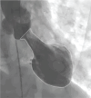
Figure 1
Figure 1
Typical shape of “octopus trap” at end-systole on invasive ventriculogram in a patient with apico-midventricular TTS. TTS, Takotsubo syndrome.
Figure 2
Figure 2
Various morphological patterns of takotsubo syndrome (TTS) on invasive left ventriculogram in the right anterior oblique view during diastole (D) and systole (S): (1) mid-apical pattern; (2) mid-ventricular pattern; (3) basal or mid-basal pattern (also termed inverted TTS); (4) focal pattern (focal anterior); (5) mid-apical pattern with apical tip-sparing namely the nipple sign (white arrow).
Figure 3
Figure 3
Challenging conditions in TTS diagnosis. *Including acute neurological conditions, sepsis, etc. that might potentially mask cardiovascular manifestations of TTS.24,25; ***Ischemic or post-ischemic myocardial stunning that might mimic a true TTS episode.28,35; TTS, Takotsubo syndrome; WMA, wall motion abnormality.
Figure 4
Figure 4
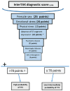
The InterTAK diagnostic score. *Excluding lead aVR; TTS, Takotsubo syndrome.
Figure 5
Figure 5
Invasive left ventriculogram and cardiac magnetic resonance imaging (CMR) in a middle-aged woman presenting with chest pain after a stressful situation. Coronary angiogram on admission revealed normal left and right coronary arteries. Invasive left ventriculogram demonstrated typical mid-apical takotsubo syndrome (TTS) with an apical tip-sparing pattern (A, B). Cardiac MRI 3 days after admission demonstrated mild improvement in left ventricular function particularly in the apical region in cine images (C, D). Native T1 mapping demonstrated significant increases in T1 values in the mid-apical regions particularly in the septal and anterior segments [(E, F), white arrows]. Extracellular volume (ECV) mapping also demonstrated significant increases in ECV values representing myocardial edema in the corresponding areas [(G, H), white arrows]. Images of late gadolinium enhancement were consistent with an increased transmural signal intensity (but no infarction changes) in the mid-apical region particularly the septal segment [(I, J), white arrows]. Consequently, this demonstrates “myocarditis-like” changes in the mid-apical (septal and anterior segments) in a typical case of TTS.
Figure 6
Figure 6
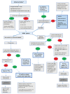
A proposed clinical algorithm for TTS diagnosis in the presence of diagnostic challenges. ACS, acute coronary syndrome; NSTEMI, non-ST segment elevation myocardial infarction; STEMI, ST segment elevation myocardial infarction; MINOCA, myocardial infarction with non-obstructive coronary arteries; TTS, Takotsubo syndrome; PE, pulmonary embolism; MRI, magnetic resonance imaging; V/P, ventilation perfusion; CAG, coronary angiogram; LV, left ventricle; PCI, percutaneous coronary intervention; WMA, wall motion abnormality; CCTA, coronary computed tomography angiography; CAD, coronary artery disease; ECG, electrocardiogram. *: ACS presentation denotes classical symptoms of ACS (chest pain, dyspnea, etc.) and significant increases in serum levels of myocardial enzymes (including troponins) with or without ischemic ECG changes. Decision making for the initial diagnostic modality (invasive CAG vs CCTA) is generally based on the presence of ST segment elevation, high-risk clinical features and the likelihood of TTS (InterTAK score ≤ 70 vs. > 70).6,13,17,23 **: High risk features include persistent chest pain, hemodynamic instability and malignant arrhythmogenesis. ***: Invasive CAG in this setting may be warranted in the presence of ACS signs on CCTA (thrombus, focal WMA within the territory of a severely stenotic coronary artery, etc.). Invasive CAG may confirm an existing ACS, and enables further management (including PCI). ****: Occlusive CAD usually signifies a stenosis severity of > 50%.33 &: Characteristic WMA pattern denotes a circumferential apical, midventricular or basal (or adjacent combination of these) LV involvement usually accompanied by hypercontraction of the non-affected segments. Global LV or biventricular WMAs may also arise following stressful triggers, and may suggest TTS in the absence of any other condition accounting for such extensive myocardial involvement including myocarditis, septic cardiomyopathy and pre-existing cardiomyopathy.2-10 Cardiac MRI may be indicated even after full recovery of a global or biventricular dysfunction to differentiate TTS from similar conditions including myocarditis. ß: ACS signs (including plaque rupture, thrombus on CAG) involving the coronary artery remote from the territory of focal WMA pattern or ACS signs in the presence of a global (or biventricular) WMA pattern may also suggest a co-existing ACS, and need to be carefully evaluated and managed accordingly. ßß: A high interTAK score denotes a score of > 70.13,36 ßßß: Specific biomarker ratios include N-terminal proBNP/troponin T (NT-proBNP/TnT).37 µClassical TTS signifies the widely recognized TTS form with a characteristic circumferential WMA pattern and with a normal or non-occlusive coronary anatomy in the presence of an ACS presentation. A high interTAK score may further support the diagnosis of classical TTS. However, a low score can not definitely exclude the diagnosis in this context.1-9,13 €: Recovery of WMAs is expected to be complete at 3 months in the setting of TTS.7 Therefore, the decision-making for incomplete or failed recovery is usually implemented at 3 months following discharge.13 Cardiac MRI is the preferred modality for the identification of the persistent WMA trigger in this context.6,13,17,23,28 Cardiac MRI and intracoronary imaging may be performed before discharge if the alternative diagnosis is highly likely based on clinical features (including signs of myocarditis, low interTAK score, ambiguous coronary artery findings suggestive of MINOCA).13,28 €€: Diagnostic accuracy of hemodynamic tests including fractional flow reserve may be hampered by the TTS-related microvascular dysfunction during the acute stage.82
Figure 7
Figure 7
A summary of the therapeutic challenges in patients with uncomplicated TTS. TTS, Takotsubo syndrome.

Dataset described in

References

    1. Sato H, Tateishi H, Uchida T. Takotsubo-type cardiomyopathy due to multivessel spasm. In: Kodama K, Haze K, Hon M, et al. (editors). Clinical Aspect of Myocardial Injury: From Ischemia to Heart Failure, Kagakuhyouronsha, Tokyo, 1990. pp. 56-64.
    1. Lyon AR, Citro R, Schneider B. Pathophysiology of Takotsubo Syndrome: JACC State-of-the-Art Review. J Am Coll Cardiol. 2021;77:902–921. - PubMed
    1. Omerovic E, Citro R, Bossone E. Pathophysiology of Takotsubo syndrome - a joint scientific statement from the Heart Failure Association Takotsubo Syndrome Study Group and Myocardial Function Working Group of the European Society of Cardiology - Part 1: overview and the central role for catecholamines and sympathetic nervous system. Eur J HeartFail. 2022;24:257–273. - PubMed
    1. Omerovic E, Citro R, Bossone E. Pathophysiology of Takotsubo syndrome - a joint scientific statement from the Heart Failure Association Takotsubo Syndrome Study Group and Myocardial Function Working Group of the European Society of Cardiology - Part 2: vascular pathophysiology, gender and sex hormones, genetics, chronic cardiovascular problems and clinical implications. Eur J Heart Fail. 2022;24(2):274–286. doi: 10.1002/ejhf.2368. - DOI - PubMed
    1. Ghadri JR, Wittstein IS, Prasad A. International Expert Consensus Document on Takotsubo Syndrome (Part I): Clinical Characteristics, Diagnostic Criteria, and Pathophysiology. Eur Heart J. 2018;39(22):2032–2046. doi: 10.1093/eurheartj/ehy076. - DOI - PMC - PubMed

MeSH terms

LinkOut - more resources