. 2024 Dec;79(12):3521-3525.
doi: 10.1111/all.16352. Epub 2024 Oct 17.

Transcriptomic evidence for T cell-fibroblast-keratinocyte axis via IL-13-periostin-integrin in atopic dermatitis

Affiliations

Transcriptomic evidence for T cell-fibroblast-keratinocyte axis via IL-13-periostin-integrin in atopic dermatitis

Nguyen Quoc Vuong Tran et al. Allergy. 2024 Dec.
No abstract available


Conflict of interest statement

The authors declare no conflicts of interest.

Figures
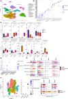
FIGURE 1
FIGURE 1
scRNA‐seq analysis of healthy, atopic dermatitis, and psoriasis samples. (A) UMAP of cell clusters integrated from healthy skin (n = 20), lesional skin from AD (n = 9), and lesional skin from psoriasis (n = 22) samples according to similarity of transcriptome. In total, seventeen cell clusters were identified. (B) Dot plot displaying average expression and frequency of canonical cell‐type markers for each cluster. (C) Boxplots represent the proportion of keratinocytes, fibroblast, and immune cell clusters (calculated from Table S2) in healthy, AD, and psoriasis skin. *p ≤ .05, **p ≤ .01, ***p ≤ .001, ****p ≤ .0001 Kruskal‐Wallis one‐way ANOVA with Dunn's post‐hoc test. (D) Dot plot displaying average expression and frequency of POSTN, ITGAV, ITGB3, ITGB5, IL‐4, IL‐13, and IL‐17 for each cluster. (E) Violin plot displaying expression levels for POSTN, ITGAV, ITGB3, and ITGB5 for fibroblast and keratinocytes. (F) UMAP for subcluster analysis of fibroblasts (left panel). Dot plot (middle panel) and violin plot for (right panel) the expression of POSTN, COL6A5, and MFAP5 in the subclusters of fibroblasts.
FIGURE 2
FIGURE 2
Cell–cell communication analysis for keratinocytes, fibroblast, and immune cells from the integrated scRNA‐seq. (A) Circle plot demonstrates the connections via POSTN‐ITGAV/ITGB5 (upper panel) and heatmap shows the relative importance (lower panel) of each cell group for the Periostin signaling network in healthy, AD, and psoriasis. In circle plot, each cell group is assigned a color together with edge color to illustrate the signal started from that cell group. Edge width represents the relative communication probability. (B) Circle plot demonstrates the connections via IL‐13‐IL4R/IL13RA1 complex and IL‐13–IL13RA1 and heatmap shows the relative importance of each cell group for IL‐4 signaling network specific for AD. (C) Circle plot demonstrates the connections via IL‐17‐IL17RA/IL17RC complex and heatmap shows the relative importance of each cell group for IL‐17 signaling network specific for psoriasis. (D) Boxplots represent the proportion of POSTN + IL4R + IL13RA1 + fibroblasts (calculated from Table S2) in healthy, AD, and psoriasis skin. Kruskal‐Wallis one‐way ANOVA with Dunn's post‐hoc test. (E) Scatter plots and correlations for the expression of POSTN with IL4R and IL13RA1 in POSTN + IL4R + IL13RA1 + fibroblasts. Spearman correlation coefficient (R) and p‐value are shown. Different colors indicated conditions as shown in the legend.

References

    1. Kou K, Okawa T, Yamaguchi Y, et al. Periostin levels correlate with disease severity and chronicity in patients with atopic dermatitis. Br J Dermatol. 2014;171(2):283‐291. doi:10.1111/bjd.12943 - DOI - PubMed
    1. Masuoka M, Shiraishi H, Ohta S, et al. Periostin promotes chronic allergic inflammation in response to Th2 cytokines. J Clin Invest. 2012;122(7):2590‐2600. doi:10.1172/JCI58978 - DOI - PMC - PubMed
    1. Nunomura S, Uta D, Kitajima I, et al. Periostin activates distinct modules of inflammation and itching downstream of the type 2 inflammation pathway. Cell Rep. 2023;42(1):111933. doi:10.1016/j.celrep.2022.111933 - DOI - PMC - PubMed
    1. Zhou J, Liang G, Liu L, et al. Single‐cell RNA‐seq reveals abnormal differentiation of keratinocytes and increased inflammatory differentiated keratinocytes in atopic dermatitis. J Eur Acad Dermatol Venereol. 2023;37(11):2336‐2348. doi:10.1111/jdv.19256 - DOI - PubMed
    1. Izuhara K, Arima K, Ohta S, Suzuki S, Inamitsu M, Yamamoto K. Periostin in allergic inflammation. Allergol Int. 2014;63(2):143‐151. doi:10.2332/allergolint.13-RAI-0663 - DOI - PubMed

LinkOut - more resources