. 2024 Oct 17;22(10):e3002786.
doi: 10.1371/journal.pbio.3002786. eCollection 2024 Oct.

Chromatin remodeller Chd7 is developmentally regulated in the neural crest by tissue-specific transcription factors

Affiliations

Chromatin remodeller Chd7 is developmentally regulated in the neural crest by tissue-specific transcription factors

Ruth M Williams et al. PLoS Biol. .

Abstract

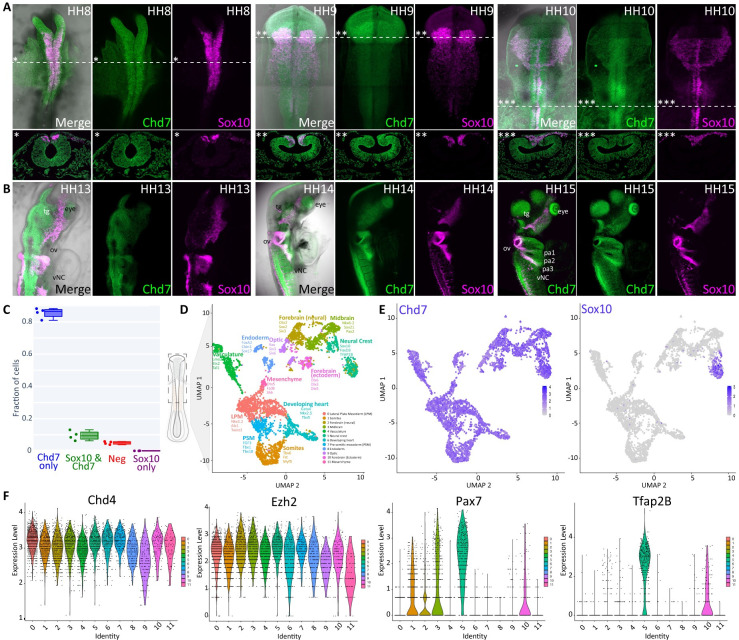
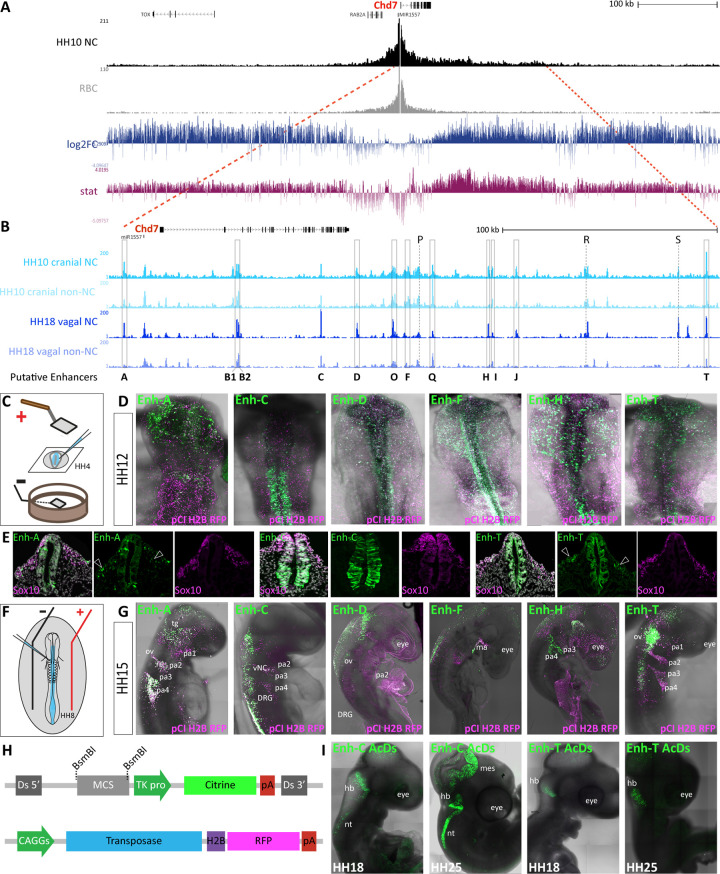
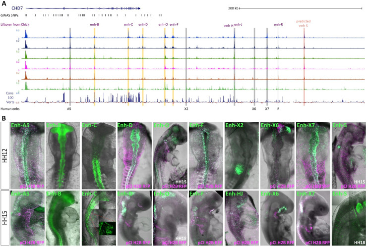
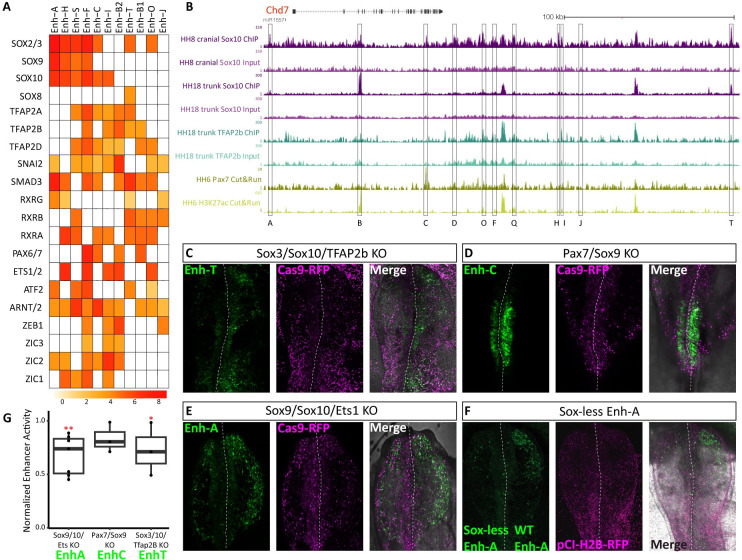
Neurocristopathies such as CHARGE syndrome result from aberrant neural crest development. A large proportion of CHARGE cases are attributed to pathogenic variants in the gene encoding CHD7, chromodomain helicase DNA binding protein 7, which remodels chromatin. While the role for CHD7 in neural crest development is well documented, how this factor is specifically up-regulated in neural crest cells is not understood. Here, we use epigenomic profiling of chick and human neural crest to identify a cohort of enhancers regulating Chd7 expression in neural crest cells and other tissues. We functionally validate upstream transcription factor binding at candidate enhancers, revealing novel epistatic relationships between neural crest master regulators and Chd7, showing tissue-specific regulation of a globally acting chromatin remodeller. Furthermore, we find conserved enhancer features in human embryonic epigenomic data and validate the activity of the human equivalent CHD7 enhancers in the chick embryo. Our findings embed Chd7 in the neural crest gene regulatory network and offer potentially clinically relevant elements for interpreting CHARGE syndrome cases without causative allocation.


Conflict of interest statement

The authors have declared that no competing interests exist.

Figures

Fig 1
Fig 1. Chd7 expression during early chick development.
(A, B) In vivo Chd7 expression (green) determined using HCR. Sox10 (magenta) is used as a neural crest marker. (A) Chd7 is expressed in neural crest cells and throughout the neural tube, as well as the surrounding ectoderm and underlying mesoderm at HH8-10. (B) Chd7 is more broadly expressed at HH13-HH15 including head and hindbrain structures. From HH13 through HH15 Chd7 expression is detected in the otic vesicle (ov), trigeminal ganglia (tg) eye, and vagal neural crest (vNC). At HH15 Chd7 is also expressed in the pharyngeal arches 1–4 (pa1-4) and dorsal root ganglion (drg). (C) Quantification of HCR signal from Chd7 and Sox10 transcripts in stage HH10 chick embryos (n = 4). (D) UMAP representation of 5,669 single cells resolved into 12 clusters of based on shared transcriptional identities. (E) Feature plots of Chd7 and Sox10 expression across scRNA-seq clusters. (F) Violin plots of selected chromatin remodellers and transcription factors expressed across scRNA-seq clusters. HCR, hybridisation chain reaction.
Fig 2
Fig 2. Chd7 enhancer prediction from neural crest Capture-C and ATAC data.
(A, B) UCSC genome browser view of the chick Chd7 locus in galGal5. (A) Capture-C tracks from cranial neural crest at HH10 and control RBC, showing the Chd7 TAD. Differential interactions were determined using DESeq2, hypothesis tested with Wald test and corrected for multiple testing using the Benjamin–Hochberg method. Wald statistics track (stat, in pink) represents ratio of LogFoldChange values and their standard errors. (B) ATAC-seq data from HH10 cranial [32] and HH18 vagal [37] neural crest cells and non-neural crest control cells collected by FACS using FoxD3 enhancer NC1 (cranial) and FoxD3/Ednrb enhancers NC2/E1, respectively. Putative enhancers within the Chd7 TAD are indicated by grey boxes. (C) Schematic representation of ex ovo chicken embryo electroporation technique used to deliver enhancer reporter constructs. (D) Fluorescent reporter activity (green) recorded from indicated enhancers at HH12 following ex ovo electroporation at HH4 with a ubiquitous electroporation control pCI-H2B-RFP (magenta). (E) Transverse sections of embryos at approximately HH12 showing enhancer activity in green and Sox10 expression in magenta, detected by HCR. Migrating neural crest cells are indicated with arrows. (F) Schematic representation of in ovo chicken embryo electroporation. (G) Enhancer activity (green) recorded from indicated enhancers at HH15 following in ovo electroporation into the neural tube at HH9 with a ubiquitous electroporation control pCI-H2B-RFP (magenta). pa; pharyngeal arch, tg; trigeminal ganglia, ov; otic vesicle, ma, mandibula arch. (H) Schematics of AcDs constructs used to integrate the enhancer reporter cassette into the genome to observe sustained enhancer activity in vivo. (I) Enhancer activity of indicated enhancers at later stages following in ovo electroporation of AcDs enhancer reporter plasmids, nt; neural tube, mes; mesencephalon, hb; hindbrain. HCR, hybridisation chain reaction; RBC, red blood cell; TAD, topologically associating domain.
Fig 3
Fig 3. Epigenomic landscape of human CHD7 locus.
(A) CHD7 locus in human genome (hg38, chr8:60,665,230–61,241,522), showing ATAC profiles of 6 clusters from Multiome data generated from human embryonic cranial samples (see Methods). Chick enhancers lifted over from galGal5 are shown in orange boxes; novel human enhancers are shown in grey boxes. GWAS SNPs from the NHGRI-EBI GWAS Catalog is also indicated. (B) Human enhancer activity (green) in vivo at HH12 following ex ovo electroporation and HH15 following in ovo electroporation. Ubiquitous electroporation control pCI-H2B-RFP is shown in magenta. GWAS, genome-wide association studies.
Fig 4
Fig 4. Upstream transcription factors controlling Chd7 enhancer activity.
(A) Heatmap showing predicted TF binding motifs within chick Chd7 enhancers. (B) Tracks from UCSC genome browser showing biotin-ChIP data for Sox10 (purple), Tfap2B (teal), Pax7 (yellow), and H3K27ac. (C–E) Chicken embryos showing indicated enhancer activity (GFP) and Cas9 (RFP) following bilateral electroporation of Cas9 and guide RNAs targeting selected TFs on the left (experimental) and scrambled guide RNA with Cas9 on the right (control). (F) Chicken embryo bilaterally electroporated with a construct containing enh-A where all the Sox sites have been mutated on the left and wild-type enh-A construct on the right, both driving GFP expression. pCI-H2B-RFP was co-electroporated on both sides as a control. (G) Loss of enhancer activity (GFP intensity) following TF knockout experiments in (C–E) was quantified left versus right, relative to control RFP intensity, two-tailed paired T test, enh-A; p = 0.023**, n = 7 embryos, enh-C; p = 0.114, ns n = 3, enh-T; p = 0.047*, n = 4. TF, transcription factor.

References

    1. Gorlin RJ, Cohen MM, Hennekam RCM. Syndromes of the head and neck. 4th ed. Oxford England; New York: Oxford University Press; 2001. xiv, p. 1283.
    1. Pagon RA, Graham JM, Jr., Zonana J, Yong SL. Coloboma, congenital heart disease, and choanal atresia with multiple anomalies: CHARGE association. J Pediatr. 1981;99(2):223–7. Epub 1981/08/01. doi: 10.1016/s0022-3476(81)80454-4 . - DOI - PubMed
    1. Zentner GE, Layman WS, Martin DM, Scacheri PC. Molecular and phenotypic aspects of CHD7 mutation in CHARGE syndrome. Am J Med Genet A. 2010;152A(3):674–86. Epub 2010/02/27. doi: 10.1002/ajmg.a.33323 ; PubMed Central PMCID: PMC2918278. - DOI - PMC - PubMed
    1. Martucciello G, Bicocchi MP, Dodero P, Lerone M, Silengo Cirillo M, Puliti A, et al.. Total colonic aganglionosis associated with interstitial deletion of the long arm of chromosome 10. Pediatr Surg Int. 1992;7(4):308–10. doi: 10.1007/BF00183991 - DOI
    1. Heuckeroth RO. Hirschsprung disease—integrating basic science and clinical medicine to improve outcomes. Nat Rev Gastroenterol Hepatol. 2018;15(3):152–67. Epub 2018/01/05. doi: 10.1038/nrgastro.2017.149 . - DOI - PubMed

MeSH terms