Review
. 2024 Dec;44(6):720-731.
doi: 10.1055/s-0044-1791666. Epub 2024 Oct 17.

The Pathophysiology and Biomarkers of Delirium

Affiliations
Review

The Pathophysiology and Biomarkers of Delirium

Camryn J Smith et al. Semin Neurol. 2024 Dec.

Abstract

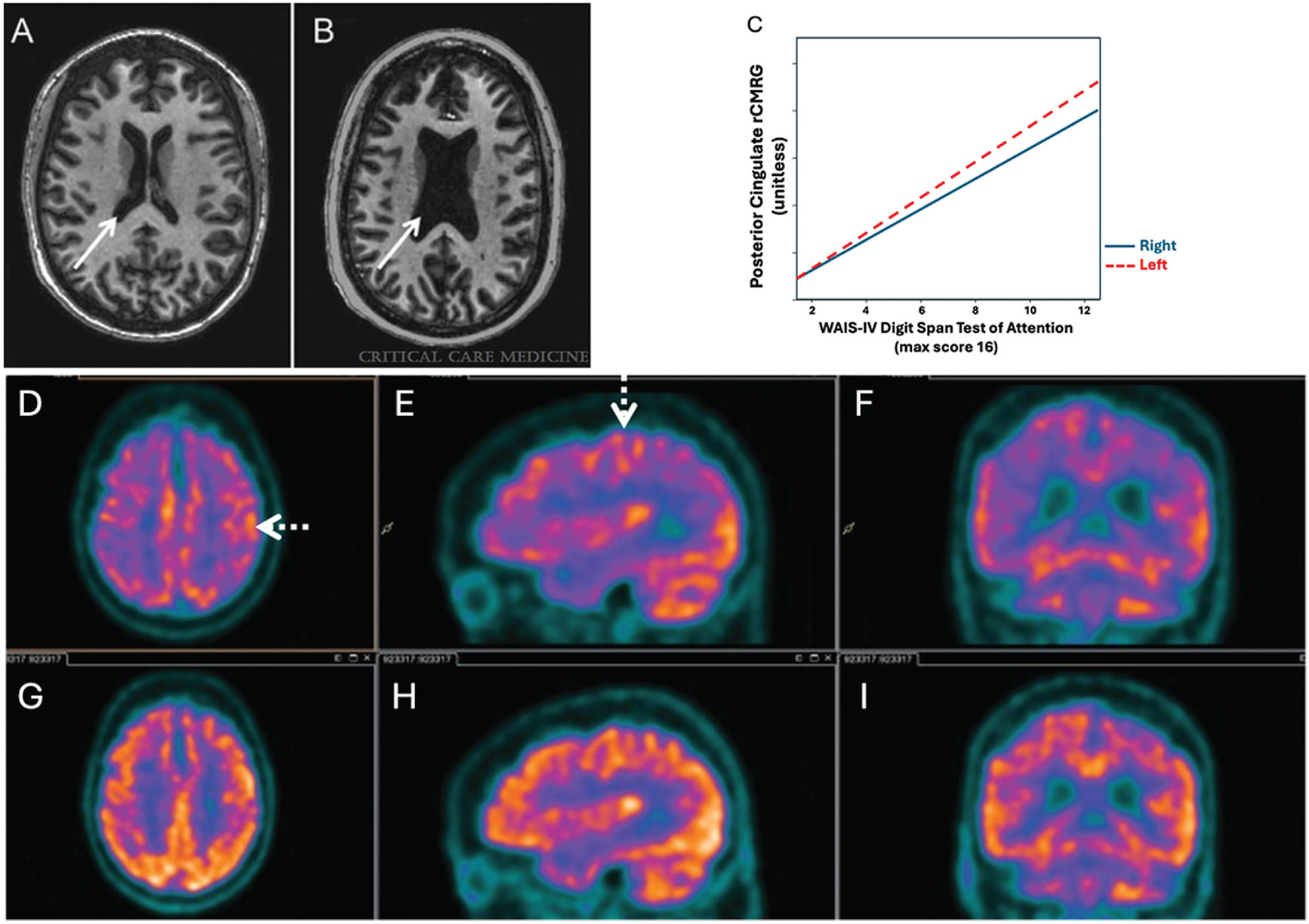
Delirium is a major disturbance in the mental state characterized by fluctuations in arousal, deficits in attention, distorted perception, and disruptions in memory and cognitive processing. Delirium affects approximately 18% to 25% of hospital inpatients, with even higher rates observed during critical illness. To develop therapies to shorten the duration and limit the adverse effects of delirium, it is important to understand the mechanisms underlying its presentation. Neuroimaging modalities such as magnetic resonance imaging (MRI), positron emission tomography, functional MRI, and near-infrared spectroscopy point to global atrophy, white matter changes, and disruptions in cerebral blood flow, oxygenation, metabolism, and connectivity as key correlates of delirium pathogenesis. Electroencephalography demonstrates generalized slowing of normal background activity, with pathologic decreases in variability of oscillatory patterns and disruptions in functional connectivity among specific brain regions. Elevated serum biomarkers of inflammation, including interleukin-6, C-reactive protein, and S100B, suggest a role of dysregulated inflammatory processes and cellular metabolism, particularly in perioperative and sepsis-related delirium. Emerging animal models that can mimic delirium-like clinical states will reveal further insights into delirium pathophysiology. The combination of clinical and basic science methods of exploring delirium shows great promise in elucidating its underlying mechanisms and revealing potential therapeutic targets.


Conflict of interest statement

None declared.

Figures

Fig. 1
Fig. 1
Structural and metabolic changes associated with delirium. (A, B) Representative examples of lateral ventricle size in two intensive care unit (ICU) survivors with no preexisting cognitive impairment (by patient and surrogate reports and review of records). Axial T1-weighted brain images at the time of hospital discharge. (A) Relatively normal ventricular volume (solid arrows) in a 46-year-old female with respiratory and cardiac failure, who required mechanical ventilation in ICU and did not experience delirium. (B) Enlarged ventricles in a 42-year-old female with community-acquired pneumonia, who required mechanical ventilation in the ICU and whose ICU course was complicated by 12 days of delirium. (C) Correlation between posterior cingulate cortex regional cerebral metabolic rate and WAIS-IV digit span score, a measure of attention, during delirium. (D-I) FDG-PET hypometabolism in bilateral frontal, parietal, and temporal cortices during delirium (D–F) with relative sparing of the sensorimotor cortex (dotted arrows), and reversal after delirium resolution (G–I). Panels D and G are axial slices; E and H are sagittal slices; F and I are coronal slices. (Panels A, B: reprinted with permission from Gunther et al, Crit Care Med 2012; C: modified with permission from Haggstrom et al., J Cereb Blood Flow Metab; D–I: reprinted with permission from Haggstrom et al, J Cereb Blood Flow Metab 2017.)
Fig. 2
Fig. 2
Typical EEG patterns observed during wakefulness, sleep, and delirium. (A–C) Representative 2-second EEG recordings from the left frontal, central, parietal, and occipital regions. Channel labels to the left of each tracing indicate the region recorded. (Refer to the left panel for a pictorial index of regions.) (D–F) Representative spectrograms indicating dynamic changes in frequency composition of the EEG over a 5-minute period. Warmer colors indicate a greater contribution of a given frequency range. During wakefulness, there is a prominent contribution of alpha (8–13 Hz) frequencies. During sleep, there is prominent delta (<4 Hz) activity, with intermittent spindle (12–15 Hz) activity. During delirium, there is prominent delta activity with a paucity of variability over time. All recordings are in the absence of sedative medications.
Fig. 3
Fig. 3
A schematic representation of inducing delirium in animal models (left half) and outcomes to measure a delirium-like state in animal models (right half). Surgical methods include cecal ligation puncture, tibial fracture, and ischemia-reperfusion, while injection methods include cecal slurry, LPS, atropine, cytokines (IL-1β, TNF-α), and neurotoxins. Environmental factors to induce a delirium-like state in animal models include sleep deprivation through light, sound, and physical disturbance. Molecular markers encompass biomarkers such as inflammatory markers (IL-6, CRP, S100B, IGF) and oxidative stress markers (reactive oxygen species, lipid peroxidation products). Functional markers include behavior (cognitive deficits, activity, anxiety- and depressive-like behavior), imaging modalities (fMRI, MRI, PET, NIRS), and EEG patterns (increase in delta and theta activity, decrease in alpha and beta, increase in episodic bursts of spikes).

References

    1. Detke HC, Goadsby PJ, Wang S, Friedman DI, Selzler KJ, Aurora SK. Galcanezumab in chronic migraine: the randomized, double-blind, placebo-controlled REGAIN study. Neurology 2018;91 (24):e2211–e2221 - PMC - PubMed
    1. Haggstrom L, Welschinger R, Caplan GA. Functional neuroimaging offers insights into delirium pathophysiology: a systematic review. Australas J Ageing 2017;36(03):186–192 - PubMed
    1. Jiang S, Efron PA, Oh ES, DeKosky ST. Optical neuroimaging in delirium. Photonics 2023;10(12):1334
    1. Maldonado JR. Delirium pathophysiology: an updated hypothesis of the etiology of acute brain failure. Int J Geriatr Psychiatry 2018;33(11):1428–1457 - PubMed
    1. Brown CH IV, Faigle R, Klinker L, et al. The association of brain MRI characteristics and postoperative delirium in cardiac surgery patients. Clin Ther 2015;37(12):2686–2699.e9 - PMC - PubMed