. 2024 Nov;300(11):107892.
doi: 10.1016/j.jbc.2024.107892. Epub 2024 Oct 16.

PP2A activation overcomes leptomeningeal dissemination in group 3 medulloblastoma

Affiliations

PP2A activation overcomes leptomeningeal dissemination in group 3 medulloblastoma

Nazia Nazam et al. J Biol Chem. 2024 Nov.

Abstract

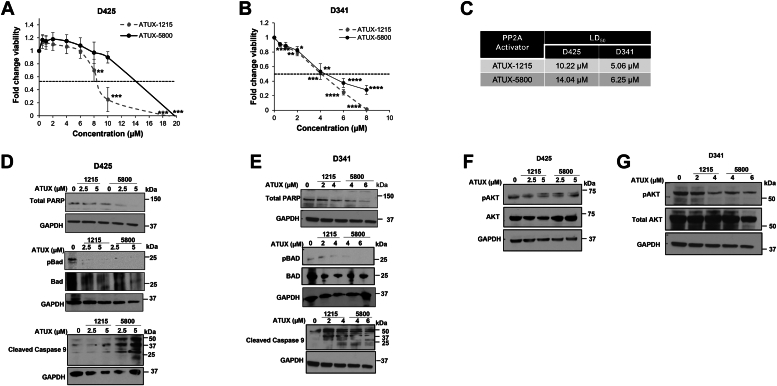
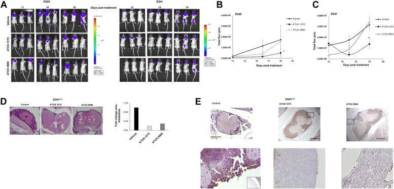
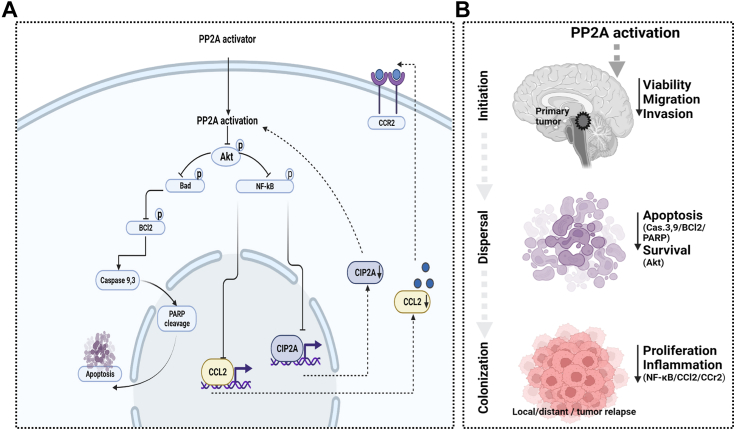
Leptomeningeal dissemination (LMD) is the primary cause of treatment failure in children with group 3 medulloblastoma (MB). Building on our previous work on protein phosphatase 2A (PP2A) activation in MB, here we present preclinical and molecular data on the effects of two novel classes of PP2A activators on disease processes of LMD in group 3 MB. The PP2A activators used in this study are ATUX-6156 and ATUX-6954 (diarylmethylcycloamine sulfonylureas), and ATUX-1215 and ATUX-5800 (diarylmethyl-4-aminotetrahydropyran-sulfonamides). Treatment with these compounds led to suppression of the endogenous PP2A inhibitor, cancerous inhibitor of PP2A (CIP2A), enhanced phosphatase activity (10-60%), and reduced MB viability, migration, and invasion, prerequisites for MB cells to access the cerebrospinal fluid, affecting the initiation stage of LMD. PP2A activator treatment of MB cells led to apoptosis mediated via caspase 9/PARP signaling due to decreased phosphorylation of Bad, impeding the dispersal stage of LMD. Cell proliferation and LMD-driving cellular traits and molecules pertinent to the third stage, colonization, were also affected. Treatment with ATUX-1215 or ATUX-5800 prevented LMD in an intraventricular murine model of MB, possibly mediated by disruption of the CCL2-CCR2 axis by altered NF-kB phosphorylation via disrupted AKT signaling. The present investigation offers proof-of-principle data for PP2A-based reactivation therapy for Group 3 MB and provides the first indications that PP2A reactivation may challenge the current paradigm in targeting the 3-stage process of MB LMD. Further investigations of PP2A activators are warranted as these compounds may prove beneficial as therapeutics for MB.

Keywords: PP2A; diarylmethyl cycloamine sulfonylureas; leptomeningeal dissemination; medulloblastoma; pyran sulfonamides.


Conflict of interest statement

Conflict of interest Patent applications WO2023/023594 and WO2021/188949, inventor MO, are assigned to Atux Iskay LLC. The other authors declare that they have no conflict of interest with the contents of this article.

Figures

Figure 1
Figure 1
Microsome stability and mouse pharmacokinetic parameters and brain exposure for PP2A activating compounds.A, the chemical structures of PP2A activators belonging to the diarylmethyl-pyran-sulfonamides group (ATUX-1215 and ATUX-5800) (top panel) and the diarylmethyl cycloamine sulfonylurea (ATUX-6156 and ATUX-6954) (bottom panel) compounds used in the present study are shown. H represents hydrogen and F represents fluorine. B, the comparative in vitro microsome stability studies for ATUX-1215, ATUX-5800, ATUX-6156, and ATUX-6954 showed a short half-life (T1/2) and high clearance (CL) of ATUX-5800 and ATUX-6954. Substituting fluorine for hydrogen leads to increased stability with respect to oxidative metabolism resulting in longer T1/2 and lower CL rendering ATUX-1215 and ATUX-6156 more stable than ATUX-5800 or ATUX-6954, respectively. C and D, the pharmacokinetic (PK) parameters of ATUX-1215, ATUX-5800, and ATUX-6156 are provided after oral dosing in mice. ATUX-1215 in mouse plasma samples post oral (PO) dosing show higher and more sustained plasma levels than ATUX-5800. E, brain-to-plasma concentration ratios detected 2 h post oral dosing of 30 mg/kg are provided. These PP2A activators demonstrated detectable brain-to-plasma concentration ratio of 1.33 ± 0.31 in ATUX-5800, 0.42 ± 0.27 in ATUX-1215, and 0.02 ± 0.01 in ATUX-6156, demonstrating poor penetrance by ATUX-6156. PP2A, protein phosphatase 2A.
Figure 2
Figure 2
ATUX-1215 and ATUX-5800 activate PP2A in human group 3 MB PDX cells.A, D425 cells (1 × 106) were treated with ATUX-1215 or ATUX-5800 at increasing concentrations (0, 2.5, and 5 μM) for 4 h. PP2A activation was measured with a kit. PP2A activity was increased in the D425 cells with all compounds but did not reach statistical significance. B, D341 cells (1 × 106) were treated with ATUX-1215 or ATUX-5800 at increasing concentrations (0, 2, 4 μM of ATUX-1215 or 0, 4, 6 μM ATUX-5800) for 4 h and PP2A activity was measured. Both drugs increased PP2A activation significantly in the D341 cells. (C) immunoblotting of D425 and (D) D341 whole cell lysates demonstrate decreased CIP2A expression with ATUX-1215 and ATUX-5800. I2PP2A expression increased with ATUX-1215 or ATUX-5800 treatment. Vinculin and GAPDH served as internal loading controls. (CIP2A control GAPDH same blot as 3E, total Parp and cleaved caspase 9). Data are normalized to no treatment group (100) and presented as mean ± standard deviation (SD). Each dot represents an independent biologic replicate. Statistical comparison was completed with two-tailed Student’s t test. ∗∗∗p = 0.001, ∗∗∗∗p = 0.0001. CIP2A, cancerous inhibitor of PP2A; I2PP2A, inhibitor 2 of protein phosphatase 2A; MB, medulloblastoma; PDX, patient-derived xenograft; PP2A, protein phosphatase 2A.
Figure 3
Figure 3
ATUX-1215 and ATUX-5800 impairs viability in human group 3 MB PDX cells.AC, group 3 MB PDX cells were treated with increasing concentrations of PP2A activators (ATUX-1215 and ATUX-5800) for 24 h (D425) or 72 h (D341), and cell viability was measured using alamarBlue assay. A and B, decreased cell viability was dose responsive with increasing PP2A concentrations. Dose-response curves demonstrate variation in lethal dose 50% (LD50) ranging between 5.06 and 14.04 μM for ATUX-1215 and ATUX-5800 in D341 and D425 cells. C, LD50 data from three independent biologic replicates are presented in tabular form. DG, D425 and D341 cells were treated with ATUX-1215 or ATUX-5800 at increasing concentrations (0–6 μM) for 24 h (D425) or 72 h (D341). Immunoblotting of whole cell lysates was completed. D and E, indication of apoptosis in treated D425 and D341 cells includes decreased total PARP and increased cleaved caspase 9. (Total Parp and cleaved caspase 9C control GAPDH same blot as 2D CIP2A). When investigating mechanisms for increased apoptosis we found that treatment with the ATUX compounds decreased phosphorylation of Bad (pBad) in D425 and D341 cells. (BAD control GAPDH same blot as 7B, NF-κB). F and G, phosphorylation of AKT is decreased with increasing concentrations of PP2A activating small molecules, indicating a potential mechanism for decreased cell viability and apoptosis. (BAD control GAPDH same blot as 7B, CCL2) β-actin, vinculin, and GAPDH provided an internal control for equal protein loading. Data are presented as mean ± standard error of the mean (SEM) and represent at least three independent biologic replicates. Statistical comparison was completed with two-tailed Student’s t test. ∗∗p = 0.01, ∗∗∗p = 0.001, ∗∗∗∗p = 0.0001. CIP2A, cancerous inhibitor of PP2A; MB, medulloblastoma; PDX, patient-derived xenograft; PP2A, protein phosphatase 2A.
Figure 4
Figure 4
PP2A activators decrease group 3 MB PDX cell migration and invasion.AF, migration and invasion were determined using Transwell inserts with 8 μm pores, coated on the bottom with laminin with the addition of matrigel to the top side for invasion. Inserts were stained, photographed, and number of cells migrated or invaded were counted. A, C, E, and G, D425 cells (5 × 105) were pretreated with ATUX-1215 or ATUX-5800 (0–5 μM) for 24 h and allowed to migrate or invade for 24 h. There was a significant decrease in the number of cells (A) migrating or (E) invading following treatment. Representative photomicrographs of (C) migration and (G) invasion plates for D425 studies are presented. B, D, F, and H, D341 (5 × 105 cells) were pretreated with ATUX-1215 or ATUX-5800 (0–6 μM) for 24 h and allowed to migrate or invade for 24 h. There was a significant decrease in (B) migration and (F) invasion of the D341 cells with treatment. Representative photomicrographs of the (D) migration and (H) invasion inserts of D341 cells with ATUX-1215 or ATUX-5800 compounds are provided. Points on graphs represent data from independent biologic replicates with data reported as mean ± SD. Data are normalized and compared to no treatment (1.0). Scale bars represent 300 μm (magnification 10×). Box inset represents magnified area to delineate insert pores (small, closed arrow), pores with cells in them (straight line), and migrated or invaded cells (large, closed arrow). Each dot represents an independent biologic replicate. Statistical comparison was completed with two-tailed Student’s t test. ∗p = 0.05, ∗∗p = 0.01, ∗∗∗p = 0.001, ns = not significant, treated versus control. MB, medulloblastoma; PDX, patient-derived xenograft; PP2A, protein phosphatase 2A.
Figure 5
Figure 5
PP2A activators decrease tumorsphere formation of group 3 MB PDX cells.AC, D425 and D341 human group 3 MB PDX cells were plated in 96-well plates under nonadherent conditions at increasing cell concentrations per well (1–5000). Cells treated with ATUX-1215 or ATUX-5800 (0, 5 μM) were assessed for sphere formation after 7 days. Treatment of (A) D425 or (B and C) D341 cells significantly decreased tumorsphere forming ability. ELDA data represents the mean from three independent biologic triplicate experiments. Data were analyzed using extreme limiting dilution analysis software (http://bioinf.wehi.edu.au/software/elda/). ∗∗∗∗p < 0.0001, treated versus control. ELDA, extreme limiting dilution analysis; MB, medulloblastoma; PDX, patient-derived xenograft; PP2A, protein phosphatase 2A.
Figure 6
Figure 6
ATUX-1215 and ATUX-5800 significantly decrease leptomeningeal dissemination in an intraventricular murine model of MB.A and B, luciferase expressing D425Luc or D341Luc cells (5 × 105 in 5 μl methylcellulose) were injected into the right lateral ventricle of athymic nude mice. Beginning on the day of injection, mice were randomized (n = 10 per group) to receive either vehicle or PP2A activators, ATUX-1215, or ATUX-5800 (75 mg/kg), twice per day via oral gavage. Disease progression was followed by bioluminescence imaging and bioluminescence data were analyzed and reported as total flux in photons/second (p/s). A, representative (three animals per group) bioluminescence images 2 to 3 weeks post treatment with ATUX-1215 and ATUX-5800 in D425 (A, right inset) and D341 (A, left inset) are shown. B and C, there was decreased tumor burden in PP2A activator treated versus vehicle-treated animals. B, there was significantly less bioluminescence signal in animals bearing D425Luc tumors treated with ATUX-1215 compared to vehicle-treated controls at 19 days. Bioluminescence trended toward a decrease in D425Luc animals treated with ATUX-5800. C, there was significantly less bioluminescence signal in D341Luc animals at 15 days post treatment. D, H&E staining confirmed histology consistent with MB metastasis in D341Luc tumors (left panels). Metastasis was outlined on photographic images of histologic sections of controls, ATUX-1215 and ATUX-5800 treated animals (two animals in each group) for quantification of metastasis (right panel, dotted black lines). Data including all animals are presented in graphic form showing a decrease in fold change of the area of metastasis upon treatment with ATUX-1215 or ATUX-5800 compared to vehicle-treated controls. E, immunohistochemical staining for CIP2A revealed strong staining in leptomeningeal metastasis in vehicle-treated control animals (left panels) and no staining in the metastasis of animals treated with ATUX-1215 (middle panels) or ATUX-5800 (right panels). Negative control rabbit IgG staining was included (inset, lower left panel). Magnification of 4× for top panels and 40× for bottom panels. Bottom panels represent the area outlined by the black boxes in the upper photomicrographs. All scale bars represent 1 mm. Data are presented as mean ± SEM. Statistical comparison was completed with two tailed Student’s t test comparing vehicle-treated controls to ATUX-1215 or ATUX-5800 treated animals. ∗p = 0.05, ∗∗p = 0.01. CIP2A, cancerous inhibitor of PP2A; Luc, luciferase; MB, medulloblastoma; PP2A, protein phosphatase 2A.
Figure 7
Figure 7
ATUX-1215 and ATUX-5800 treatment disrupts CCL2/CCR2 molecular axis. D425 and D341 PDX cells were treated with increasing concentrations of ATUX-1215 or ATUX-5800, and whole cell lysates were evaluated with immunoblotting. A, in D425 cells, treatment with ATUX-1215 or ATUX-5800 led to decreased CCL2, decreased CCR2 and variable effects on phosphorylation of NF-κB. B, in D341 cells, treatment with ATUX-1215 or ATUX-5800 resulted in decreased CCR2, small decrease in CCL2, and decreased phosphorylation of NF-κB, indicating attenuated NF-κB activation. (CCL2 control GAPDH same blot as 3G, BAD) (NF-κB control GAPDH same blot as 3E, BAD) GAPDH served as an internal protein loading control. PDX, patient-derived xenograft.
Figure 8
Figure 8
PP2A activation overcomes 3-stage process of LMD in group 3 MB.A, small molecule based PP2A activation via reduced CIP2A which downregulates PI3K/Akt survival signals leading to apoptotic induction and reduced CCL2/CCR2 via NF-kB signaling and (B) therefore a decrease in the 3-stage process of LMD via this axis. CIP2A, cancerous inhibitor of PP2A; LMD, leptomeningeal dissemination; MB, medulloblastoma; PP2A, protein phosphatase 2A.

References

    1. Sung H., Ferlay J., Siegel R.L., Laversanne M., Soerjomataram I., Jemal A., et al. Global cancer statistics 2020: GLOBOCAN estimates of incidence and mortality Worldwide for 36 cancers in 185 countries. CA Cancer J. Clin. 2021;71:209–249. - PubMed
    1. Taylor M.D., Northcott P.A., Korshunov A., Remke M., Cho Y.J., Clifford S.C., et al. Molecular subgroups of medulloblastoma: the current consensus. Acta Neuropathol. 2012;123:465–472. - PMC - PubMed
    1. Kijima N., Kanemura Y. Molecular classification of medulloblastoma. Neurol. Med. Chir (Tokyo) 2016;56:687–697. - PMC - PubMed
    1. Thompson E.M., Hielscher T., Bouffet E., Remke M., Luu B., Gururangan S., et al. Prognostic value of medulloblastoma extent of resection after accounting for molecular subgroup: a retrospective integrated clinical and molecular analysis. Lancet Oncol. 2016;17:484–495. - PMC - PubMed
    1. Yeo K.K., Margol A.S., Kennedy R.J., Hung L., Robison N.J., Dhall G., et al. Prognostic significance of molecular subgroups of medulloblastoma in young children receiving irradiation-sparing regimens. J. Neurooncol. 2019;145:375–383. - PMC - PubMed

Publication types

MeSH terms

Substances

LinkOut - more resources