. 2024 Dec 16;13(12):1198-1212.
doi: 10.1093/stcltm/szae074.

Exploring P2X7 receptor antagonism as a therapeutic target for neuroprotection in an hiPSC motor neuron model

Affiliations

Exploring P2X7 receptor antagonism as a therapeutic target for neuroprotection in an hiPSC motor neuron model

Alexandra E Johns et al. Stem Cells Transl Med. .

Abstract

ATP is present in negligible concentrations in the interstitium of healthy tissues but accumulates to significantly higher concentrations in an inflammatory microenvironment. ATP binds to 2 categories of purine receptors on the surface of cells, the ionotropic P2X receptors and metabotropic P2Y receptors. Included in the family of ionotropic purine receptors is P2X7 (P2X7R), a non-specific cation channel with unique functional and structural properties that suggest it has distinct roles in pathological conditions marked by increased extracellular ATP. The role of P2X7R has previously been explored in microglia and astrocytes within the context of neuroinflammation, however the presence of P2X7R on human motor neurons and its potential role in neurodegenerative diseases has not been the focus of the current literature. We leveraged the use of human iPSC-derived spinal motor neurons (hiPSC-MN) as well as human and rodent tissue to demonstrate the expression of P2X7R on motor neurons. We extend this observation to demonstrate that these receptors are functionally active on hiPSC-MN and that ATP can directly induce death via P2X7R activation in a dose dependent manner. Finally, using a highly specific P2X7R blocker, we demonstrate how modulation of P2X7R activation on motor neurons is neuroprotective and could provide a unique pharmacologic target for ATP-induced MN death that is distinct from the role of ATP as a modulator of neuroinflammation.

Keywords: ALS; ATP; P2X7; hiPSC; purinergic signaling.


Conflict of interest statement

A.E.J and N.J.M are unpaid collaborators with Janssen Pharmaceuticals. The other authors declared no potential conflicts of interest.

Figures

Graphical Abstract
Graphical Abstract
Figure 1.
Figure 1.
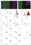
Immunohistochemical labeling and snRNA sequencing analysis showing P2X7R expression in the human cervical spinal cord. All snRNA sequencing data was collected and previously analyzed by Gautier et al., 2023. Alpha and gamma motor neuron clusters were identified by authors based on the enrichment of known marker genes from the mouse. All plots show normalized expression data. (A) Human cervical spinal cord sections showing P2X7R expression co-localized to ChAT+ MN in the ventral horn. White arrows indicate representative spinal MN that are enlarged in the lower panel. Scale bar = 50µM. (B) Mouse cervical spinal cord sections showing a similar expression pattern as observed in human with P2X7R co-localizing with ChAT+ MN in the ventral horn. (C and D) Feature plot showing expression of P2RX7 and P2RX4 in alpha and gamma motor neuron clusters from snRNA sequencing data from the human cervical spinal cord. (E and F) Ridge plots comparing P2RX7 and P2RX4 gene expression in alpha and gamma motor neurons highlighting alpha motor neurons with greater P2RX7 expression compared to gamma neurons. (G and H) Violin plots showing the distribution of motor neurons that express P2RX7 and those that express P2RX4. (I) Feature plots comparing normalized expression of all P2RX genes in human motor neurons showing that P2RX7 is expressed more strongly and in a greater proportion of motor neurons compared to all other P2RX receptors.
Figure 2.
Figure 2.
P2X7R expression on spinal hiPSC-MN with intracellular compared to extracellular epitope-directed antibodies. (A) Graphical depiction of the differentiation protocol used to generate spinal motor neurons from hiPSC. (B) Immunohistochemical labeling of spinal hiPSC-MN with P2X7R antibodies directed to either an intracellular or extracellular portion of P2X7R showing differential staining patterns. Scale bar = 20 µm. (C) Confocal imaging comparing extracellular and intracellular epitope-directed P2X7R antibodies relative to DiI-labeled membranes on hiPSC-MN. (D) Confocal 3D rendering of CM-DiI labeled hiPSC-MN showing colocalization of P2X7R (extracellular epitope) with DiI-positive areas on the cell body (white arrow) and the neurites. Scale bar = 50 µm. Detail of confocal imaging showing co-localization of punctate P2X7R (extracellular epitope) expression along hiPSC-MN neurites. (E) Western blots showing P2X7R expression in hiPSC-MN, hiPSC-spinal astrocytes. Samples 1-4 represent distinct hiPSC lines from control subjects. (F) The membrane protein fraction of hiPSC-MN after streptavidin-biotin pulldown contains P2X7R (marked by extracellular P2X7R antibody) and membrane N-Cadherin but not cytoplasmic GAPDH. Each band represents a separate protein sample from the CS8PAA hiPSC line. (G) P2X7R expression in human cervical spinal cord (CSC), human brain and mouse cervical spinal cord. All western blots were conducted using the intracellular epitope-directed P2X7R antibody.
Figure 3.
Figure 3.
Treatment of control hiPSC-MN with BzATP leads to P2X7R-mediated calcium influx blocked by JNJ-5567. (A) Colorimetric image showing the change in Fura-2 AM ratio following the application of 300 µM of BzATP compared to vehicle. Scale bar = 20 µm. (B) Fura-2 AM signal in response to 300 µM of BzATP following prior incubation in either 3 µM or 300 nM of JNJ-5567 showing a reduction in calcium influx that is dependent on the concentration of JNJ-5567. (C) Representative Fura-2 AM signals from 3 technical replicates from a single hiPSC-MN cell line treated with 300 µM of BzATP showing a stereotypic response. (D) Quantification of the maximum change in intracellular calcium following BzATP application for the experiments shown in (B). Significance values indicate **P < 0.01, ***P < 0.001, n = 3 biological replicates (cell lines CS8PAA, CS9XH7, CS0002) and n = 3 technical replicates per condition.
Figure 4.
Figure 4.
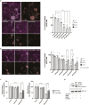
Treatment of control hiPSC-MN with BzATP leads to P2X7R-mediated, dose dependent, caspase-mediated, motor neuron loss rescued by by JNJ-5567. (A) Immunocytochemistry and motor motor neuron quantification showing survival of hiPSC-MN treated with BzATP for 2 weeks. (B) The P2X7R antagonist JNJ-5567 provides neuroprotection of hiPSC-MN following BzATP incubation. Scale bar = 50 µm. (C) Neurotoxicity by BzATP and neuroprotection by JNJ-5567 were confirmed using alamarBlue (resazurin) assay, where fluorescence and absorbance of treated and untreated conditions where compared to vehicle condition. (D) Western blot results showing increased caspase 3 expression in hiPSC-MN treated for 2 hours in 1 mM of BzATP which could be reduced with prior incubation in 3 µM of JNJ-5567. Significance values indicate *P < .05, **P < .01, ***P < .001, ****P < .0001. For (A) and (B) n = 4 biological replicates represented as different shaped data points, and n = 3 technical replicates per condition. For (C), a single cell line (CS8PAA) and n = 12 technical replicates were used.

References

    1. Cotrina ML, Nedergaard M.. Physiological and pathological functions of P2X7 receptor in the spinal cord. Purinergic Signal. 2009;5:223-232. https://doi.org/10.1007/s11302-009-9138-2 - DOI - PMC - PubMed
    1. Davalos D, Grutzendler J, Yang G, et al.ATP mediates rapid microglial response to local brain injury in vivo. Nat Neurosci. 2005;8:752-758. https://doi.org/10.1038/nn1472 - DOI - PubMed
    1. Burnstock G. Introduction to purinergic signalling in the brain. Adv Exp Med Biol. 2020;1202:1-12. https://doi.org/10.1007/978-3-030-30651-9_1 - DOI - PubMed
    1. Burnstock G. The past, present and future of purine nucleotides as signalling molecules. Neuropharmacology. 1997;36:1127-1139. https://doi.org/10.1016/s0028-3908(97)00125-1 - DOI - PubMed
    1. Butt AM. ATP: a ubiquitous gliotransmitter integrating neuron-glial networks. Semin Cell Dev Biol. 2011;22:205-213. https://doi.org/10.1016/j.semcdb.2011.02.023 - DOI - PubMed

Substances