Review
. 2024 Dec;54(13):2175-2184.
doi: 10.1007/s00247-024-06068-3. Epub 2024 Oct 18.

Has the cat got your tongue, or is something obstructing your throat? A review of imaging of ingested and aspirated foreign bodies in the paediatric population

Affiliations
Review

Has the cat got your tongue, or is something obstructing your throat? A review of imaging of ingested and aspirated foreign bodies in the paediatric population

Ola Kvist et al. Pediatr Radiol. 2024 Dec.

Abstract

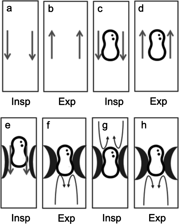
Children frequently swallow or inhale foreign objects, a situation that can be life-threatening. Radiographic imaging plays a lead role in the early identification and location of inhaled or swallowed objects is essential. Promptly identifying and locating inhaled or swallowed objects are essential, as some items require immediate removal. For example, button batteries in the throat can cause grave harm; magnets can attract each other through the gut and cause perforations; and other objects can obstruct the airway or intestinal tract. Radiologists must understand how these objects appear in images to assist doctors in treating patients effectively. Recognising signs of inhaled objects on radiographs is also crucial, as symptoms may not always be clear, and many inhaled objects are not visible on radiographs. Radiographs are the primary means of checking for swallowed or inhaled objects, although other tests like fluoroscopy and computed tomography may be used in complex cases. Doctors working with children should be acquainted with the appearance of these common objects on images and their clinical importance.

Keywords: Child; Computed tomography; Fluoroscopy; Foreign bodies; Radiography; Ultrasonography.


Conflict of interest statement

Declarations. Competing interests: The authors declare no competing interests.

Figures

Fig. 1
Fig. 1
Radiographs of a 2-year-old boy who has choked whilst consuming carrots. a The anteroposterior (AP) view shows an expanded and hyperlucent left lung with a mediastinal shift to the right hemithorax. b Right lateral decubitus view shows a reduced aeration in the right lung, i.e. no sign of air trapping. c The left lateral decubitus view demonstrates the aeration in the left lung is maintained and the lung volume is almost constant to the contralateral side as well as unchanged compared to the AP view. The findings are conclusive with air trapping indicating a foreign body in the left main bronchus
Fig. 2
Fig. 2
a, b Normal inspiration (a) and expiration (b) in the absence of a foreign body. c, d Normal inspiration (c) and expiration (d) in the presence of an airway foreign body. e, f A foreign body that permits air to pass by on inspiration (e) but is trapped during expiration (f) causing obstructive emphysema. g, h Complete obstruction during inspiration (g) and expiration (h)
Fig. 3
Fig. 3
Radiographs of an 18-month-old girl. a The anteroposterior (AP) view indicates the left lung appears more radiolucent than the right, with a slight diminution in lung markings. There is a suspicion of a foreign body in the left main bronchus causing a so-called interrupted bronchus sign. b Coronal view computed tomography (CT) with contrast, in lung window, confirms the presence of an inhaled foreign object in the left main bronchus, which has resulted in air trapping in the left upper lobe
Fig. 4
Fig. 4
a Coronal view computed tomography (CT) without contrast, in lung window, of a 7-year-old girl with a piece of carrot in the right bronchus (white arrow). b Displays a coronal view CT without contrast, in lung window, of a 2-year-old with a Perler bead in the left main bronchus (black arrow). Both examinations had an effective dose of 0.04 mSv. Republished from Diagnosis of a foreign body aspiration with ultralow-dose CT using a tin filter: a comparison study, Courtesy of Dr Lena Gordon Murkes [14]
Fig. 5
Fig. 5
Lung radiographs, anteroposterior (AP) views of foreign bodies in the mediastinum. a A coin which appears as a solid structure in a 3-year-old boy. b A button battery with a visible double ring (arrow) which is pathognomonic, in a 2-year-old girl
Fig. 6
Fig. 6
A 2-year-old girl presenting with haematemesis. a An anteroposterior radiograph displays a button battery within the stomach. b Magnified view of (a) shows small erosions (solid white arrows). c Axial view computed tomography (CT) with contrast, in soft tissue window, shows a pseudoaneurysm from the aortic arch, adjacent to the oesophagus (open white arrow). d Axial view CT with contrast, in soft tissue window, shows fluid accumulation between the oesophagus and the pseudoaneurysm, which was confirmed to be necrosis caused by the button battery (asterisk)
Fig. 7
Fig. 7
Anteroposterior (a), lateral (b) and left lateral decubitus (c) abdominal radiographs of a 1-year-old boy who presented at the emergency department with vomiting and a clinical suspicion of ileus or gastroenteritis. The radiographs reveal a radiopaque pearl bracelet in the bowel. The boy’s guardian denied known ingestion of foreign bodies
Fig. 8
Fig. 8
Magnification of the right decubitus view radiograph (1-year-old boy) shows a small quantity of free air between the liver and the diaphragm (arrow). Surgery verified perforation of four small bowel loops and 25 small magnetic balls

References

    1. Karakoç F, Karadağ B, Akbenlioğlu C et al (2002) Foreign body aspiration: what is the outcome? Pediatr Pulmonol 34:30–36 - PubMed
    1. Ngamsanga S, Vathanophas V, Ungkanont K et al (2023) Pediatric respiratory tract foreign bodies in children: a systematic review. Auris Nasus Larynx 50:607–613 - PubMed
    1. Passàli D, Lauriello M, Bellussi L et al (2010) Foreign body inhalation in children: an update. Acta Otorhinolaryngol Ital 30:27–32 - PMC - PubMed
    1. Chik KK, Miu TY, Chan CW (2009) Foreign body aspiration in Hong Kong Chinese children. Hong Kong Medical J=Xianggang Yi Xue Za Zhi 15:6–11 - PubMed
    1. Barrios Fontoba JE, Gutierrez C, Lluna J et al (1997) Bronchial foreign body: should bronchoscopy be performed in all patients with a choking crisis? Pediatr Surg Int 12:118–120 - PubMed

Publication types

LinkOut - more resources