. 2024 Oct 17;51(1):1062.
doi: 10.1007/s11033-024-10005-w.

Skeletal muscle fibre type-dependent effects of atorvastatin on the PI3K/Akt/mTOR signalling pathway and atrophy-related genes in rats

Affiliations

Skeletal muscle fibre type-dependent effects of atorvastatin on the PI3K/Akt/mTOR signalling pathway and atrophy-related genes in rats

Anna Gawedzka et al. Mol Biol Rep. .

Abstract

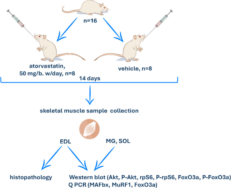
Background: One of the probable causes of statin myotoxicity is an imbalance between protein synthesis and degradation. These processes are regulated by the PI3K/Akt/mTOR pathway and the ubiquitin‒proteasome system (UPS). The aim of this study was to assess whether the effects of atorvastatin on PI3K/Akt/mTOR pathway downstream proteins, the FoxO3a transcription factor and the UPS genes, i.e., MuRF-1 and MAFbx, depend on muscle fibre type.

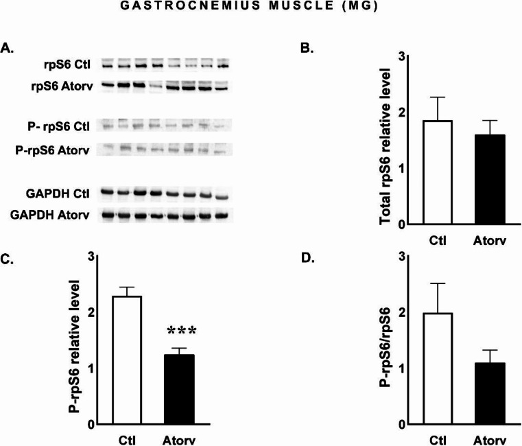
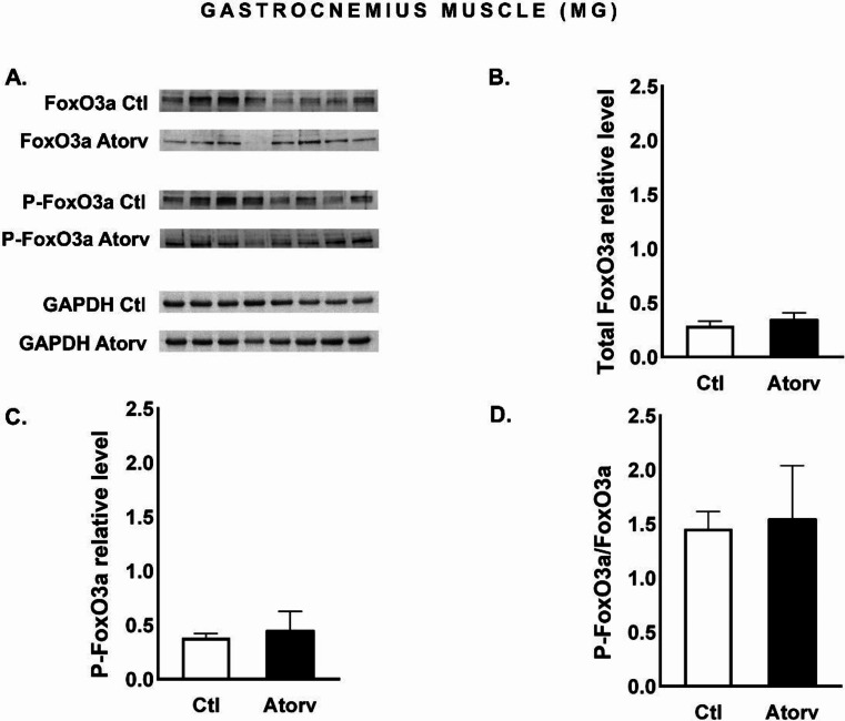
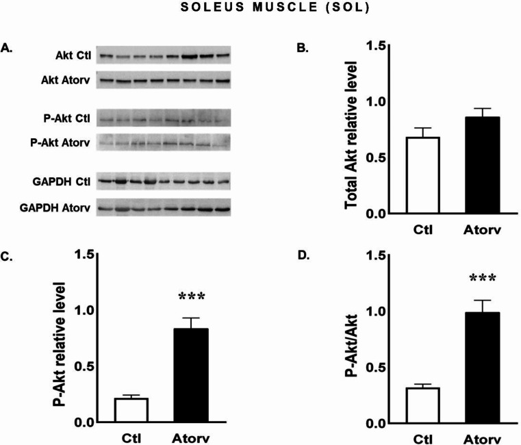
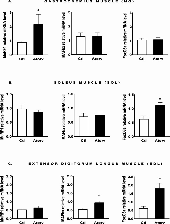
Methods and results: Atorvastatin (50 mg/kg) was administered to Wistar rats. The levels of selected PI3K/Akt/mTOR pathway proteins were assayed via Western blotting, whereas MuRF-1, MAFbx and FoxO3a mRNA levels were measured using reverse transcription quantitative polymerase chain reaction (RT‒qPCR). Gomöri trichrome staining was performed to assess skeletal muscle pathology. A decrease in the P-Akt/Akt ratio was observed in the gastrocnemius muscle (MG), whereas an increase in the P-Akt/Akt ratio was observed in the soleus muscle (SOL). FoxO3a gene expression increased in the SOL and extensor digitorum longus (EDL) muscles. MuRF-1 gene expression increased in the MG, and MAFbx expression increased in the EDL. No histopathological changes were observed in any of the tested muscles.

Conclusions: In the absence of overt muscle damage, atorvastatin decreased the P-Akt/Akt ratio in the MG, indicating an increase in inactive Akt. Consistent with the decrease in Akt activation, rpS6 phosphorylation decreased. In SOL, atorvastatin increased the P-Akt/Akt ratio, indicating Akt activation. P-FoxO3a and the P-FoxO3a/FoxO3a ratio increased, suggesting that FoxO3a inactivation occurred. Moreover, in the SOL, atorvastatin did not affect the expression of atrophy-related genes. These findings indicate that atorvastatin has no adverse effect on the Akt pathway in the SOL. Our results showed that the effects of atorvastatin on the Akt signalling pathway and atrophy-related gene expression depend on muscle type.

Keywords: Akt pathway; Atorvastatin; Atrophy-related genes; Skeletal muscle fibres.


Conflict of interest statement

The authors declare no competing interests.

Figures

Fig. 1
Fig. 1
Experimental design; EDL- extensor digitorum longus; MG- gastrocnemius muscle; SOL- soleus muscle
Fig. 2
Fig. 2
Semi-quantitative analysis of atorvastatin effects on Akt and P-Akt protein levels and the P-Akt/Akt ratio in the MG performed via Western blotting. A: Representative immunoblots for Akt, P-Akt and GAPDH; B: Relative Akt protein levels; C: Relative P-Akt protein levels; D: P-Akt/Akt ratio. Abbreviations Ctl, control group; Atorv, atorvastatin-treated group. The data are presented as the means ± SEMs of n = 8 animals per group. *p < 0.05
Fig. 3
Fig. 3
Semi-quantitative analysis of atorvastatin effects on rpS6 and P-rpS6 protein levels and the P-rpS6/rpS6 ratio in the MG performed via Western blotting. A: Representative immunoblots for rpS6, P-rpS6 and GAPDH; B: Relative rpS6 protein levels; C: Relative P-rpS6 protein levels; D: P-rpS6/rpS6 ratio. Abbreviations Ctl, control group; Atorv, atorvastatin-treated group. The data are presented as the means ± SEMs of n = 8 animals per group. ***p < 0.001
Fig. 4
Fig. 4
Semi-quantitative analysis of atorvastatin effects on FoxO3a and P-FoxO3a protein levels and the P-FoxO3a/FoxO3a ratio in the MG performed via Western blotting. A: Representative immunoblots for FoxO3a, P-FoxO3a and GAPDH; B: Relative FoxO3a protein levels; C: Relative P-FoxO3a protein levels; D: P-FoxO3a/FoxO3a ratio. Abbreviations Ctl, control group; Atorv, atorvastatin-treated group. The data are presented as the means ± SEMs of n = 8 animals per group
Fig. 5
Fig. 5
Semi-quantitative analysis of atorvastatin effects on Akt and P-Akt protein levels and the P-Akt/Akt ratio in the SOL muscle performed via Western blotting. A: Representative immunoblots for Akt, P-Akt and GAPDH; B: Relative Akt protein levels; C: Relative P-Akt protein levels; D: P-Akt/Akt ratio. Abbreviations Ctl, control group; Atorv, atorvastatin-treated group. The data are presented as the means ± SEMs of n = 8 animals per group. ***p < 0.001
Fig. 6
Fig. 6
Semi-quantitative analysis of atorvastatin effects on rpS6 and P-rpS6 protein levels and the P-rpS6/rpS6 ratio in the SOL muscle performed via Western blotting. A: Representative immunoblots for rpS6, P-rpS6 and GAPDH; B: Relative rpS6 protein levels; C: Relative P-rpS6 protein levels; D: P-rpS6/rpS6 ratio. Abbreviations Ctl, control group; Atorv, atorvastatin-treated group. The data are presented as the means ± SEMs of n = 8 animals per group
Fig. 7
Fig. 7
Semi-quantitative analysis of atorvastatin effects on FoxO3a and P-FoxO3a protein levels and the P-FoxO3a/FoxO3a ratio in the SOL muscle performed via Western blotting. A: Representative immunoblots for FoxO3a, P-FoxO3a and GAPDH; B: Relative FoxO3a protein levels; C: Relative P-FoxO3a protein levels; D: P-FoxO3a/FoxO3a ratio. Abbreviations Ctl, control group; Atorv, atorvastatin-treated group. The data are presented as the means ± SEMs of n = 8 animals per group. *p < 0.05
Fig. 8
Fig. 8
Semi-quantitative analysis of atorvastatin effects on Akt and P-Akt protein levels and the P-Akt/Akt ratio in the EDL muscle performed via Western blotting. A: Representative immunoblots for Akt, P-Akt and GAPDH; B: Relative Akt protein levels; C: Relative P-Akt protein levels; D: P-Akt/Akt ratio. Abbreviations Ctl, control group; Atorv, atorvastatin-treated group. The data are presented as the means ± SEMs of n = 8 animals per Ctl group and n = 7 animals per Atrov group
Fig. 9
Fig. 9
Semi-quantitative analysis of atorvastatin effects on rpS6 and P-rpS6 protein levels and the P-rpS6/rpS6 ratio in the EDL muscle performed via Western blotting. A: Representative immunoblots for rpS6, P-rpS6 and GAPDH; B: Relative rpS6 protein levels; C: Relative P-rpS6 protein levels; D: P-rpS6/rpS6 ratio. Abbreviations Ctl, control group; Atorv, atorvastatin-treated group. The data are presented as the means ± SEMs of n = 8 animals per Ctl group and n = 7 animals per Atorv group
Fig. 10
Fig. 10
Semi-quantitative analysis of atorvastatin effects on FoxO3a and P-FoxO3a protein levels and the P-FoxO3a/FoxO3a ratio in the EDL muscle performed via Western blotting. A: Representative immunoblots for FoxO3a, P-FoxO3a and GAPDH; B: Relative FoxO3a protein levels; C: Relative P-FoxO3a protein levels; D: P-FoxO3a/FoxO3a ratio. Abbreviations Ctl, control group; Atorv, atorvastatin-treated group. The data are presented as the means ± SEMs of n = 8 animals per Ctl group and n = 7 animals per Atorv group
Fig. 11
Fig. 11
qPCR analysis of atorvastatin effects on MuRF-1, MAFbx and FoxO3a mRNA levels in the A: gastrocnemius (MG), B: soleus (SOL) and C: extensor digitorum longus (EDL) muscles. Abbreviations Ctl, control group; Atorv, atorvastatin-treated group. The data are presented as the means ± SEMs of n = 8 animals per group for MG and SOL and of n = 7 animals per group for EDL. *p < 0.05

References

    1. Ward NC, Watts GF, Eckel RH (2019) Statin toxicity. Circ Res 124:328–350. 10.1161/CIRCRESAHA.118.312782 - PubMed
    1. Matyori A, Brown CP, Ali A, Sherbeny F (2023) Statins utilization trends and expenditures in the U.S. before and after the implementation of the 2013 ACC/AHA guidelines. Saudi Pharm J 31:795–800. 10.1016/j.jsps.2023.04.002 - PMC - PubMed
    1. Hoste E, Haufroid V, Deldicque L, Balligand JL, Elens L (2024) Atorvastatin-associated myotoxicity: a toxicokinetic review of pharmacogenetic associations to evaluate the feasibility of precision pharmacotherapy. Clin Biochem 124:110707. 10.1016/j.clinbiochem.2024.110707 - PubMed
    1. Mueller AM, Liakoni E, Schneider C, Burkard T, Jick SS, Krähenbühl S, Meier CR, Spoendlin J (2021) The risk of muscular events among new users of hydrophilic and lipophilic statins: an Observational Cohort Study. J Gen Intern Med 36(9):2639–2647. 10.1007/s11606-021-06651-6 - PMC - PubMed
    1. Bonifacio A, Sanvee GM, Bouitbir J, Krähenbühl S (2015) The AKT/mTOR signaling pathway plays a key role in statin-induced myotoxicity. Biochim Biophys Acta 1853:1841–1849. 10.1016/j.bbamcr.2015.04.010 - PubMed

MeSH terms

LinkOut - more resources