. 2024 Oct 17;12(1):61.
doi: 10.1038/s41413-024-00353-5.

Osteopetrosis-like disorders induced by osteoblast-specific retinoic acid signaling inhibition in mice

Affiliations

Osteopetrosis-like disorders induced by osteoblast-specific retinoic acid signaling inhibition in mice

Siyuan Sun et al. Bone Res. .

Abstract

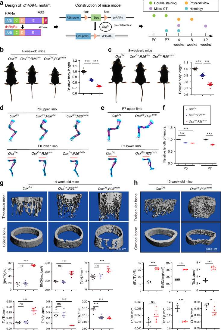
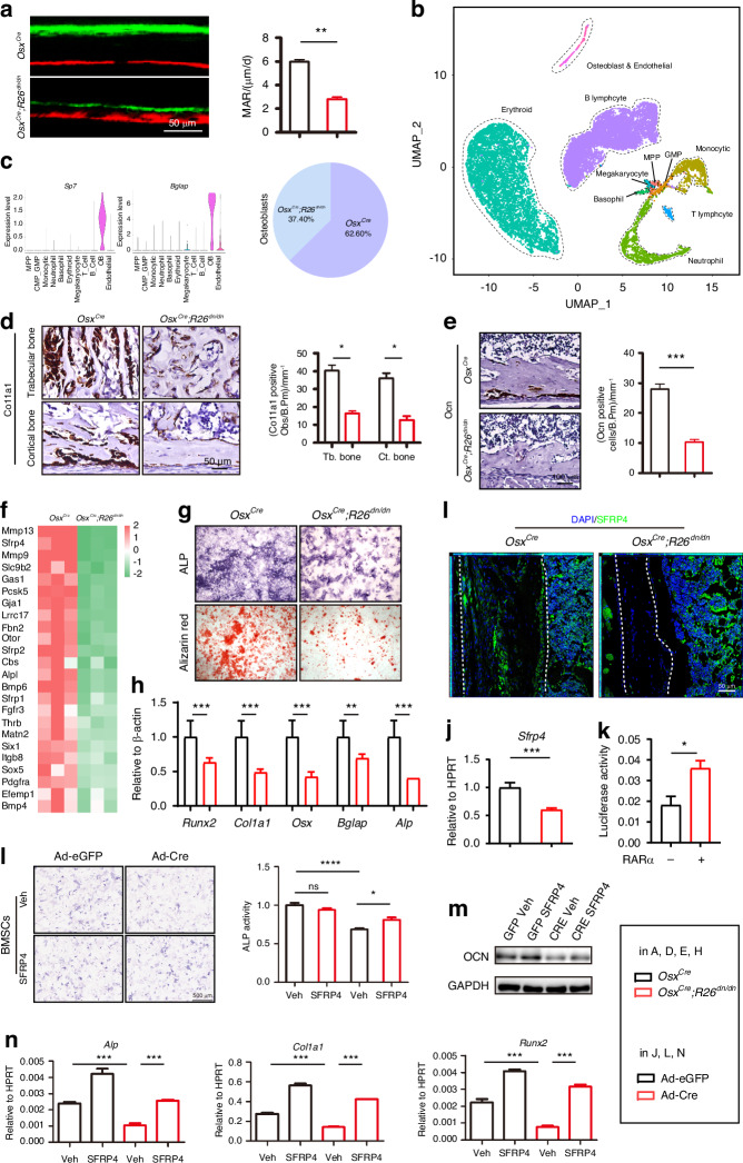
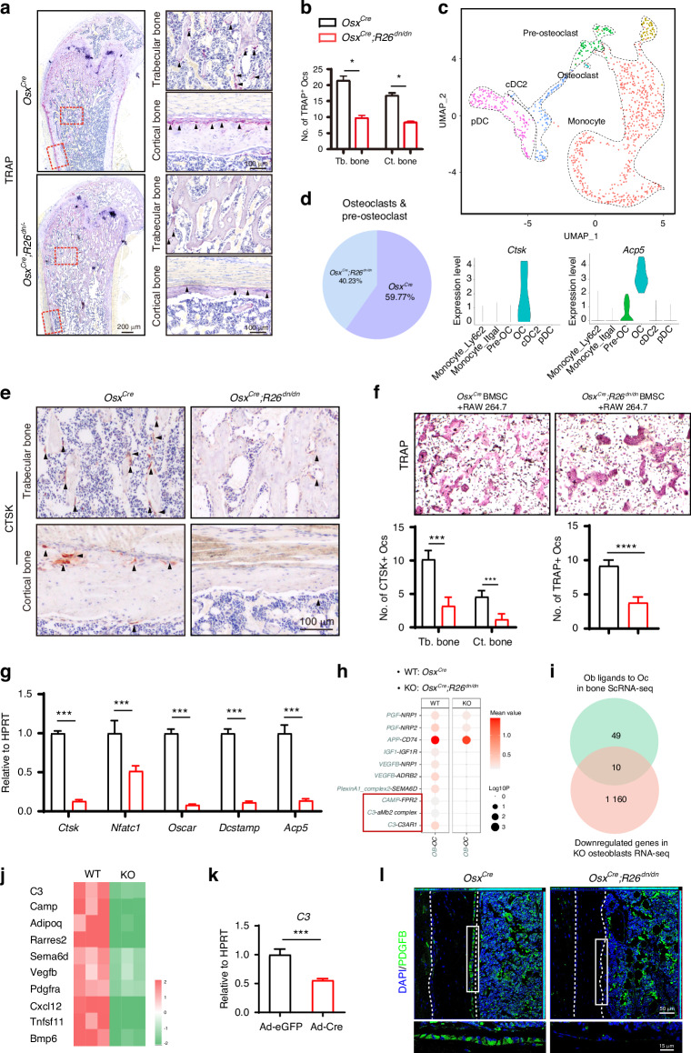
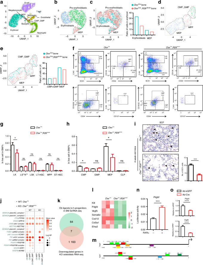
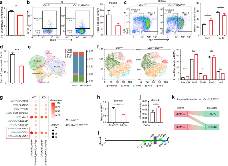
Osteopetrosis is an inherited metabolic disease, characterized by increased bone density and narrow marrow cavity. Patients with severe osteopetrosis exhibit abnormal bone brittleness, anemia, and infection complications, which commonly cause death within the first decade of life. Pathologically, osteopetrosis impairs not only the skeletal system, but also the hemopoietic and immune systems during development, while the underlying osteoimmunological mechanisms remain unclear. Osteoclastic mutations are regarded as the major causes of osteopetrosis, while osteoclast non-autonomous theories have been proposed in recent years with unclear underlying mechanisms. Retinoic acid (RA), the metabolite of Vitamin A, is an essential requirement for skeletal and hematopoietic development, through the activation of retinoic acid signaling. RA can relieve osteopetrosis symptoms in some animal models, while its effect on bone health is still controversial and the underlying mechanisms remain unclear. In this study, we constructed an osteoblast-specific inhibitory retinoic acid signaling mouse model and surprisingly found it mimicked the symptoms of osteopetrosis found in clinical cases: dwarfism, increased imperfectly-formed trabecular bone deposition with a reduced marrow cavity, thin cortical bone with a brittle skeleton, and hematopoietic and immune dysfunction. Micro-CT, the three-point bending test, and histological analysis drew a landscape of poor bone quality. Single-cell RNA sequencing (scRNA-seq) of the femur and RNA-seq of osteoblasts uncovered an atlas of pathological skeletal metabolism dysfunction in the mutant mice showing that osteogenesis was impaired in a cell-autonomous manner and osteoclastogenesis was impaired via osteoblast-osteoclast crosstalk. Moreover, scRNA-seq of bone marrow and flow cytometry of peripheral blood, spleen, and bone marrow uncovered pathology in the hematopoietic and immune systems in the mutant mice, mimicking human osteopetrosis. Results showed that hematopoietic progenitors and B lymphocyte differentiation were affected and the osteoblast-dominated cell crosstalk was impaired, which may result from transcriptional impairment of the ligands Pdgfd and Sema4d. In summary, we uncovered previously unreported pathogenesis of osteopetrosis-like disorder in mice with skeletal, hematopoietic, and immune system dysfunction, which was induced by the inhibition of retinoic acid signaling in osteoblasts, and sheds new insights into a potential treatment for osteopetrosis.


Conflict of interest statement

The authors declare no competing interests.

Figures

Fig. 1
Fig. 1
Inhibition of retinoic acid signaling in osteoblasts results in abnormally increased trabecular bone density. a Illustration of the dnRARα mutant design, mouse model construction and phenotype analysis. b Representative views of 4-week-old OsxCre, OsxCre;R26dn/- and OsxCre;R26dn/dn mice. c Representative views of 8-week-old OsxCre mice, OsxCre;R26dn/- mice and OsxCre;R26dn/dn mice. d Upper and lower limb from OsxCre, OsxCre;R26dn/- and OsxCre;R26dn/dn newborns were double-stained with alcian blue and alizarin red S. e Upper and lower limb from OsxCre and OsxCre;R26dn/dn 7-day-old mice were double-stained with alcian blue and alizarin red S. f Quantitative analysis of panels d and e. g Micro-CT images of trabecular bone and cortical bone from the femurs of 4-week-old OsxCre, OsxCre;R26dn/- and OsxCre;R26dn/dn mice and their quantitative analysis, including bone volume per tissue volume (BV/TV), trabecular thickness (Tb.Th.), trabecular number (Tb.N.), trabecular space (Tb.Sp.), and cortical thickness (Ct.Th). h Micro-CT images of trabecular bone and cortical bone from the femurs of 12-week-old OsxCre and OsxCre;R26dn/dn mice and quantitative analysis. Error bars are represented as mean±S.D. ns = not significant. **P < 0.01. ***P < 0.001. Five pairs of mice were tested
Fig. 2
Fig. 2
Inhibition of retinoic acid signaling in osteoblasts led to a brittle bone with narrow marrow cavity. a Illustration of the three-point bending test. Representative load-deflection diagram from a three-point bending test performed on femora from 4-week-old OsxCre and OsxCre;R26dn/dn mice. The maximum load measured during the test. n = 3, Error bars are represented as mean ± S.D., ***P < 0.001. b Ribs preparations from 7-day-old male OsxCre and OsxCre;R26dn/dn mice were double-stained with alcianblue and alizarin red S. Red arrows indicated pathological fracture callus. c Micro-CT and 3-dimensional reconstruction of femur from 4-week-old OsxCre and OsxCre;R26dn/dn mice. d H&E staining of the femurs from 4-week-old male OsxCre and OsxCre;R26dn/dn mice. e H&E staining of the SOC (upper panel), trabecular bone (middle panel) and cortical bone (lower panel) from 4-week-old male OsxCre and OsxCre;R26dn/dn mice. f Perilipin staining of the femurs from 4-week-old male OsxCre and OsxCre;R26dn/dn mice. g H&E staining, Alizarin Red staining and Oil Red staining of the trabecular bone from 7-day-old male OsxCre and OsxCre;R26dn/dn mice
Fig. 3
Fig. 3
Inhibition of retinoic acid signaling in osteoblasts impairs bone anabolism. a Representative images of calcein-alizarin red S double labeling of the femurs from 4-week-old male OsxCre and OsxCre;R26dn/dn mice and quantitative parameters mineral apposition rate (MAR). b A total of 11 060 cells from 2-week-old OsxCre mice and a total of 10 552 cells from 2-week-old OsxCre;R26dn/dn mice altogether identified by scRNA-seq were visualized with UMAP. Eleven cell populations were defined and distinguished by color. Each point represents an individual cell. (C) Feature plots of the expression levels of marker genes for osteoblasts. Ratio of OsxCre and OsxCre;R26dn/dn mice-derived-osteoblasts in total osteoblasts. d In situ hybridization for Col1α1 in the trabecular and cortical bone of femurs from 7-day-old male OsxCre and OsxCre;R26dn/dn mice and quantitation of Col1α1-positive cell number. e In situ hybridization for Ocn in the cortical bone of femurs from 7-day-old male OsxCre and OsxCre;R26dn/dn mice and quantitation of Ocn-positive cell number. f Total RNA was isolated from osteoblasts from 4-week-old male OsxCre and OsxCre;R26dn/dn mice, followed by RNA sequencing analysis. Heatmap analysis of osteogenesis-related genes in the OsxCre and OsxCre;R26dn/dn sets. g ALP staining and alizarin red S staining of OsxCre and OsxCre;R26dn/dn mouse osteoblasts after culture in osteogenic medium for 7 days and 14 days separately. h The relative mRNA levels of the osteogenesis-related genes Runx2, Col1a1, Osx, Bglap, and Alp in osteoblasts from OsxCre and OsxCre;R26dn/dn mice after culture in osteogenic medium for 7 days. i SFRP4 expression in the femur bone of 4-week-old OsxCre and OsxCre;R26dn/dn mouse by immunofluorescence staining. j The relative mRNA levels of Sfrp4 in adenovirus (eGFP or Cre) infected R26dn/dn osteoblasts after culture in osteogenic medium for 7 days. k Luciferase activity analysis for the effect of RARα on the Sfrp4 promoter activity in the 293 T cell line (n = 3). l ALP staining of the Ad-eGFP or Ad-Cre infected and 0.1 μg/mL SFRP4 recombinant protein rescued BMSCs from R26dn/dn mouse after culture in osteogenic medium for 7 days. m The OCN protein expression in the Ad-eGFP or Ad-Cre infected and 0.1 μg/mL SFRP4 recombinant protein rescued BMSCs from R26dn/dn mouse after culture in osteogenic medium for 7 days by western blotting. n The mRNA expression of Alp, Col1a1 and Runx2 in the Ad-eGFP or Ad-Cre infected and 0.1 μg/mL SFRP4 recombinant protein rescued BMSCs from R26dn/dn mouse after culture in osteogenic medium for 7 days by RT-qPCR Error bars are represented as mean ± SD. *P < 0.05. **P < 0.01, ***P < 0.001
Fig. 4
Fig. 4
Inhibition of retinoic acid signaling in osteoblasts impairs bone catabolism. a TRAP staining of the trabecular and cortical bone from the femurs of 4-week-old OsxCre and OsxCre;R26dn/dn mice. b Quantitation of TRAP-positive cell number in Fig. 5a. c Subclustering of the “Monocytic” lineage cells from Fig. 4d, visualized as a UMAP plot. d Feature plots of the expression levels of marker genes from pre-osteoclasts and osteoclasts. Ratio of OsxCre and OsxCre;R26dn/dn mice-derived-osteoclasts and pre-osteoclasts in total osteoclasts and pre-osteoclasts. e CTSK immunohistochemical staining of the trabecular and cortical bone of femurs from 4-week-old OsxCre and OsxCre;R26dn/dn mice. f TRAP staining of RAW264.7 cells after coculturing with 4-week-old OsxCre and OsxCre;R26dn/dn mouse osteoblasts for 7 days. g The relative mRNA levels of osteoclast-specific genes Ctsk, Nfatc1, Oscar, Dcstamp and Acp5 in RAW264.7 cells after coculturing for 7 days. h Cell communication analysis between osteoblasts and osteoclasts and pre-osteoclasts in OsxCre and OsxCre;R26dn/dn mice. i Number of downregulated osteoblastic ligands screened by ScRNA-seq and RNA-seq. j Heatmap analysis of the screened osteoblastic ligands in the OsxCre and OsxCre;R26dn/dn osteoblast RNA-seq sets. k The relative mRNA levels of the screened osteoblastic ligands C3 in adenovirus (eGFP or Cre) infected R26dn/dn osteoblasts after culture in osteogenic medium for 7 days. l PDGFB expression in the femoral bone of OsxCre and OsxCre;R26dn/dn mouse by immunofluorescence staining. Error bars are represented as mean±S.D. ns = not significant. *P < 0.05. ***P < 0.001. ****P < 0.000 1
Fig. 5
Fig. 5
Disorders of hematopoiesis were caused by inhibition of retinoic acid signaling in osteoblasts through impairing hematopoietic progenitors differentiation. a A total of 6 827 cells from 2-week-old OsxCre mice and a total of 9 151 cells from 2-week-old OsxCre;R26dn/dn mice were identified by scRNA-seq and visualized with UMAP. Eleven cell populations were defined and distinguished by color. Each point represents an individual cell. b Subclustering of the “Erythroid” lineage cells from Fig. 6a visualized as a UMAP plot. c The ratio of MEP in the “Erythroid” lineage cells from OsxCre and OsxCre;R26dn/dn mice. d Population of hematopoietic progenitors visualized as a UMAP plot after merging the MEP, MPP and CMP_GMP subclusters. e The ratio of CMP_GMP and MEP in the hematopoietic progenitors from OsxCre and OsxCre;R26dn/dn mice, correspondingly. fh The frequencies of LK, LSlowKlow, LSK, LT-HSC, MPP, ST-HSC, CMP, GMP, MEP and CLP cells in bone marrow from OsxCre and OsxCre;R26dn/dn mice (n = 4 for each group). i SCF immunohistochemical staining of the bone marrow of femurs from 4-week-old OsxCre and OsxCre;R26dn/dn mice and quantitation of SCF-positive cell number. j Cell communication analysis between osteoblasts and hematopoietic progenitors in OsxCre and OsxCre;R26dn/dn mice. k Number of downregulated osteoblastic ligands screened by ScRNA-seq and RNA-seq. l Heatmap analysis of the screened osteoblastic ligands related to hematopoietic progenitor differentiation in the OsxCre and OsxCre;R26dn/dn osteoblasts RNA-seq sets. m Predicted RAR family binding sites on the screened osteoblastic ligands Pdgfd and Vegfb promoters. n Luciferase activity analysis of the effect of RARα on Pdgfd promoter activity in the 293 T cell line (n = 3). o The relative mRNA levels of the screened osteoblastic ligands Pdgfd and Vegfb in adenovirus (eGFP or Cre) infected R26dn/dn osteoblasts after culture in osteogenic medium for 7 days. Error bars are represented as mean±S.D. ns = not significant. *P < 0.05. **P < 0.01. ****P < 0.000 1
Fig. 6
Fig. 6
Lymphopenia caused by inhibition of retinoic acid signaling in osteoblasts through impairing B lymphocyte differentiation. a Number of lymphocyte in the peripheral blood (PB) of OsxCre and OsxCre;R26dn/dn mice using hematological analysis. b The frequencies of plasma in B lymphocytes in PB from OsxCre and OsxCre;R26dn/dn mice (n = 4 for each group). c The frequencies of immature B and mature B cells in B lymphocytes from the spleens of OsxCre and OsxCre;R26dn/dn mice (n = 4 for each group). d Ratio of B lymphocytes from the total cells in BM from OsxCre and OsxCre;R26dn/dn mice (n = 4 for each group). e Subclustering of the “B lymphocyte” lineage cells from Fig. 6a visualized as a UMAP plot, and the ratio of pro B, pre B, immature B, and mature B cells in the “B lymphocyte” lineage cells. f Flow cytometry analysis showing the ratio of prepro B, pro B, pre B, immature B and mature B from the B lymphocytes in BM, visualized with tSNE (n = 4 for each group). g Cell communication analysis between osteoblasts and pro B and pre B cells in OsxCre and OsxCre;R26dn/dn mice. h The relative mRNA levels of Sema4d in the BMSCs of OsxCre and OsxCre;R26dn/dn mice after culture in osteogenic medium for 7 days. i Predicted RAR family binding sites on the screened Semaphorin family (Sema4d) human promoters. j Luciferase activity analysis for the effect of RARα on the Sema4d promoter activity in the 293 T cell line (n = 3). k The impaired interaction between osteoblasts and B lymphocytes in OsxCre;R26dn/dn mice. Error bars are represented as mean±S.D. ns = not significant. *P < 0.05. **P < 0.01. ***P < 0.001. ****P < 0.000 1

References

    1. Yaga, U. & Panta, P. Osteopetrosis. N. Engl. J. Med.376, e34 (2017). - PubMed
    1. Tolar, J., Teitelbaum, S. L. & Orchard, P. J. Osteopetrosis. N. Engl. J. Med.351, 2839–2849 (2004). - PubMed
    1. Loría-Cortés, R., Quesada-Calvo, E. & Cordero-Chaverri, C. Osteopetrosis in children: a report of 26 cases. J. Pediatr.91, 43–47 (1977). - PubMed
    1. Fasth, A. & Porras, O. Human malignant osteopetrosis: pathophysiology, management and the role of bone marrow transplantation. Pediatr. Transpl.3, 102–107 (1999). - PubMed
    1. Gerritsen, E. J. et al. Autosomal recessive osteopetrosis: variability of findings at diagnosis and during the natural course. Pediatrics93, 247–253 (1994). - PubMed

LinkOut - more resources