. 2024 Oct 17;15(1):8974.
doi: 10.1038/s41467-024-53306-1.

Reverse hierarchical DED assembly in the cFLIP-procaspase-8 and cFLIP-procaspase-8-FADD complexes

Affiliations

Reverse hierarchical DED assembly in the cFLIP-procaspase-8 and cFLIP-procaspase-8-FADD complexes

Chao-Yu Yang et al. Nat Commun. .

Erratum in

Abstract

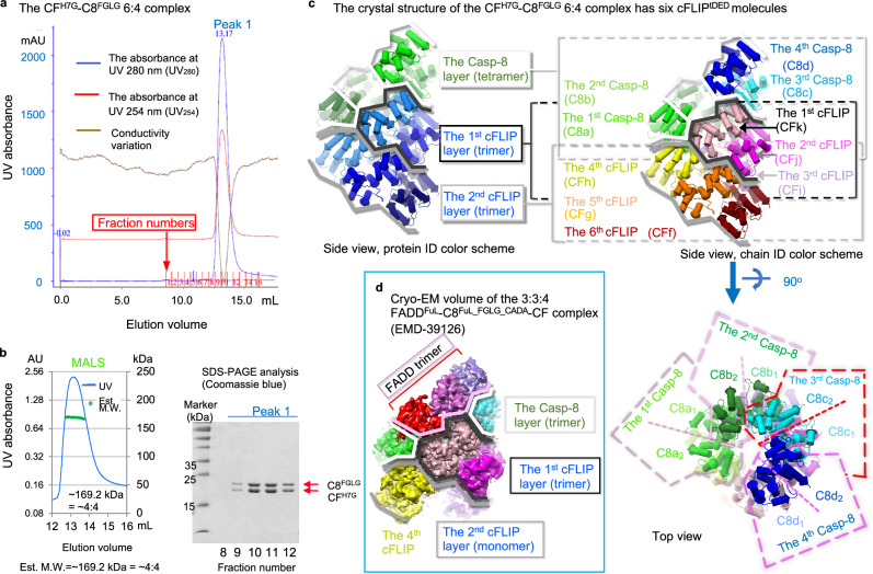
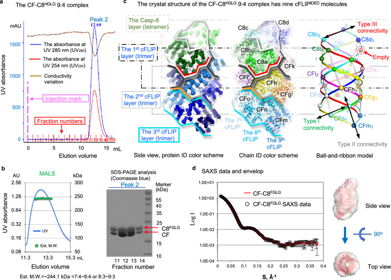
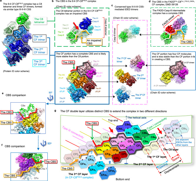
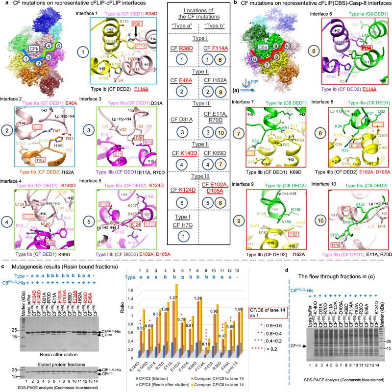
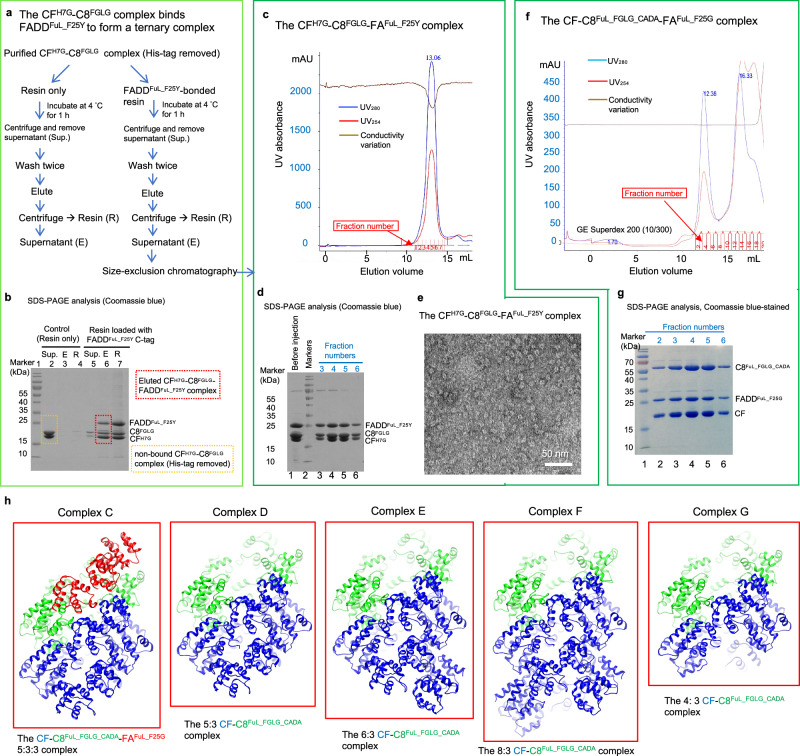
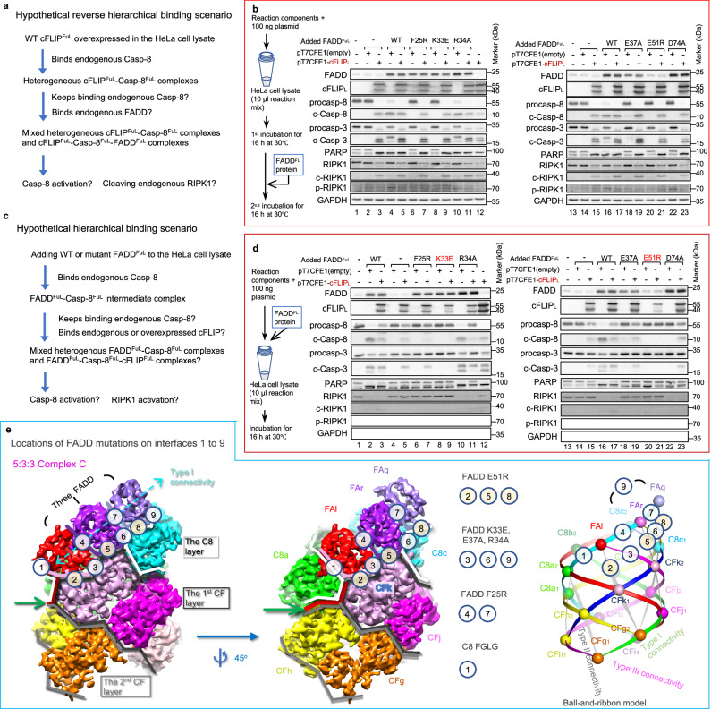
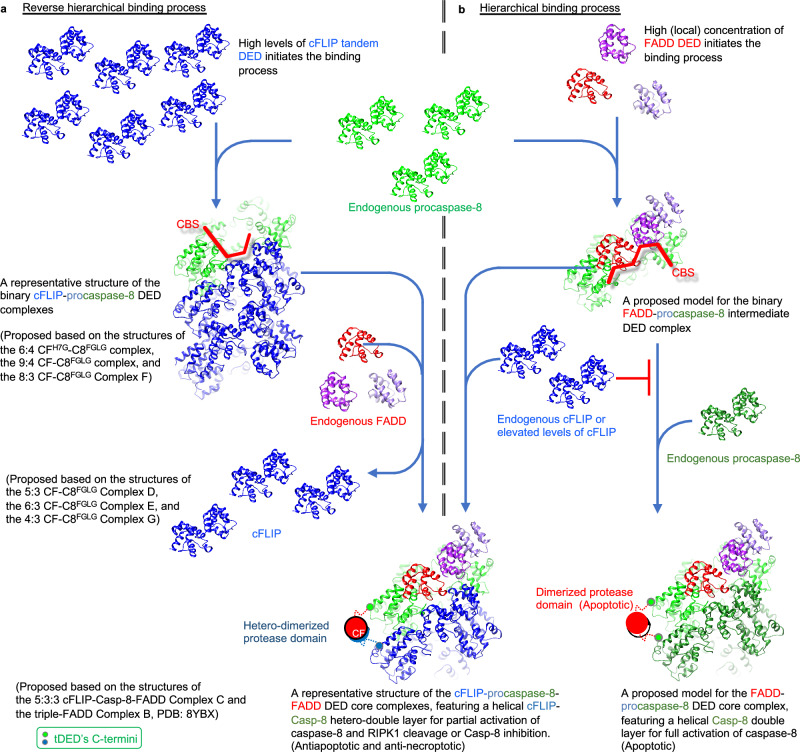
cFLIP, a master anti-apoptotic regulator, targets the FADD-induced DED complexes of procaspase-8 in death receptor and ripoptosome signaling pathways. Several tumor cells maintain relatively high levels of cFLIP in achieving their immortality. However, understanding the three-dimensional regulatory mechanism initiated or mediated by elevated levels of cFLIP has been limited by the absence of the atomic coordinates for cFLIP-induced DED complexes. Here we report the crystal plus cryo-EM structures to uncover an unconventional mechanism where cFLIP and procaspase-8 autonomously form a binary tandem DED complex, independent of FADD. This complex gains the ability to recruit FADD, thereby allosterically modulating cFLIP assembly and partially activating caspase-8 for RIPK1 cleavage. Our structure-guided mutagenesis experiments provide critical insights into these regulatory mechanisms, elucidating the resistance to apoptosis and necroptosis in achieving immortality. Finally, this research offers a unified model for the intricate bidirectional hierarchy-based processes using multiprotein helical assembly to govern cell fate decisions.


Conflict of interest statement

The authors declare no competing interests.

Figures

Fig. 1
Fig. 1. CFH7G and C8FGLG form a binary oligomeric complex in solution and crystals.
a, b SEC-MALS analysis of the binary CFH7G-C8FGLG complex, with corresponding SDS-PAGE analysis in (b) for the peak fractions in (a). These results demonstrate that cFLIPtDED and Casp-8tDED form an oligomeric complex in solution. The MALS data suggests that the complex is smaller in solution than in crystals, indicating that the end molecules, likely cFLIPtDED, have high dissociation constants similar to the DD complex. Est. M.W., estimated molecular weight. The experiments were repeated twice with similar results. Source data are provided as a Source Data file. c The pipes-and-planks diagrams illustrate the crystal structure of the binary CFH7G-C8FGLG complex from different perspectives. In the protein ID color scheme of Figs. 1–3, Casp-8 and cFLIP are colored in different shades of green and blue, respectively, while in the chain ID color scheme, the molecules in Figs. 1–3 are colored as their counterparts in Fig. 3g. Additionally, the C8FGLG layer/tetramer, the 1st CFH7G layer/trimer, and the 2nd CFH7G layer/trimer are consistently highlighted by white, dark gray, and gray lines, respectively. C8d1 and C8d2 denote DED1 and DED2 of Casp-8tDED chain d, respectively, while CFf represents cFLIPtDED chain f. The dashed boxes in the top view indicate the positions of Casp-8 molecules as well as the DED1 and DED2 domains. d Depicts the cryo-EM volume of our previous ternary 3:3:4 FADDFuL-C8FuL_FGLG_CADA-CF complex for comparison (EMDB: EMD-39126). The FADD trimer is highlighted by pink lines, while the C8FGLG trimer is highlighted by white lines.
Fig. 2
Fig. 2. The binary complex has more WT CF molecules than the ternary complex.
a, b SEC-MALS analysis of the binary CF-C8FGLG complex, with corresponding SDS-PAGE analysis in (b) for the peak fractions in (a). The experiments were repeated twice with similar results. Source data are provided as a Source Data file. c The pipes-and-planks and ribbon diagrams illustrate the crystal structure of the binary CF-C8FGLG complex. The C8FGLG layer/tetramer and the 1st, 2nd, and 3rd WT CF layers/trimers are highlighted by white, dark gray, gray, and cyan lines, respectively, while the CBS for binding the cFLIP molecule CFo and the Casp-8 molecule C8a are highlighted by orange and red lines, respectively. The ball-and-ribbon diagram depicts the spatial relationship of individual DEDs with the type I, II, and III connectivity, represented by ribbons generated by the PyMOL program version 1.8.2.1, connecting the spatially conserved Cα atoms of every two adjacent DEDs. Each conserved Cα atom, representing DEDs, is shown as a ball, including those of Casp-8 residues L62 and L162, and cFLIP residues L55 and L152. A red arrow indicates a space left by the absence of FADD DED, predicted as a dashed ball. Please note that all the spatially conserved tDEDs in different representations, including ribbons, balls, and hexagons, in different Figs., would receive the same chain ID and color. Please refer to the legend of Fig. 1c for details on the molecular color schemes used. C8d1 denotes DED1 of Casp-8tDED chain d, while CFf represents cFLIPtDED chain f. d The crystal structure of the CF-C8FGLG complex exhibits a size and shape comparable to the envelop derived from the SAXS data of the same complex.
Fig. 3
Fig. 3. cFLIPtDED double-layer intermediate complex utilizes different CBS to recruit Casp-8tDED and cFLIPtDED.
a Crystal structure of the binary CF-C8FGLG complex. Please refer to the legend of Fig. 1c for details on the molecular color schemes used. b Different CBS in the binary CF-C8FGLG complex. The green dashed box shows the C8 tetramer portion, while the blue box shows the CF portion. Orange lines highlight the impaired CBS on the C8FGLG tetramer due to a space, indicated by a red arrow, created by the absence of FADD DED. Red lines indicate the potential CBS on the WT CF double-layer intermediate complex. The CF and C8FGLG portions are separated for clarity in visualizing the CBS. c Displays the conserved type III-II-III CSS-mediated CF and C8FGLG trimers from (b) and (d), respectively. Molecules with the same chain ID in (b) to (d) are oriented similarly. d The green dashed box highlights the CBS, represented by orange lines, on the FADD-Casp-8 intermediate complex of our previous ternary complex in Fig. 1d for comparing with the CBS in (b). For clarity, the FADD-C8FGLG and CF portions are separated. The CF portion is in the blue box. e, f Red lines and orange lines highlight the CBS for the Casp-8 molecule C8a and the cFLIP molecule CFo, respectively, on the WT CF double-layer intermediate complex in (b), viewed from different angles. g 2D representation of the binary complexes illustrates distinct CBS, extending the complex along the C8 (top) end and CF (bottom) end. A notable distinction between the CFH7G-C8FGLG and CF-C8FGLG complexes is the presence of additional cFLIPtDED molecules CFo, CFm, and CFn in the latter. Each hexagon represents a DED domain, while the dashed ones represent the adjacent tDEDs from the other layers. As in (b), a red arrow indicates a space created by the absence of FADD DED. Red lines and orange lines highlight different CBS described in (e) and (f). Pink and green angled lines are detailed in Supplementary Fig. 3. C8d1 denotes DED1 of Casp-8tDED chain d, while CFf represents cFLIPtDED chain (f).
Fig. 4
Fig. 4. cFLIPtDED self-assembly is crucial for the formation of the CFH7G-C8FGLG complex.
a Illustration of five representative interfaces involved in the cFLIP-cFLIP interaction in the CFH7G-C8FGLG complex. Interface residues and the cFLIP mutations generated in this study are depicted. Thick green lines highlight the type III-II-III interface, while thick blue lines highlight the representative interfaces between two type III-II-III CSS-mediated cFLIPtDED trimers. Residues used in mutagenesis studies are highlighted with red boxes, while glycine residues are indicated by red dots. ‘Lp’ denotes loop regions. The list in the black box outlines the positions of CF mutations on the surfaces displayed in (a) and (b). The molecules are colored as their counterparts in Fig. 3g. b Illustration of five interfaces involved in the interaction between the cFLIP double-layer intermediate complex and Casp-8 within the same complex. Thick red lines highlight the Casp-8-recruiting CBS on the cFLIP double-layer intermediate complex. Interface residues and the cFLIP mutations generated in this study are depicted. c Pulldown assay of CFH7G and mutant CFH7G by His-tagged C8FGLG demonstrating the importance of cFLIPtDED self-assembly in the formation of the binary complex. Resin-bound fractions are divided into the eluted protein fractions and the resin after elution fractions, each analyzed by SDS-PAGE to assess the amount of bound CF and His-tagged C8FGLG. Protein bands were quantified using ImageJ version 1.50i (https://imagej.net/ij/). The bar chart made by Excel version 16.54 shows the quantified mutagenesis results of (c) (therefore, n = 1), with the ratios of cFLIP to Casp-8 plotted as blue and light blue bars for the eluted protein and resin fractions after elution, respectively. Each ratio is normalized to that of lane 14, with normalized results plotted as orange and yellow bars, respectively, and the ratio indicated on top. “Type a” and “type b” cFLIP mutations are indicated. d SDS-PAGE analysis result of the flow through fractions from (c). The experiments of (c) and (d) were repeated twice with similar results. Source data are provided as a Source Data file.
Fig. 5
Fig. 5. The binary CFH7G-C8FGLG complex binds FADD to form the ternary complex in reverse order.
a Procedure of reconstituting the ternary CFH7G-C8FGLG-FAFuL_F25Y complex in reverse order, utilizing resin-bound FADDFuL_F25Y to pulldown tag-removed binary CFH7G-C8FGLG complex, with resin-only serving as a negative control. b SDS-PAGE analysis of the unbound supernatant fraction (Sup.), eluted bound protein fraction (E), and resin after elution (R) from (a). c Gel filtration profile of the ternary CFH7G-C8FGLG-FAFuL_F25Y complex, using the sample from the eluted bound protein fraction (E) in (a) and (b), analyzed with a Superdex 200 increase (10/300 GL) column. d SDS-PAGE analysis of the peak fractions from (c). e Negative-stain electron microscopy (EM) analysis of the peak fractions in (c), demonstrating the globular shape of the ternary CFH7G-C8FGLG-FAFuL_F25Y complex obtained in reverse order. Scale bar = 50 nm. f Gel filtration profile of the CFH7G-C8FuL_FGLG_CADA-FAFuL_F25G complex obtained in reverse order following the procedure in (a). g SDS-PAGE analysis of the peak fractions from (f). The experiments of (b), (d), (e), and (g) were repeated twice with similar results. Source data are provided as a Source Data file. h Ribbon diagrams illustrating the atomic coordinates of five cryo-EM structures obtained from the cryo-EM sample of the CFH7G-C8FuL_FGLG_CADA-FAFuL_F25G complex sample in (f). Blue, green, and red ribbons represent cFLIP, Casp-8, and FADD, respectively.
Fig. 6
Fig. 6. Expressed cFLIP inhibits FADD-induced Casp-8 activation but partially activates Casp-8.
a Illustration of possible reactions in HeLa cell lysate under a hypothetical reverse hierarchical binding scenario. Overexpression of cFLIPFuL could trigger reverse binding by forming a binary complex with endogenous Casp-8, which can then recruit FADD. Possible biochemical reactions, demonstrated in (c) and (d), include partial Casp-8 activation and RIPK1 cleavage. b Illustration of possible reactions in HeLa cell lysate under a hypothetical hierarchical binding scenario. The addition of FADD protein could initiate the formation of an intermediate complex with endogenous Casp-8, which could then recruit either Casp-8 or cFLIP. Added FADD could also bind RIPK1. Possible biochemical reactions, demonstrated in (c) and (d), include full Casp-8 activation and RIPK1 activation. c Cell-lysate-based mutagenesis results. cFLIP-expressing or control plasmids were added to HeLa cell lysate for a 16-h incubation, followed by the addition of FADD protein for another 16-h incubation. Results were analyzed by western blotting. The western blotting data were repeated twice with similar results. d Cell-lysate-based mutagenesis results with simultaneous addition of cFLIP-expressing plasmids and FADD protein to HeLa cell lysate for a 16-h incubation. The effects of FADD mutations were examined by western blotting. Source data are provided as a Source Data file. e Illustration of seven interfaces between FADD and the binary CF-C8 sub-complex and two interfaces between adjacent FADD molecules in the triple-FADD 5:5:3 Complex C. The complex is shown as molecular surfaces using cryo-EM envelops and as a ball-and-ribbon model. Locations of various FADD mutations on different interfaces are indicated. Green arrows point to the CBS, illustrated by red lines, for recruiting Casp-8 molecule C8a. White and gray lines indicate the Casp-8 layer or cFLIP layers. Interface residues are shown in Supplementary Fig. 8b. The molecules are colored as their counterparts in Figs. 1d and 3g. C8d1 denotes DED1 of Casp-8tDED chain d, while CFf represents cFLIPtDED chain (f).
Fig. 7
Fig. 7. Proposed bidirectional DED assembly mechanisms in forming the ternary complex of cFLIP, FADD, and procaspase-8 in determining cell fate.
a Proposed DED assembly mechanism in the reverse hierarchical binding process. When cFLIP levels are elevated, some cFLIP molecules could initiate the formation of a transient cFLIP double-layer intermediate complex. This intermediate complex recruits endogenous Casp-8 and cFLIP to form a stable binary complex. The resultant cFLIP-Casp-8 binary complex subsequently utilizes the CBS to recruit endogenous FADD and loses excess cFLIP molecule, resulting in a ternary cFLIP-Casp-8-FADD complex. This complex, similar to the one formed through the hierarchical binding process, provides resistance to apoptosis and necroptosis. Together with the hierarchical binding process shown in (b), the reverse hierarchical binding process completes the bidirectional DED assembly mechanism. b Proposed DED assembly mechanism in the hierarchical binding process. Upon high levels of FADD or a high local concentration of FADD, FADD initiates the formation of a transient binary intermediate complex with endogenous Casp-8. cFLIP targets the CBS of this intermediate complex to block apoptotic signaling, resulting in a ternary FADD-Casp-8-cFLIP complex. When cFLIP is depleted, the intermediate complex could recruit endogenous procaspase-8 to initiate apoptosis. Blue and green ribbons represent cFLIP and Casp-8, respectively. FADD molecules are colored as their counterparts in Fig. 1d.

References

    1. Tang, D., Kang, R., Berghe, T. V., Vandenabeele, P. & Kroemer, G. The molecular machinery of regulated cell death. Cell Res29, 347–364 (2019). - PMC - PubMed
    1. Singh, R., Letai, A. & Sarosiek, K. Regulation of apoptosis in health and disease: the balancing act of BCL-2 family proteins. Nat. Rev. Mol. Cell Biol.20, 175–193 (2019). - PMC - PubMed
    1. Peter, M. E. et al. The CD95 receptor: apoptosis revisited. Cell129, 447–450 (2007). - PubMed
    1. Wilson, N. S., Dixit, V. & Ashkenazi, A. Death receptor signal transducers: nodes of coordination in immune signaling networks. Nat. Immunol.10, 348–355 (2009). - PubMed
    1. Hughes, M. A. et al. Co-operative and hierarchical binding of c-FLIP and caspase-8: a unified model defines how c-FLIP isoforms differentially control cell fate. Mol. Cell61, 834–849 (2016). - PMC - PubMed

Publication types

MeSH terms

Substances

LinkOut - more resources