. 2024 Oct 17;15(1):8966.
doi: 10.1038/s41467-024-53284-4.

Parallel genome-scale CRISPR-Cas9 screens uncouple human pluripotent stem cell identity versus fitness

Affiliations

Parallel genome-scale CRISPR-Cas9 screens uncouple human pluripotent stem cell identity versus fitness

Bess P Rosen et al. Nat Commun. .

Abstract

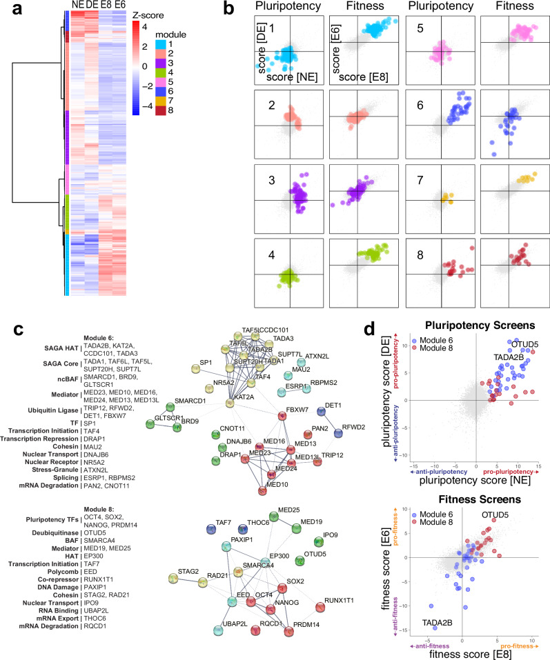
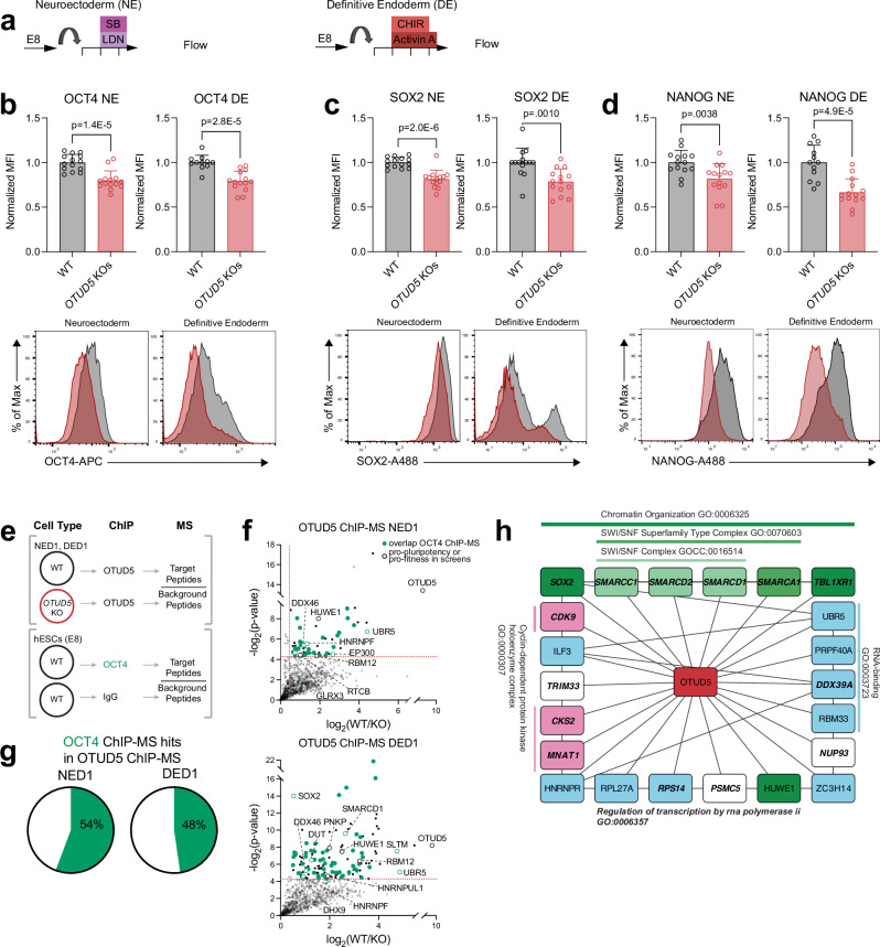
Pluripotent stem cells have remarkable self-renewal capacity: the ability to proliferate indefinitely while maintaining the pluripotent identity essential for their ability to differentiate into almost any cell type in the body. To investigate the interplay between these two aspects of self-renewal, we perform four parallel genome-scale CRISPR-Cas9 loss-of-function screens interrogating stem cell fitness in hPSCs and the dissolution of primed pluripotent identity during early differentiation. These screens distinguish genes with distinct roles in pluripotency regulation, including mitochondrial and metabolism regulators crucial for stem cell fitness, and chromatin regulators that control pluripotent identity during early differentiation. We further identify a core set of genes controlling both stem cell fitness and pluripotent identity, including a network of chromatin factors. Here, unbiased screening and comparative analyses disentangle two interconnected aspects of pluripotency, provide a valuable resource for exploring pluripotent stem cell identity versus cell fitness, and offer a framework for categorizing gene function.


Conflict of interest statement

The authors declare no competing interests.

Figures

Fig. 1
Fig. 1. NE and DE screens for exit of pluripotency reveal common programs.
a Schematic of NE and DE CRISPR screen comparison. b NE and DE Screen results by pluripotency score per gene = log10(RRA score OCT4-GFPhi enrichment) − log10(RRA score OCT4-GFPlo enrichment), top 150 pro-pluripotency (ranked by RRA score OCT4-GFPlo enrichment) and top 150 anti-pluripotency (ranked by RRA score OCT4-GFPhi enrichment) hits from each screen labeled, RRA scores determined by MAGeCK. c Overlap of top 150 pro-pluripotency genes identified in NE vs. DE context screens, and top 150 anti-pluripotency genes identified in NE vs. DE context screens, two-tailed Fisher’s exact test used for comparison. d GSEA for enrichment of pro- and anti-pluripotency top hits in screen results ranked by pluripotency score, NE vs. DE screens. e Bar graphs show relative MFI of OCT4-GFP following gRNA targeting of H1 iCas9 OCT4GFP/+ hESCs during NE differentiation. Relative MFI OCT4-GFP = (MFI per gRNA)/(MFI non-targeting controls). n = 9 independent experiments. 2 non-targeting controls analyzed per experiment. Data represented as mean. Error bars indicate s.d. Statistical analysis was performed by unpaired two-tailed Student’s t test. P values indicated. f Bar graphs show relative MFI of OCT4-GFP following gRNA targeting of H1 iCas9 OCT4GFP/+ hESCs during DE differentiation. Relative MFI OCT4-GFP = (MFI per gRNA/ MFI in-well tdT uninfected control)/(MFI non-targeting controls/ MFI in-well tdT uninfected control). n = 5 independent experiments. 2 non-targeting controls analyzed per experiment. Data represented as mean. Error bars indicate s.d. Statistical analysis was performed by unpaired two-tailed Student’s t test. P values indicated. g Comparison NE screen pluripotency score vs. NE validation mean relative intensity OCT4-GFP by gene. Statistical analysis by Pearson correlation test. h Comparison DE screen pluripotency score vs. DE validation mean relative intensity OCT4-GFP by gene. Statistical analysis by Pearson correlation test. Source data are provided as a Source Data file.
Fig. 2
Fig. 2. Comparison pluripotency and cell fitness screens reveal distinct hits.
a Schematic E8 and E6 fitness screens. b E8 and E6 Screen results by fitness score per gene = log10(RRA score Day 10 enrichment) − log10(RRA score Day 0 enrichment), top 150 pro-fitness (ranked by RRA score Day 0 enrichment) and top 150 anti-fitness (ranked by RRA score Day 10 enrichment) hits from each screen labeled. c E8 and E6 screen fitness hits in NE and DE screens. d NE and DE Screen results by pluripotency score per gene = log10(RRA score OCT4-GFPhi enrichment) − log10(RRA score OCT4-GFPlo enrichment), top 150 pro-pluripotency (ranked by RRA score OCT4-GFPlo enrichment) and top 150 anti-pluripotency (ranked by RRA score OCT4-GFPhi enrichment). e NE and DE screen pluripotency hits in E8 and E6 screens. f Upset plot of overlap NE, DE pro-pluripotency hits and E8, E6 pro-fitness, and Upset plot overlap NE, DE anti-pluripotency and E8, E6 anti-fitness hits. g GSEA for enrichment of pro-pluripotency and pro-fitness gene sets from NE, DE, E8, E6 screens, previous screens by Yilmaz et al., Mair et al., and Ihry et al., as re-analyzed by Mair et al., and previous pluripotency screen as published in Ihry et al.. Gene sets from previous fitness screens are top 150 genes per screen as ranked by Bayes factor score, calculated by BAGEL analysis. Gene sets from previous pluripotency screen are top 150 genes per screen as ranked by RSA score. Pluripotency screens labeled in red text, fitness screens labeled in orange text. h STRING database analysis pro-pluripotency and pro-fitness hits from NE, DE, E8, and E6 screens, strength = log10(observed/expected) for enrichment of gene types within a set. i Proportion of common essentiality genes as defined by DepMap data sets,, identified as top pro-pluripotency or pro-fitness hits in NE, DE, E8, and E6 screens.
Fig. 3
Fig. 3. Defining gene modules based on screening results.
a Hierarchical clustering by z-score-per-gene of pro-pluripotency hits from NE and DE screens, and pro-fitness hits from E8 and E6 screens. Relative levels of LFC represented as column z-score. b Modules by pluripotency score in NE and DE screens, and fitness score in E8 and E6 screens. c Annotation of all genes within modules 6 and 8, and interactome indicating known associations between members of module gene sets identified through STRING analysis using default parameters for high-confidence interactions. Each gene-set was further clustered using k-means clustering (k = 5), and dotted lines indicate connections between clusters. d Modules 6 and 8 in NE and DE screens by pluripotency score, and E8 and E6 screens by fitness score with archetypal genes TADA2B and OTUD5 labeled.
Fig. 4
Fig. 4. TADA2B KOs and OTUD5 KOs have opposing fitness phenotype.
a Schematic of TADA2B including location of gRNA targeting and sequences of knockouts (KOs). Boxes indicate TADA2B exons, and filled gray boxes indicate the coding sequence of TADA2B. b Western blot of TADA2B confirming KOs in HUES8 iCas9 WT1, WT2, WT3, and HUES8 iCas9 TADA2B-/- KO1, KO2, and KO3 clonal lines. Representative from 2 independent experiments. c Schematic of OTUD5 targeting and KOs. d Western blot of OTUD5 confirming KOs in HUES8 iCas9 WT2, WT3, and HUES8 iCas9 OTUD5-/- KO1, and KO2 clonal lines. Representative from 2 independent experiments. e Growth curve and area under growth curve (AUC) of WT and TADA2B KOs in E8 maintenance conditions and (f) E6 challenge conditions. 3 WT and 3 KO lines analyzed per experiment. n = 3 independent experiments. Data represented as mean. Error bars indicate s.d. Statistical analysis was performed by unpaired two-tailed Student’s t test. P values for significant comparisons indicated. g Growth curve and AUC of WT and OTUD5 KOs in E8 maintenance conditions. 2 WT and 2 KO lines analyzed per experiment. n = 5 independent experiments. Data represented as mean. Error bars indicate s.d. Statistical analysis was performed by unpaired two-tailed Student’s t-test. No significant comparisons were found. h Growth curve and AUC of WT and OTUD5 KOs in E6 challenge conditions and (i) after plating without Y-27632 (ROCKi), a standard component of hESC culture. 2 WT and 2 KO lines analyzed per experiment. n = 3 independent experiments. Data represented as mean. Error bars indicate s.d. Statistical analysis was performed by unpaired two-tailed Student’s t test. P values for significant comparisons indicated. j Cell competition assay WT vs. OTUD5 KOs. Individual cell lines were labeled with LARRY barcodes, and pooled. Pooled cells were expanded in E8 conditions for 28 days. Pools were collected and sequenced for representation of barcodes every 7 days. WT parental line and KO1, KO2 lines analyzed per experiment. n = 5 independent experiments. Data represented as mean. Error bars indicate s.d. Statistical analysis was performed by unpaired two-tailed Student’s t test. P values for significant comparisons indicated. Source data are provided as a Source Data file.
Fig. 5
Fig. 5. TADA2B KOs display reduced markers of apoptosis and increased markers of differentiation.
a Flow cytometry quantification and representative dot-plots cleaved caspase 3 in WT and TADA2B KO hESCs. n = 3 independent experiments, 3 WT and 3 KO lines were analyzed per experiment (HUES8 iCas9 WT1, WT2, WT3, and HUES8 iCas9 TADA2B-/- KO1, KO2, and KO3 clonal lines). Data represented as mean. Error bars indicate s.d. Statistical analysis was performed by unpaired two-tailed Student’s t test. P values indicated. b Flow cytometry quantification and representative histograms Annexin-V WT and TADA2B KO hESCs. n = 3 independent experiments, 3 WT and 3 KO lines were analyzed per experiment (HUES8 iCas9 WT1, WT2, WT3, and HUES8 iCas9 TADA2B-/- KO1, KO2, and KO3 clonal lines). Data represented as mean. Error bars indicate s.d. Statistical analysis was performed by unpaired two-tailed Student’s t-test. P-values indicated. c Flow cytometry quantification and representative histograms. Phosphohistone H3 WT and TADA2B KO hESCs. n = 3 independent experiments 2 WT and 3 KO lines were analyzed per experiment (HUES8 iCas9 WT2, WT3, and HUES8 iCas9 TADA2B-/- KO1, KO2, and KO3 clonal lines). Data represented as mean. Error bars indicate s.d. Statistical analysis was performed by unpaired two-tailed Student’s t-test. P-values indicated. Source data are provided as a Source Data file. d Volcano plot showing differential expression in TADA2B KO vs. WT hESCs. 3 WT and 3 KO lines analyzed by RNA-Seq. Statistical analysis with DeSeq2. e GSEA shows negative enrichment of pluripotency markers,, and (f) positive enrichment of early differentiation gene sets in TADA2B KO vs. WT differential expression.
Fig. 6
Fig. 6. OTUD5 KOs identify role for OTUD5 in the regulation pluripotency.
a Schematic of NE and DE differentiations for flow cytometry. b Flow cytometry quantification and representative histograms OCT4, (c) SOX2, and (d) NANOG in WT and OCT4 KO hESCs in NE and DE differentiations. HUES8 iCas9 WT2, WT3, and HUES8 iCas9 OTUD5-/- KO1, and KO2, lines analyzed in each experiment. n = 7 independent experiments. Data represented as mean. Error bars indicate s.d. Statistical analysis was performed by unpaired two-tailed Student’s t-test. P-values are indicated. Source data are provided as a Source Data file. e Schematic of OTUD5 and OCT4 ChIP-MS. f Plots showing proteins identified in OTUD5 ChIP-MS in NE Day 1 and DE Day 1. Dotted lines indicate the cut-offs (log2(Fold Change OTUD5/IgG)) > 0.5, –log2 (P) > 4.32) for significantly enriched proteins. Proteins that are also identified by OCT4 ChIP-MS in hESCs are indicated with green circles. Pro-pluripotency and pro-fitness screen hits labeled. OTUD5 ChIP-MS n = 3 independent experiments using HUES8 iCas9 WT2, and HUES8 iCas9 OTUD5-/- KO2. OCT4 ChIP-MS n = 2 independent experiments using HUES8 iCas9 parental hESCs. P values assessed using a heteroscedastic t-test. g Proportion of proteins identified by OCT4 ChIP-MS in OTUD5 ChIP-MS hits. h Cytoscape network visualization displays the interactions between 22 hits identified by OTUD5 ChIP-MS. To select these hits, we merged the top 15 hits by p value in NE and DE OTUD5 ChIP-MS, and only included those with interaction scores >0.4 with at least one other hit based on STRING analysis. Edges and GO terms determined by STRING analysis. Edges visualizing the connection with OTUD5 from the ChIP-MS added.
Fig. 7
Fig. 7. The pull of differentiation signaling is distinct from the push of pluripotency loss.
Schematic of the push-pull model of the dissolution of pluripotency during differentiation. Waddington landscape generated using the waddingtonplot R-package.

Update of

References

    1. Nichols, J. et al. Formation of pluripotent stem cells in the mammalian embryo dependes on the POU transcription factor Oct4. Cell95, 379–391 (1998). - PubMed
    1. Niwa, H., Miyazaki, J. & Smith, A. G. Quantitative expression of Oct-3/4 defines differentiation, dedifferentiation or self-renewal of ES cells. Nat. Genet.24, 372–376 (2000). - PubMed
    1. Mitsui, K. et al. The homeoprotein Nanog is required for maintenance of pluripotency in mouse epiblast and ES cells. Cell113, 631–642 (2003). - PubMed
    1. Avilion, A. A. et al. Multipotent cell lineages in early mouse development on SOX2 function. Genes Dev.17, 126–140 (2003). - PMC - PubMed
    1. Chambers, I. et al. Functional expression cloning of Nanog, a pluripotency sustaining factor in embryonic stem cells. Cell113, 643–655 (2003). - PubMed

Publication types

Associated data

LinkOut - more resources