Meta-Analysis
. 2024 Oct 17;14(1):24341.
doi: 10.1038/s41598-024-75383-4.

A systematic review and meta-analysis of GFAP gene variants in Alexander disease

Affiliations
Meta-Analysis

A systematic review and meta-analysis of GFAP gene variants in Alexander disease

Alice Grossi et al. Sci Rep. .

Abstract

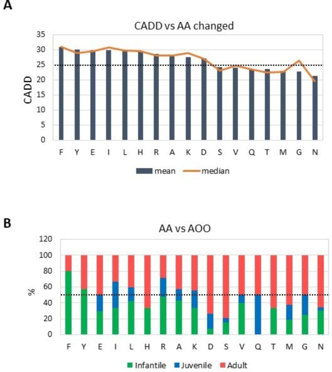
Alexander disease (ALXDRD) is a rare neurodegenerative disorder of astrocytes resulting from pathogenic variants in the GFAP gene. The genotype-phenotype correlation remains elusive due to the variable expressivity of clinical manifestations. In an attempt to clarify the effects of GFAP variants in ALXDRD, numerous studies were collected and analyzed. In particular, we systematically searched for GFAP variants associated with ALXDRD and collected information on the location within the gene and protein, prediction of deleteriousness/pathogenicity, occurrence, sex and country of origin of patients, DNA source, genetic testing, and clinical signs. To identify possible associations, statistical analyses and meta-analyses were applied, thus revealing a higher than expected percentage of adult patients with ALXDRD. Furthermore, substitution of Arginine, the most frequently altered residue among the 550 predominantly missense causative GFAP variants collected, were mostly de novo and more prevalent in early-onset forms of ALXDRD. The effect of defective splicing in modifying the impact of GFAP variants on the age of onset of ALXDRD was also postulated after evaluating the distribution of the corresponding deleterious predictive values. In conclusion, not only previously unrecognized genotype-phenotype correlations were revealed in ALXDRD, but also subtle mechanisms could explain the variable manifestations of the ALXDRD clinical phenotype.

Keywords: Alexander Disease; GFAP; Genotype-phenotype correlation; Meta-analysis; Variant effect.


Conflict of interest statement

The authors declare no competing interests.

Figures

Fig. 1
Fig. 1
PRISMA workflow. The PRISMA workflow outlines the systematic approach for conducting a meta-analysis. The diagram provides a visual representation of the paper’s selection process and outlines the study identification, screening, eligibility, and inclusion steps.
Fig. 2
Fig. 2
Relations between GFAP variants, Age of onset, CADD score and inheritance. (A) The barplot illustrates the amino acid predominantly affected in each of the three age of onset (AOO) categories, represented by light red for adult, light blue for juvenile, and light green for infantile. (B) The radial plot compares the mutated amino acids, reported as Arg versus non-Arg variants, in terms of associated AOO and occurrence. Almost half are arginine variants, prevalent in infantile cases, followed by adults and juveniles, with some missing data on age of onset. (C) Barplots of the six most prevalent variants at the Arg codon and their prevalence in different ALXDRD AOO. (D) Clinical data for 161 patients with the six prevalent Arg variants, categorized by EMBL-EBI Ontology (1. Neurodevelopmental abnormality; 2. Abnormality of higher mental function; 3. Seizure; 4. Abnormal central motor function; 5. Bulbar signs; 6. Other brainstem signs; 7. Somatic sensory dysfunction and abnormality of the autonomic nervous system; 8. Phenotypic abnormality). Notably, the distribution of symptoms varies among the different 6 most prevalent variants. (E) The rate of the de novo variants progressively increases with increasing CADD score, as shown in the bar graph for three different CADD ranges; conversely, inherited variants are progressively less represented with increasing CADD score range. (F) The distribution of CADD values with respect to age of onset shows that infantile and juvenile variants, represented in the box plots, have similar scores, on average higher than adults. (G) The mean (blue) and median (orange) CADD values were also stratified according to the position of the corresponding variants along the GFAP gene from N-terminal to C-terminal ends. Lines highlights the up-down alternating trend between secondary structured regions (1A-1B-2A-2B), belonging to the rod domain, and non-structured regions (C- and N-term and the L1, L12, L2 linkers). (H) Patients were also stratified by age of onset and position of the corresponding variant along the GFAP gene: infantile and adult cases present multiple variants in regions 1A and 2A, and in regions 2B and C-terminal, respectively.
Fig. 3
Fig. 3
Focus on AA changes and their relations with CADD and Age of onset. (A) The graph represents the mean (dark blue bars) and median (orange line) CADD values for each amino acid (AA) that is affected in the surveyed variants of the GFAP gene. Values are represented from highest to lowest. The dotted line indicates the CADD 25 cut-off. (B). In the graph, each bar corresponds to an AA affected by a GFAP variant. For each bar, the percentage of infantile (light green), juvenile (light blue), and adult (light red) patients carrying a variant that modifies this AA is represented. The dotted line indicates the percentage of 50% of total cases.
Fig. 4
Fig. 4
Forest Plots for GFAP variants vs. age of onset associations using the REML model. (A) The meta-analysis compares studies reporting the p.Arg416Trp variant between adult, juvenile, and infantile patients. (B) Results from the meta-analysis comparing studies reporting the p.Arg88Cys and p.Arg88Ser variants between adult, juvenile, and infantile patients. (C) Comparison of the effects induced by Arg (arginine) and non-Arg variants focused mainly on disease progression.

References

    1. Russo, L. S., Aron, A. & Anderson, P. J. Alexander’s disease: a report and reappraisal. Neurol. 26, 607–614 (1976). - PubMed
    1. Springer, S. et al. Alexander disease–classification revisited and isolation of a neonatal form. Neuropediatrics. 31, 86–92 (2000). - PubMed
    1. Prust, M. et al. GFAP mutations, age at onset, and clinical subtypes in Alexander disease. Neurol. 77, 1287–1294 (2011). - PMC - PubMed
    1. Yoshida, T. et al. Nationwide survey of Alexander disease in Japan and proposed new guidelines for diagnosis. J. Neurol. 258, 1998–2008 (2011). - PubMed
    1. Nielsen, A. L., Jørgensen, P. & Jørgensen, A. L. Mutations associated with a childhood leukodystrophy, Alexander disease, cause deficiency in dimerization of the cytoskeletal protein GFAP. J. Neurogenet. 16, 175–179 (2002). - PubMed

Substances

LinkOut - more resources