. 2024 Oct 17;14(1):24343.
doi: 10.1038/s41598-024-75207-5.

In vivo imaging identified efficient antimicrobial treatment against Mycobacterium marinum infection in mouse footpads

Affiliations

In vivo imaging identified efficient antimicrobial treatment against Mycobacterium marinum infection in mouse footpads

Kentaro Yamamoto et al. Sci Rep. .

Abstract

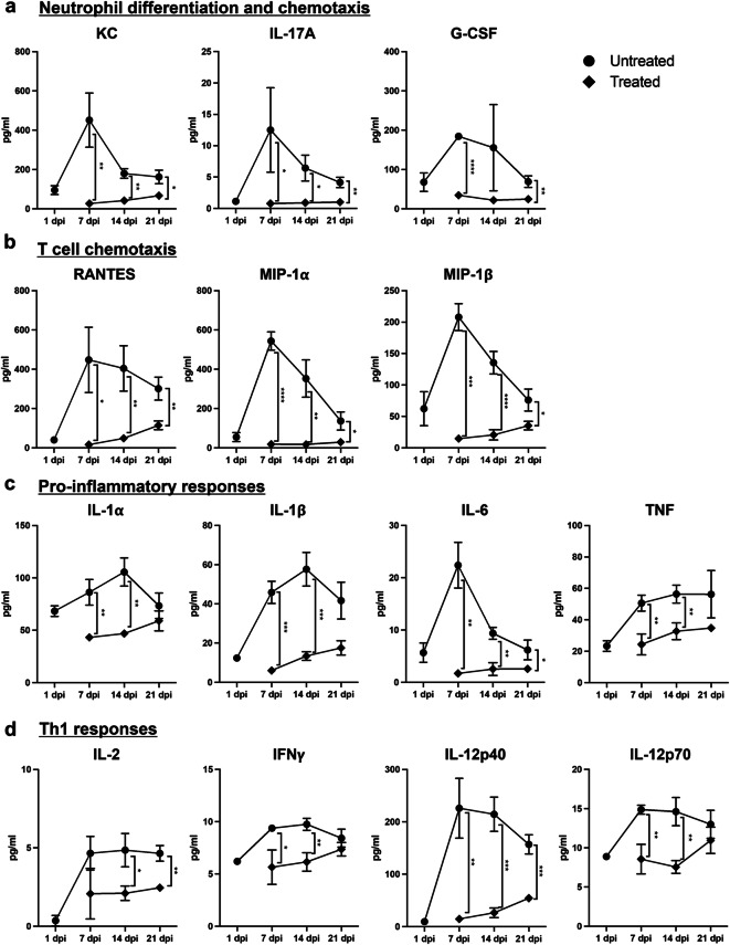
Mycobacterium marinum (M. marinum) is the most common causative bacteria of cutaneous non-tuberculous mycobacterial (NTM) infections, including fish tank granuloma. Treating M. marinum-caused infection takes longer than other NTM diseases because M. marinum is less susceptible to antimicrobial agents. A standard treatment regimen for M. marinum infection has not been established yet, and few in vivo experiments have been performed in mammals to evaluate the bactericidal effects of antimicrobials. In this study, we developed a noninvasive in vivo imaging method to assess the therapeutic efficacy of antimicrobials against M. marinum infection. The data obtained using fluorescent protein or bioluminescence from luciferase will offer valuable insights into bacteria visualization across various bacterial infections. Furthermore, through this imaging technique, we demonstrated that combining clarithromycin, rifampicin, ethambutol, and minocycline effectively cleared M. marinum from the footpad. Granulomas with necrotic abscesses formed on the footpad of M. marinum-infected mice, primarily due to neutrophils involved in the host's cell-mediated immune response. Inflammatory cytokine and chemokine levels significantly increased 7 days post-infection, aligning with the footpad swelling and granuloma formation observed in the untreated group. Interestingly, immune mediators and cells induced by M. marinum footpad infection were crucial factors associated with hypersensitivity and granuloma formation, as seen in pulmonary tuberculosis. This novel imaging analysis using a cutaneous NTM mouse model might be a powerful tool for the comprehensive analysis of mycobacterial infections.


Conflict of interest statement

The authors declare no competing interests.

Figures

Figure 1
Figure 1
In vivo imaging of M. marinum-infected BALB/c-nu/nu mouse footpad using various labeling tools. (a) Representative series of bacterial fluorescence and bioluminescence signals on a color scale superimposed on a grayscale image of a single mouse at indicated time points. The color scale displays radiant efficiency (photons/sec/cm2/steradian)/(µW/cm2) for fluorescent imaging and radiance (photons/sec/cm2/steradian) for bioluminescent imaging. (b) Calculation and plot of bacterial fluorescence and bioluminescence signals at each time point expressed in average radiant efficiency (photons/sec/cm2/steradian)/(µW/cm2) for fluorescent imaging and total flux (photons/sec) for bioluminescent imaging. (c) The mean CFU/footpad of each imaging tool was plotted at 0 and 28 dpi. No significant differences were observed between each increasing CFU.
Figure 2
Figure 2
Observation of pathology of M. marinum-infected BALB/c mouse footpad using in vivo imaging. (a) Representative series of bacterial fluorescence of iRFP670 (top) and bioluminescence signals of the Lux system (bottom) at each time point. Gross pathology images corresponding to each in vivo imaging are also presented. The color scale displays radiant efficiency (photons/sec/cm2/steradian)/(µW/cm2) for fluorescent imaging and radiance (photons/sec/cm2/steradian) for bioluminescent imaging. The mean CFU/footpad (b) and the differences in footpad thickness using preinfection thickness as a reference (c) were plotted at each time point. (d) Calculation and plot of Lux bioluminescent intensity at each time point expressed in photons/s (p/s). (e) Histological analysis of footpad tissues infected with M. marinum expressing the LuxCDABE gene using H&E and FF staining. The left panel (4×) presents a wide-angle view of the tissue, and the right panel (20×) is a magnified view of the tissue surrounding granulomas. Scale bar, 200 μm (4×) and 50 μm (20×). Dotted lines indicate the border of the granuloma; asterisks indicate the center of the granuloma; arrowheads indicate neutrophils.
Figure 3
Figure 3
Evaluation of antimicrobial efficacy in M. marinum infection using in vivo imaging. (a) Representative series of bacterial bioluminescence signals of the Lux system and gross pathology images in infected footpads from mice untreated and treated with antimicrobials at the indicated time points. The color scale indicates radiance (photons/sec/cm2/steradian). Calculated Lux bioluminescent intensity (b), the mean CFU/footpad (c), and the differences in footpad thickness (d) were plotted at each time point. Diamonds and circles represent the antimicrobial treatment and the control (untreated) groups, respectively. Statistical analysis by ANOVA with Sidack’s multiple comparison test: * P  < 0.05; **** P  < 0.0001. ND, not detected.
Figure 4
Figure 4
Profile of inflammatory cytokines and chemokines in footpads infected with M. marinum. (a) KC, IL-17A, G-CSF (b) RANTES, MIP-1α, MIP-1β (c) IL -1α, IL-1β, IL-6, TNF (d) IL-2, IFN-γ, IL-12p40, and IL-12p70 were detected in M. marinum -infected footpads with/without antimicrobial treatment at 1, 7, 14, and 21 dpi. The unpaired two-tailed Student’s t -test was used for statistical analysis. * P  < 0.05; ** P  < 0.01; *** P  < 0.001; **** P  < 0.0001.

References

    1. Schlossberg, D. Tuberculosis and Nontuberculous Mycobacterial Infections. Vol. 6th editionAmerican Society for Microbiology, (2011).
    1. Aronson, J. D. Spontaneous tuberculosis in Salt Water Fish. J. Infect. Dis.39, 315–320. 10.1093/infdis/39.4.315 (1926).
    1. Linell, F. & Norden, A. Mycobacterium Balnei, a new acid-fast bacillus occurring in swimming pools and capable of producing skin lesions in humans. Acta Tuberculosea Scand.33, 1–84 (1954). - PubMed
    1. Ang, P., Rattana-Apiromyakij, N. & Goh, C. L. Retrospective study of Mycobacterium marinum skin infections. Int. J. Dermatol.39, 343–347. 10.1046/j.1365-4362.2000.00916.x (2000). - PubMed
    1. Sia, T. Y. et al. Clinical and Pathological Evaluation of Mycobacterium marinum Group Skin Infections Associated with Fish Markets in New York City. Clin. Infect. Diseases: Official Publication Infect. Dis. Soc. Am.62, 590–595. 10.1093/cid/civ937 (2016). - PubMed

MeSH terms

Supplementary concepts

LinkOut - more resources