. 2024 Nov;21(11):2013-2016.
doi: 10.1038/s41592-024-02472-7. Epub 2024 Oct 17.

In vivo imaging of the human brain with the Iseult 11.7-T MRI scanner

Affiliations

In vivo imaging of the human brain with the Iseult 11.7-T MRI scanner

Nicolas Boulant et al. Nat Methods. 2024 Nov.

Abstract

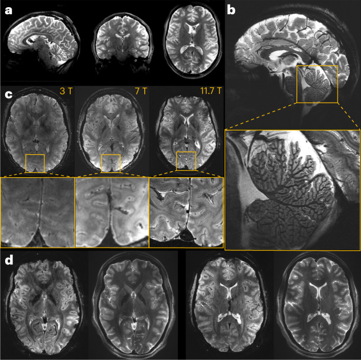
The understanding of the human brain is one of the main scientific challenges of the twenty-first century. In the early 2000s, the French Atomic Energy Commission launched a program to conceive and build a human magnetic resonance imaging scanner operating at 11.7 T. We have now acquired human brain images in vivo at such a magnetic field. We deployed parallel transmission tools to mitigate the radiofrequency field inhomogeneity problem and tame the specific absorption rate. The safety of human imaging at such high field strength was demonstrated using physiological, vestibular, behavioral and genotoxicity measurements on the imaged volunteers. Our technology yields T2 and T2*-weighted images reaching mesoscale resolutions within short acquisition times and with a high signal and contrast-to-noise ratio.


Conflict of interest statement

N.B. (US20190252788A1, WO2022194711A1), F.M. (WO2022194711A1), V.G. (US20190252788A1, WO2022194711A1) and A.A. (US9291691B2) hold several patents related to this work (pTx). A.M. is an employee of Siemens Healthineers. The other authors declare no competing interests.

Figures

Fig. 1
Fig. 1. Iseult magnet and installation set-up.
a, Schematic of the magnet showing the main coil and the two actively shielding coils. The magnet weighs 132 tons, has an outer diameter and total length of just under 5 m and the bore diameter is 90 cm. b, Cross-section of the wire used for the magnet showing the packing of the ten strands of NbTi-Cu wire within a copper channel. c, General view of the Iseult MRI inside the NeuroSpin building: (1) magnet (2), Faraday cages, (3) satellite, (4) dump resistor, (5) magnet control command and power convertor, (6) Siemens Healthineers MRI equipment, (7) helium liquefier, (8) helium pumps, (9) compressors and (10) helium balloon. d, Left: initial installation of the magnet within a dedicated arch (10 m in diameter) at NeuroSpin. Right: current side view of the magnet within the arch, extending above the ceiling and under the floor of the patient side room, going all the way in the back of the arch.
Fig. 2
Fig. 2. In vivo images of the human brain obtained at 11.7 T.
a, In vivo 3D T2 variable flip angle turbo spin-echo acquisition at 11.7 T with parallel transmission GRAPE universal pulses (resolution 0.55 × 0.55 × 0.55 mm3, acquisition time 13 min) demonstrating the high B1+ (RF) field homogeneity reached throughout the whole brain volume. b, In vivo T2*-weighted 2D GRE sagittal slice (resolution 0.2 × 0.2 × 1 mm3, acquisition time 8 min 30 s). c, T2* weighted 2D GRE axial images acquired at 3 T (left), 7 T (middle) and 11.7 T (right) with identical acquisition times (4 min 17 s), while keeping similar contrast-to-noise ratio through adjusted acquisition parameters (FA (°), TR (ms) and TE (ms) of 27, 750 and 45; 34, 950 and 25; 27, 600 and 20 at 3, 7 and 11.7 T, respectively) and spatial resolution (0.5, 0.325 and 0.2-mm in-plane resolution for 3, 7 and 11.7 T, respectively, 1-mm thickness). d, The 11.7 T T2*-weighted 2D GRE axial images (resolution 0.19 × 0.19 × 1 mm3, acquisition time 5 min 16 s) juxtaposed to turbo spin-echo T2-weighted images (resolution 0.3 × 0.3 × 1 mm3, acquisition time 4 min 26 s).
Extended Data Fig. 1
Extended Data Fig. 1. T2*-weighted 2D GRE axial images acquired at 3T, 7T and 11.7T (different subjects).
Acquisitions were performed with resolution = 0.2 × 0.2 × 1 mm3, FA = 27°, TE = 20 ms, TR = 0.6 s, bandwidth = 40 Hz/pixel, acquisition time = 4 min 20 s.
Extended Data Fig. 2
Extended Data Fig. 2. Results of the Attentional Network Task.
The number of samples is n = 20 for both 0T and 11.7T groups. Data are presented as mean values ± 1 standard error. Panel A (left). Mean decision times as a function of Run and Group. An Analysis of Variance with the factors Group and Run show no significant effect of Group (p = 0.54) and not any interaction (p = 0.62) (Extended Data Table 2). Both groups were slower inside the scanner than outside (Effect of Run: p < 0.001). Panel B (right). Flanker cost (difference in decision-times between incongruent and congruent trials). In an Analysis of Variance, the effect of Group was non significant (p = 0.57), nor interacted with any other factor (all p-values > 0.5) (Extended Data Table 3).

References

    1. Vogt, N. Human brain mapping. Nat. Methods20, 1869 (2023). - PubMed
    1. Finn, E. S., Poldrack, R. A. & Shine, J. M. Functional neuroimaging as a catalyst for integrated neuroscience. Nature623, 263–273 (2023). - PubMed
    1. Le Bihan, D. Looking into the functional architecture of the brain with diffusion MRI. Nat. Rev. Neurosci.4, 469–480 (2003). - PubMed
    1. Assaf, Y., Johansen-Berg, H. & de Schotten, M. T. The role of diffusion MRI in neuroscience. NMR Biomed.32, e3762 (2019). - PubMed
    1. Feinberg, D. A. et al. Next-generation MRI scanner designed for ultra-high-resolution human brain imaging at 7 Tesla. Nat. Methods20, 2048–2057 (2023). - PMC - PubMed

LinkOut - more resources