. 2024 Oct 17;7(1):1345.
doi: 10.1038/s42003-024-07008-5.

Insights into the cotranscriptional and translational control mechanisms of the Escherichia coli tbpA thiamin pyrophosphate riboswitch

Affiliations

Insights into the cotranscriptional and translational control mechanisms of the Escherichia coli tbpA thiamin pyrophosphate riboswitch

Jonathan P Grondin et al. Commun Biol. .

Abstract

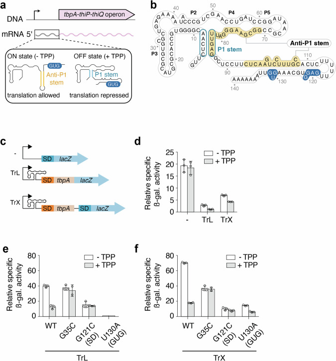
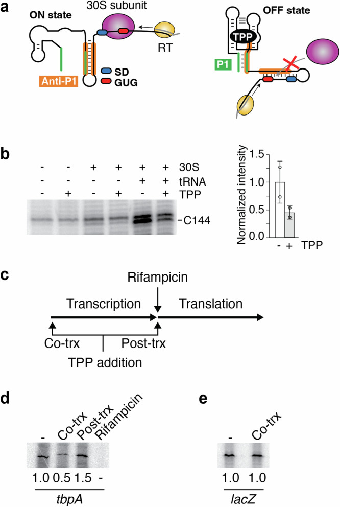
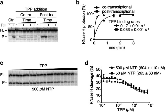
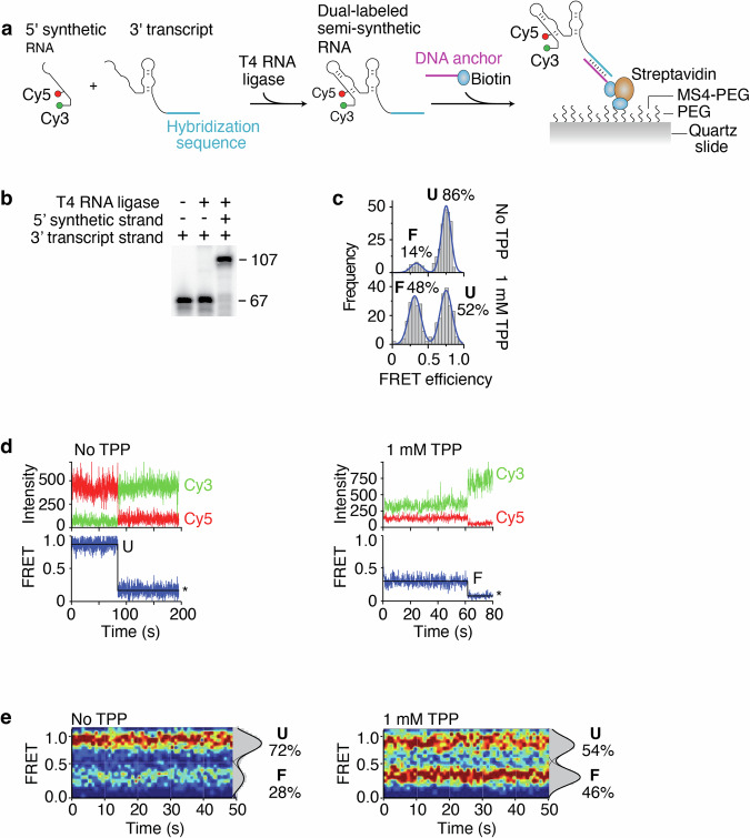
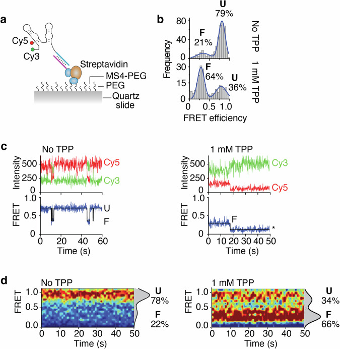
Riboswitches regulate gene expression by modulating their structure upon metabolite binding. These RNA orchestrate several layers of regulation to achieve genetic control. Although Escherichia coli riboswitches modulate translation initiation, several cases have been reported where riboswitches also modulate mRNA levels. Here, we characterize the regulation mechanisms of the thiamin pyrophosphate (TPP) tbpA riboswitch in E. coli. Our results indicate that the tbpA riboswitch modulates both levels of translation and transcription and that TPP sensing is achieved more efficiently cotranscriptionally than post-transcriptionally. The preference for cotranscriptional binding is also observed when monitoring the TPP-dependent inhibition of translation initiation. Using single-molecule approaches, we observe that the aptamer domain freely fluctuates between two main structures involved in TPP recognition. Our results suggest that translation initiation is controlled through the ligand-dependent stabilization of the riboswitch structure. This study demonstrates that riboswitch cotranscriptional sensing is the primary determinant in controlling translation and mRNA levels.


Conflict of interest statement

The authors declare no competing interests.

Figures

Fig. 1
Fig. 1. The regulation mechanism of the E. coli tbpA riboswitch.
a The tbpA riboswitch controls the expression of the tbpA-thiP-thiQ operon that is involved in the transport of thiamin and related metabolites. The riboswitch is located in the 5’ untranslated region and adopts the ON state in the absence of TPP in which the anti-P1 stem is formed, thereby allowing the initiation of translation. However, in the presence of TPP, the OFF state is adopted in which the P1 stem of the aptamer domain is stabilized, therefore repressing translation initiation. The GUG start codon is shown in blue. b Secondary structure of the tbpA riboswitch in the TPP-bound OFF structure. The anti-P1 stem is highlighted in yellow and the P1 stem is circled in blue. The Shine-Dalgarno and GUG start codon are shown in blue. c Schematics depicting the constructs used for ß-galactosidase assays. All fusions contain the natural promoter. The “- fusion” represents the construct in which the riboswitch domain is not present. The translational fusion (TrL) contains 11 codons of tbpA and is directly fused to the lacZ reporter. The transcriptional fusion (TrX) contains an additional Shine-Dalgarno (SD) sequence that allows the translation of the lacZ reporter without being affected by the structure of the riboswitch. d ß-galactosidase assays for the promoter-only (-), translational (TrL) and transcriptional (TrX) fusions. The average values of three independent experiments with standard deviations are shown. ß-galactosidase assays for constructs expressed from an arabinose-inducible promoter (pBAD). Gene expression was assessed for the wild-type (WT), G35C, G121C and U130A constructs in the context of both the translational (TrL) (e) and transcriptional (TrX) (f) fusions. The average values of three independent experiments with standard deviations are shown.
Fig. 2
Fig. 2. The tbpA riboswitch modulates translation initiation through cotranscriptional TPP sensing.
a Schematics representing the toeprint assays. Left, in the absence of TPP, the formation of the ON state allows the binding of the 30S ribosomal subunit. In such a case, the reverse transcriptase (RT) is expected to stop elongating upon reaching the bound 30S subunit. The Shine-Dalgarno (SD) and the GUG start codon are shown in blue and red, respectively. The anti-P1 stem is shown in orange. The DNA oligo used to produce the cDNA is shown in gray. Right, in the presence of TPP, the formation of the OFF state prevents the binding of the 30S subunit, thereby not producing a toeprint at the ribosome binding site. The P1 stem is shown in green. b Left, toeprint assays performed on the tbpA transcript as a function of the 30S subunit, tRNA-fMet and TPP. The toeprint at position C144 is preferentially obtained with the 30S subunit and tRNA-fMet, but is decreased upon TPP binding. Right, quantification of the toeprint efficiency. The experiments have been performed three times and the average and the standard deviations are shown. c Experimental assays monitoring TPP binding using in vitro transcription-translation assays. In these assays, transcription is first allowed to proceed until the addition of rifampicin. The second step involves the addition of amino acids to permit translation. The effect of TPP on the translation efficiency is assessed by adding it during the first (cotranscriptional) or second (post-transcriptional) step. d Transcription-translation assays assessing TPP binding to the riboswitch. Reactions were achieved in the absence (-) or presence of TPP added either cotranscriptionally (Co-trx) or post-transcriptionally (Post-trx) as indicated in c. The ratio of repression is shown below the gels. e Control experiments for transcription-translation assays were performed with lacZ. No translation repression was obtained when adding TPP cotranscriptionally. The ratio of repression is indicated below the gels.
Fig. 3
Fig. 3. TPP sensing is preferentially achieved cotranscriptionally by the tbpA riboswitch.
a RNase H probing experiments monitoring TPP binding when performed cotranscriptionally (Co-trx) or post-transcriptionally (Post-trx). Control reactions (Ctrl) done in the presence of RNase H (RH) show a cleaved product (P) and the uncleaved full-length (FL) transcript without TPP. Cotranscriptional binding was determined by adding TPP before transcription initiation and performing RNase H cleavage during transcription. Post-transcriptional measurements were achieved by performing transcription without TPP, which was followed by the addition of rifampicin, TPP and RNase H cleavage assays. b Quantifications of cotranscriptional and post-transcriptional binding assays. Experiments were fitted to a single-exponential assays and the values of the calculated rates are indicated. c Kswitch measurements for the wild-type tbpA riboswitch done in the presence of increasing TPP concentrations. The full-length (FL) and the cleave product (P) are shown on the left of the gel. d Quantification of the Kswitch experiments done when using 50 µM or 500 µM NTP. The Kswitch value is increasing upon decreasing the NTP concentrations. The average and the standard deviations are shown for each data point.
Fig. 4
Fig. 4. Single-molecule FRET studies of semi-synthetic tbpA aptamers.
a Schematic representing the procedure to obtain the semi-synthetic tbpA aptamer. A 5’ synthetic RNA strand contains Cy3 and Cy5 dyes at the 5’ extremity and position 14, respectively. The complete aptamer is reconstituted by ligating the 5’ strand and a 3’ T7 RNAP-transcribed strand. The aptamer is attached to a PEG slide using a sequence that is hybridized to a DNA anchor containing a biotin. b Ligation reactions performed in the presence of the 5’ and 3’ RNA strands. While the 3’ strand corresponds to the 67 nt product, the ligated product of both RNA molecules corresponds to the 107 nt product. c smFRET histograms of the semi-synthetic tbpA aptamer obtained in the absence and presence of 1 mM TPP. The folded (F) and unfolded (U) states are indicated. Histograms obtained without and with TPP were built using 264 molecules and 323 molecules, respectively. d smFRET time trajectories obtained in the absence (Left) and presence (Right) of TPP. The anti-correlated Cy3 donor and Cy5 acceptor emission intensities are shown with the resulting FRET trace. Photobleaching events are shown by an asterisk. e smFRET contour plots obtained in the absence (Left) and presence (Right) of TPP. Selected traces have been accumulated for the first 50 s. Histograms representing the percentual occupancy of each state are shown to the right. Contour plots obtained without and with TPP were built using 48 molecules and 51 molecules, respectively.
Fig. 5
Fig. 5. Single-molecule FRET studies of nascent tbpA aptamers obtained by stepwise transcription.
a Schematic representing the nascent RNA construct. The nascent RNA has been produced using stepwise transcription reactions and contains a 3’ sequence allowing to hybridize to a DNA anchor coupled to a biotin. b smFRET histograms of the nascent tbpA aptamer obtained in the absence and presence of 1 mM TPP. The folded (F) and unfolded (U) states are indicated. Histograms obtained without and with TPP were built using 210 molecules and 398 molecules, respectively. c smFRET time trajectories obtained in the absence (Left) and presence (Right) of TPP. The anti-correlated Cy3 donor and Cy5 acceptor emission intensities are shown with the resulting FRET trace. Photobleaching events are shown by an asterisk. d smFRET contour plots obtained in the absence (Left) and presence (Right) of TPP. Selected traces have been accumulated for the first 50 s. Histograms representing the percentual occupancy of each state are shown to the right. Contour plots obtained without and with TPP were built using 108 molecules and 117 molecules, respectively.

References

    1. Salvail, H. & Breaker, R. R. Riboswitches. Curr. Biol.33, R343–R348 (2023). - DOI - PMC - PubMed
    1. Breaker, R. R. The Biochemical Landscape of Riboswitch Ligands. Biochemistry61, 137–149 (2022). - DOI - PMC - PubMed
    1. Kavita, K. & Breaker, R. R. Discovering riboswitches: the past and the future. Trends Biochem. Sci.48, 119–141 (2023). - DOI - PMC - PubMed
    1. Ariza-Mateos, A., Nuthanakanti, A. & Serganov, A. Riboswitch Mechanisms: New Tricks for an Old Dog. Biochem. Mosc.86, 962–975 (2021). - DOI - PMC - PubMed
    1. Richards, J. & Belasco, J. G. Riboswitch control of bacterial RNA stability. Mol. Microbiol.116, 361–365 (2021). - DOI - PMC - PubMed

MeSH terms

LinkOut - more resources