Review
. 2024 Oct 18;9(1):274.
doi: 10.1038/s41392-024-01979-x.

Cold and hot tumors: from molecular mechanisms to targeted therapy

Affiliations
Review

Cold and hot tumors: from molecular mechanisms to targeted therapy

Bo Wu et al. Signal Transduct Target Ther. .

Abstract

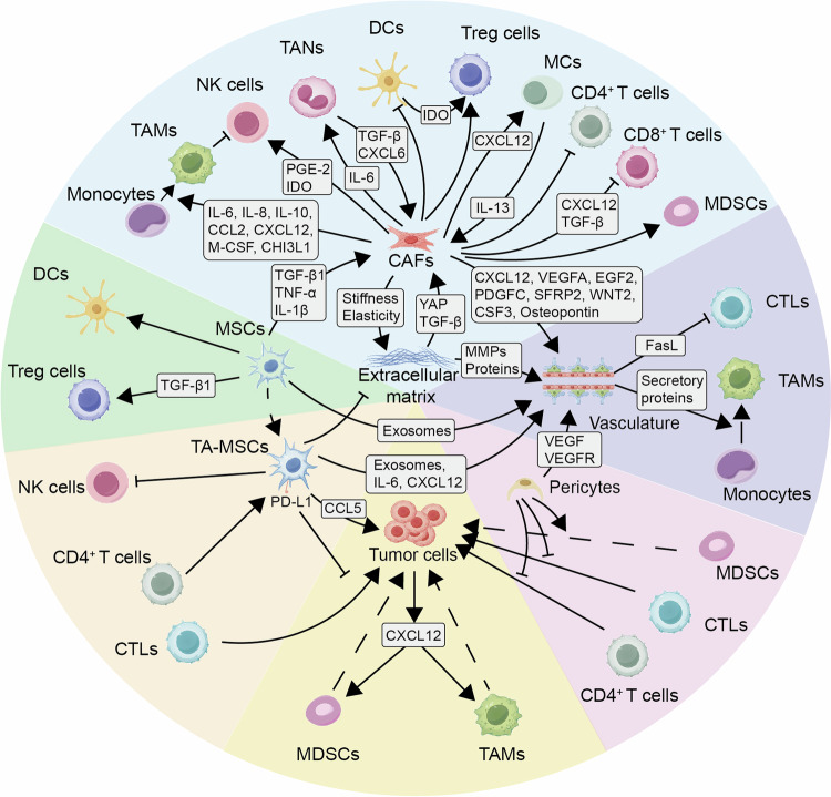
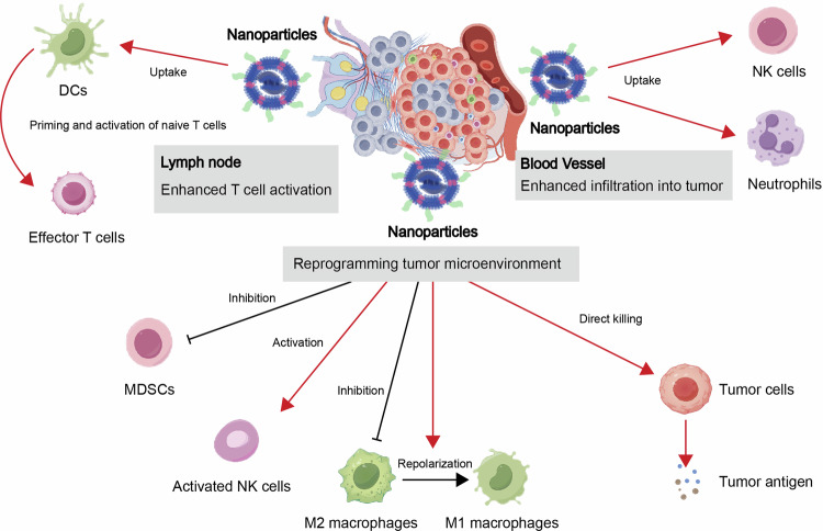
Immunotherapy has made significant strides in cancer treatment, particularly through immune checkpoint blockade (ICB), which has shown notable clinical benefits across various tumor types. Despite the transformative impact of ICB treatment in cancer therapy, only a minority of patients exhibit a positive response to it. In patients with solid tumors, those who respond well to ICB treatment typically demonstrate an active immune profile referred to as the "hot" (immune-inflamed) phenotype. On the other hand, non-responsive patients may exhibit a distinct "cold" (immune-desert) phenotype, differing from the features of "hot" tumors. Additionally, there is a more nuanced "excluded" immune phenotype, positioned between the "cold" and "hot" categories, known as the immune "excluded" type. Effective differentiation between "cold" and "hot" tumors, and understanding tumor intrinsic factors, immune characteristics, TME, and external factors are critical for predicting tumor response and treatment results. It is widely accepted that ICB therapy exerts a more profound effect on "hot" tumors, with limited efficacy against "cold" or "altered" tumors, necessitating combinations with other therapeutic modalities to enhance immune cell infiltration into tumor tissue and convert "cold" or "altered" tumors into "hot" ones. Therefore, aligning with the traits of "cold" and "hot" tumors, this review systematically delineates the respective immune characteristics, influencing factors, and extensively discusses varied treatment approaches and drug targets based on "cold" and "hot" tumors to assess clinical efficacy.


Conflict of interest statement

The authors declare no competing interests.

Figures

Fig. 1
Fig. 1
Illustrates the mechanisms underlying the anti-tumor immune response and immune evasion. The effectiveness of the anti-tumor immune response hinges on the activation, infiltration, and cytotoxic activity of effector T cells. These crucial processes encompass: a initiation of the T cell-mediated anti-tumor immune response through recognition of tumor-specific antigens (TSAs) in tumor microenvironment; (b) uptake and processing of tumor-specific antigens by dendritic cells (DCs); facilitation of cross-presentation in lymph node draining areas; (c) priming of naive T cells; recruitment of T cells by chemokines in blood vessels; (d) and identification and elimination of tumor cells in tumor microenvironment. Mechanisms of tumor immune evasion include characteristics that (a) diminish tumor immunogenicity, such as the absence of novel antigens, reduced expression of HLA molecules, or interference with antigen presentation to HLA molecules; b defects in antigen presentation possibly linked to dysfunctional DCs, affecting recruitment, activation, maturation, antigen cross-presentation, and T cell priming; c within the tumor microenvironment (TME), restrictions on T cell migration due to inadequate chemokine secretion and compromised chemotactic function of peripheral T cells are observed. Furthermore, abnormal vascular structures and a matrix rich in collagen/fibroblasts impede T cell infiltration. Genetic abnormalities in tumors also hinder T cell migration and infiltration; d Tumors and their immunosuppressive TME play a significant role in inducing T cell dysfunction and apoptosis. Immunotherapy is grounded in principles like tumor antigen release and presentation, T cell priming and activation, T cell migration and infiltration into tumors, and activation of T cell effector functions. Various therapeutic modalities, including chemotherapy, radiotherapy, targeted therapy, and anti-angiogenic therapy, aim to modulate the immune microenvironment and augment the efficacy of immunotherapy. In Fig. 1, the red dashed arrows symbolize effector T cells advancing anti-tumor responses, while the black dashed bars depict obstacles encountered by effector T cells during the anti-tumor response. This figure was created using Figdraw
Fig. 2
Fig. 2
Regulation of tumor immune efficacy by the cGAS-STING pathway. Activation of the cGAS-STING pathway in tumor cells plays a crucial role in inducing the secretion of cytokines and chemokines, thereby promoting the immune-mediated elimination of early-stage tumor cells. Additionally, tumors have the capability to produce cGAMP, which initiates the transcription of STING in neighboring cells within the tumor microenvironment (TME). Following uptake of abnormal extracellular DNA from dying tumor cells, dendritic cells (DCs) and macrophages engage directly with cGAS. This interaction results in increased expression of co-stimulatory molecules (CD80 and CD86) and MHC molecules in these immune cells, enhancing their capability to activate a cytotoxic T-cell response. By releasing type I interferons, antigen-presenting cells (APCs) augment the cytotoxic potential of natural killer (NK) cells. Furthermore, cGAMP mitigates immunosuppression by inhibiting the recruitment of M2 macrophages and myeloid-derived suppressor cells (MDSCs). Conversely, sustained activation of the STING pathway suppresses dendritic cells (DCs) while attracting myeloid-derived suppressor cells (MDSCs), thereby tilting the balance towards an immunosuppressive tumor microenvironment (TME). Moreover, the involvement of STING in stromal and endothelial cells elicits anti-tumor effects by enhancing the inflammatory milieu, attracting immune cells, and guiding tumor necrosis. The cGAS-STING signaling pathway exhibits a dual role in both promoting and inhibiting tumor growth, with its effects predominantly influenced by the intensity and duration of the stimuli. In this context, black arrows represent promotion, while black bars symbolize inhibition. This figure was created using Figdraw
Fig. 3
Fig. 3
Epigenetic modulation and tumor immune efficacy. DNMT1 and EZH2 play critical roles in DNA and histone methylation, respectively. This epigenetic modification leads to the downregulation of chemokine genes Cxcl9 and Cxcl10, impeding the recruitment of CD8+ T cells. Reduced Cxcl9 secretion by antigen-presenting cells (APCs) following interferon (IFN)-γ exposure is associated with Ccl5 methylation in cancer cells, resulting in diminished CD8+ T cell infiltration. Leukemia inhibitory factor (LIF) promotes the recruitment of EZH2 to the Cxcl9 promoter in tumor-associated macrophages (TAMs), contributing to epigenetic silencing. The formation of immunological synapses and antigen presentation is essential for mounting effective cytotoxic responses against tumors. Nevertheless, epigenetic mechanisms, notably DNA methylation, can silence this process within tumor cells. Methylation of genes in PD-1+CD8+ T cells may induce an exhaustion state, leading to resistance to therapies targeting the PD-1 pathway, such as anti-PD-1 antibodies. In this context, black arrows represent promotion, while red bars symbolize inhibition. This figure was created using Figdraw
Fig. 4
Fig. 4
Illustrates the mechanisms of metabolic regulation in tumor immune evasion. Tumor cells and immune cells adapt to the tumor microenvironment by modifying their metabolic programs in response to conditions such as hypoxia and nutrient deprivation. a Tumor oncogenic signaling pathways and transcription factors play a crucial role in regulating the expression of immune checkpoint molecules and genes associated with glycolysis, ultimately contributing to tumor immune evasion. Additionally, metabolites can directly influence the expression of immunosuppressive molecules. b Dysfunctions in immune cells may arise due to alterations in metabolites. The upregulation of glycolysis in tumor cells affects the expression levels of MHC-I and PD-L1 proteins, while glucose deprivation and increased lactate levels inhibit the function of NK and CD8+ T cells but enhance the suppressive activity of Treg cells within the tumor microenvironment. c A competition in glutamine metabolism is observed in the tumor microenvironment, where enhanced arginine-sensing mechanisms support the survival of T cells. Furthermore, lactate produced by tumors can induce macrophages to shift towards the M2 phenotype, potentially leading to arginine deprivation in T cells and NK cells. d Tumor immune cells display distinct metabolic characteristics, with Treg cells and M2 macrophages maintaining their suppressive function facilitated by fatty acid transporters like CD36, while the presence of fatty acids hinders the effector function and viability of CD8+ T cells. In this context, black arrows represent promotion, while black bars symbolize inhibition. This figure was created using Figdraw
Fig. 5
Fig. 5
illustrates the dual role of ferroptosis in the tumor microenvironment. Concerning antitumor immunity, ferroptotic tumor cells release immunostimulatory signals that facilitate dendritic cell maturation, activate M1-polarized macrophages, and enhance T cell infiltration and activity within tumors. Both CD8+ T cells and neutrophils contribute to promoting ferroptosis in tumor cells. Ferroptosis in tumor cells alleviates the inhibition of cancer-associated fibroblasts (CAFs) by reducing TGF-b1 levels. Moreover, ferroptosis induction in various immunosuppressive cells, such as tumor-infiltrating neutrophils, myeloid-derived suppressor cells (MDSCs), regulatory T (Treg) cells, and M2-polarized tumor-associated macrophages (TAMs), boosts antitumor immunity. On the other hand, in terms of immunosuppression, ferroptotic tumor cells impede dendritic cell maturation through products of phospholipid peroxidation. Additionally, CXCL10 and HMGB1 released by ferroptotic cancer cells upregulate PD-L1 expression. The release of oxidized phospholipids and prostaglandin E2 (PGE2) by ferroptotic polymorphonuclear-MDSCs suppresses the function of CD8+ T cells. Furthermore, ferroptosis induction in various antitumor immune cells, including natural killer (NK) cells, B cells, and T follicular helper (TFH) cells, leads to inhibited antitumor immunity. In this context, black arrows represent promotion, while black bars symbolize inhibition. This figure was created using Figdraw
Fig. 6
Fig. 6
illustrates the impact of chemokines on shaping the tumor microenvironment (TME). To begin with, tumor cells release tumor-specific antigens (TAAs) and newly formed antigens designed to be captured and processed by professional antigen-presenting cells (APCs). Conventional dendritic cells (cDCs) undergo maturation and upregulate CCR7, facilitating their migration to lymph nodes that drain the tumor site. The chemotactic axis of CCR7-CCL19/CCL21 guides naïve CD8+ and CD4+ T cells towards these lymph nodes. Inside the lymph nodes, naïve T cells that recognize TAAs interact with both cDC1s and cDC2s, resulting in activation of CD4+ and CD8+ T cells, along with increased expression of CXCR3. This directs the activated T cells to specific regions known as interfollicular areas (IFRs) within the lymph nodes. Within the IFRs, CD4+ T cells specific to TAAs engage with dendritic cells through CXCR3-dependent mechanisms, promoting their transformation into Th1 cells. Immunological cells with anti-tumor properties, including natural killer (NK) cells, cDC1s, Th1 cells, and CD8+ T cells, enter the tumor microenvironment (TME) guided by chemotactic gradients originating from the bloodstream. cDC1s secrete CXCL9 and CXCL10 to attract CXCR3+CD8+ T cells and enhance the functions of intra-tumoral T effector cells. Activated CD8+ T cells and Th1 cells position themselves close to tumor cells to aid in the elimination of tumors either by producing cytokines or directly killing them. Additionally, within the TME, tumor-associated macrophages (TAMs) are attracted to the tumor site as activated monocytes through the chemokine pathways of CCR5-CCL5 and CCR2-CCL2. TAMs promote tumor progression by releasing CCL17, CCL22, and CCL18 to recruit CCR4+ and CCR8+ regulatory T cells (Treg cells). Interactions between TAMs and tumor cells via the pathways of CCR2-CCL2 and CCR5-CCL5 enhance tumor stemness and metastatic potential. Furthermore, myeloid-derived suppressor cells (MDSCs) and tumor-associated neutrophils (TANs) present in the TME inhibit T cells and NK cells while attracting Treg cells by secreting chemokines such as CCL3, CCL4, CCL5 (by MDSCs), and CCL17 (by TANs). Treg cells are also lured by various chemokine systems, playing a role in tumor growth by suppressing T cell responses within the TME. This figure was created using Figdraw
Fig. 7
Fig. 7
Illustrates the tumor stroma cells and non-cellular components influencing the tumor microenvironment. The tumor comprises cancer cells and an encompassing stroma, which is a key constituent of the tumor microenvironment (TME), displaying distinct characteristics specific to the tumor type. This encompasses the extracellular matrix, a unique cancer-related vasculature, and various cellular elements such as activated cancer-associated fibroblasts, mesenchymal stromal cells, and pericytes. The cellular and non-cellular components within the tumor stroma actively engage in interdependent interactions, playing crucial roles in a finely regulated dynamic process. This collaborative mechanism promotes the evolution, progression, dissemination, and resistance to treatment of cancer. Notably, these findings underscore the integration of stromal-based cancer therapies in discourse. A profound comprehension of the dynamic interplay between stroma and cancer cells is imperative for devising innovative therapeutic approaches. In this context, black arrows represent promotion, while black bars symbolize inhibition. This figure was created using Figdraw
Fig. 8
Fig. 8
Nanoparticle-Mediated Tumor Microenvironment Intrinsic Immunomodulation Enhancing Cancer Immunotherapy. Engineered nanoparticles administered via subcutaneous or intravenous injection are internalized by either innate immune cells or tumor cells, releasing various payloads in lymph nodes, the tumor immune microenvironment, and vasculature. Nanoparticles possess the capacity to selectively target innate immune pathways, thereby augmenting innate immune responses against cancer. This is primarily due to the stimulation of innate immune cells by released agonists/antigens. This stimulation initiates the secretion of pro-immunogenic cytokines downstream, which subsequently enhances T-cell activation and infiltration in lymph node. Consequently, this process enhances the anti-tumoral immune responses of cytotoxic T lymphocytes (CTLs). Furthermore, nanoparticles effectively modulate the immunosuppressive tumor microenvironment (TME), improving tumors’ sensitivity to immunotherapy by engaging in specific interactions with innate immune cells. This includes elevating the presence of neutrophils and natural killer (NK) cells at tumor sites, diminishing the functions of M2 macrophages and myeloid-derived suppressor cells (MDSCs), transforming M2 macrophages into the M1 phenotype, and inciting the activation of NK cells. In this context, red arrows represent promotion, while black bars symbolize inhibition. This figure was created using Figdraw

References

    1. Sharma, P. et al. Immune checkpoint therapy-current perspectives and future directions. Cell186, 1652–1669 (2023). - PubMed
    1. Albelda, S. M. CAR T cell therapy for patients with solid tumours: key lessons to learn and unlearn. Nat. Rev. Clin. Oncol.21, 47–66 (2024). - PubMed
    1. Tsimberidou, A. M., Fountzilas, E., Nikanjam, M. & Kurzrock, R. Review of precision cancer medicine: Evolution of the treatment paradigm. Cancer Treat. Rev.86, 102019 (2020). - PMC - PubMed
    1. Li, Y. et al. mRNA vaccine in cancer therapy: current advance and future outlook. Clin. Transl. Med.13, e1384 (2023). - PMC - PubMed
    1. Märkl, F., Huynh, D., Endres, S. & Kobold, S. Utilizing chemokines in cancer immunotherapy. Trends Cancer8, 670–682 (2022). - PubMed

Publication types

MeSH terms

Substances

LinkOut - more resources