. 2024 Oct 18;14(1):24408.
doi: 10.1038/s41598-024-74898-0.

Pegylated gold nanoparticles interact with lipid bilayer and human serum albumin and transferrin

Affiliations

Pegylated gold nanoparticles interact with lipid bilayer and human serum albumin and transferrin

Elżbieta Okła et al. Sci Rep. .

Abstract

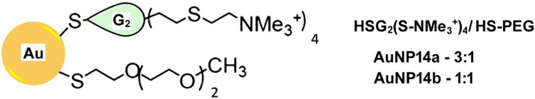
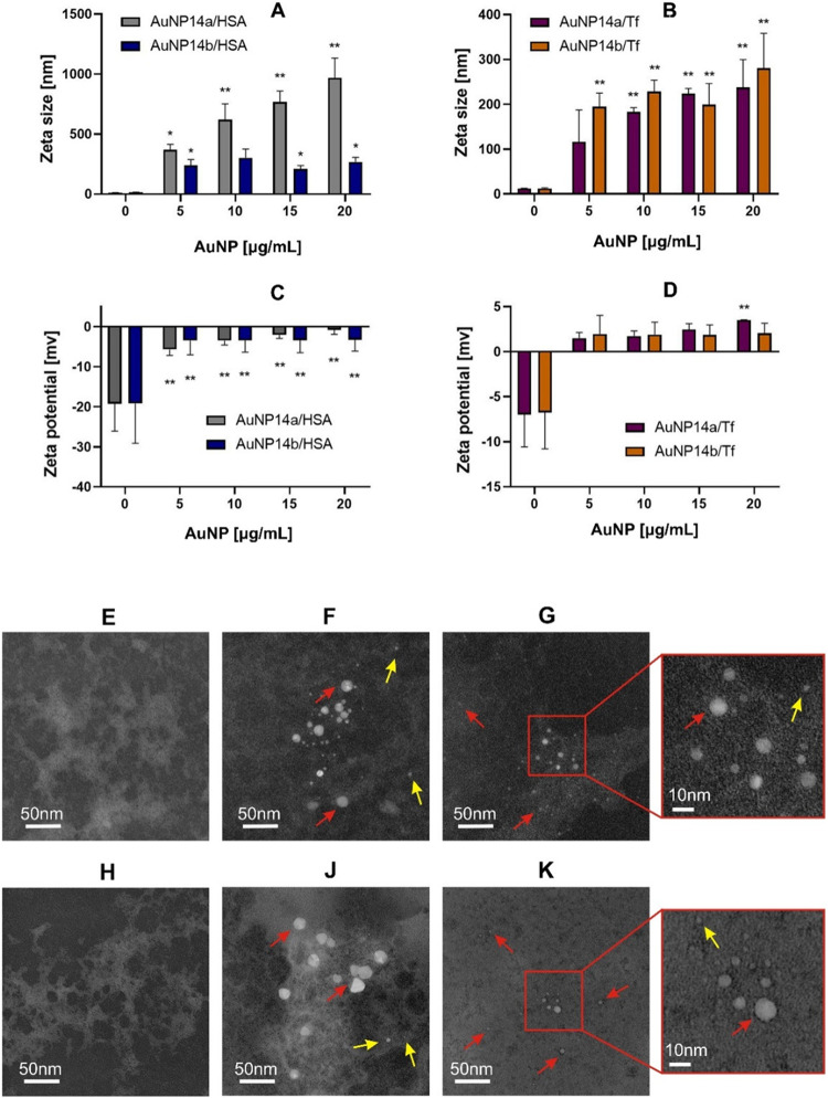
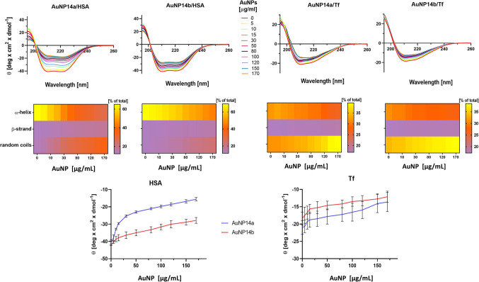
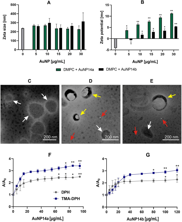
Gold nanoparticles (AuNPs) are potentially applicable in drug/nucleic acid delivery systems. Low toxicity, high stability, and bioavailability are crucial for the therapeutic use of AuNPs and they are mainly determined by their interactions with proteins and lipids on their route to the target cells. In this work, we investigated the interaction of two pegylated gold nanoparticles, AuNP14a and AuNP14b, with human serum proteins albumin (HSA) and transferrin (Tf) as well as dimyristoyl-phosphatidylcholine (DMPC) liposomes, which can be a representative of biomembranes. We showed that AuNP14a/b interacted with HSA and Tf changing their electrical, thermodynamic, and structural properties as evidenced by dynamic light scattering, zeta potential, transmission electron microscopy, circular dichroism, fluorescence quenching, and isothermal titration calorimetry. These nanoparticles penetrated the DMPC membrane suggesting their ability to reach a target inside the cell. In most of the effects, AuNP14b was more effective than AuNP14a, which might result from its more positive charge. Further studies are needed to evaluate whether the interaction of AuNP14a/b with HSA and Tf is safe for the cell/organism and whether they may safely penetrate natural membranes.

Keywords: DMPC lipid membranes; Liposomes; Pegylated gold nanoparticles; Protein corona; Serum human albumin; Transferrin.


Conflict of interest statement

The authors declare no competing interests.

Figures

Figure 1
Figure 1
Structure of pegylated gold nanoparticles of second generation (G2) with carbosilane dendron (left panel). The cationic dendron, HSG2(SNMe3+)4 and commercial PEG ligand CH3O(CH2CH2O)nCH2CH2SH, HS-PEG with the presentation of dendron/PEG ratio (right panel).
Figure 2
Figure 2
Influence of pegylated gold nanoparticles AuNP1a and AuNP14b on the hydrodynamic diameter expressed as zeta size of human serum albumin (HSA) (A) and transferrin (Tf) (B), and ζ-potential of HSA (C) and Tf (D). Graphs present mean values ± SD, * – p < 0.05, *** – p < 0.001, n = 3. Transmission electron microscopy shows the morphology of the proteins in the presence and the absence of AuNP14a/b: HSA (E), HSA and AuNP14a (F), HSA and AuNP14b (G), Tf (H) Tf and AuNP14a (J) Tf and AuNP14b (K). Red arrows point at large and yellow arrows to small nanoparticle/protein complexes.
Figure 3
Figure 3
Circular dichroism spectra of human serum albumin (HSA) and transferrin (Tf) incubated with AuNP14a/b nanoparticles expressed by ellipticity (top panel). Heat maps for the contribution of α-helix, β-strand, and random coils conformation to the overall structure of the proteins incubated with the AuNPs (middle panel). Ellipticity of HSA and Tf in the presence of AuNPs at λ 208 nm (bottom panels). Mean ± SD, n = 3.
Figure 4
Figure 4
Thermal effects of the interaction between gold nanoparticles AuNP14a/b and human serum albumin (HSA) or transferrin (Tf) evaluated by the heat (q) in isothermal titration calorimetry (left and middle panels). The right panel presents the thermal effects of the direct interaction of the proteins with AuNPs after subtracting the effects of AuNP dilution from their corresponding thermal effects of the titration of the protein solution with AuNP solution.
Figure 5
Figure 5
Zeta diameter (A) and zeta potential (B) of DMPC liposomes in the presence of gold nanoparticles AuNP14a/b. The shape of DMPC liposomes revealed by inverted transmission electron microscopy in the absence (C) and the presence of AuNP14a (D) and AuNP14b (E). White arrows point at free liposomes, red at free gold nanoparticles, and yellow at liposome/nanoparticle complexes. The mean relative fluorescence anisotropy (A/A0) of the DPH and TMA-DPH probes added to DMPC liposomes incubated with AuNP14a (F) or AuNP14b (G). Error bars denote SD, n = 3. * – p < 0.05; ** – p < 0.01 as compared with free liposomes.

References

    1. Bailly, A. L. et al. In vivo evaluation of safety, biodistribution and pharmacokinetics of laser-synthesized gold nanoparticles. Sci. Rep.9, 12890 (2019). - PMC - PubMed
    1. Lin, Q., Fathi, P. & Chen, X. Nanoparticle delivery in vivo: a fresh look from intravital imaging. EBioMedicine59, 102958 (2020). - PMC - PubMed
    1. Xu, M. et al. How entanglement of different physicochemical properties complicates the prediction of in Vitro and in vivo interactions of gold nanoparticles. ACS Nano. 12, 10104–10113 (2018). - PubMed
    1. Peña-González, C. E. et al. Gold nanoparticles stabilized by cationic carbosilane dendrons: synthesis and biological properties. Dalt Trans.46, 8736–8745 (2017). - PubMed
    1. Barrios-Gumiel, A. et al. Effect of PEGylation on the biological properties of cationic carbosilane dendronized gold nanoparticles. Int. J. Pharm.573, 118867 (2020). - PubMed

LinkOut - more resources