. 2024 Dec;20(12):1691-1700.
doi: 10.1038/s41589-024-01752-9. Epub 2024 Oct 17.

Paenilamicins are context-specific translocation inhibitors of protein synthesis

Affiliations

Paenilamicins are context-specific translocation inhibitors of protein synthesis

Timm O Koller et al. Nat Chem Biol. 2024 Dec.

Abstract

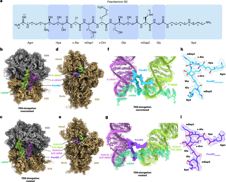
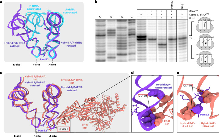
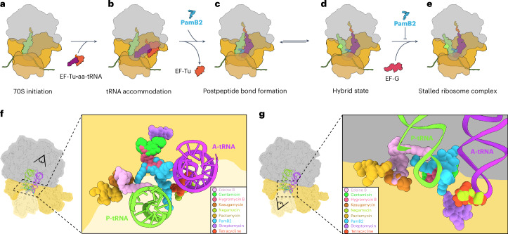
The paenilamicins are a group of hybrid nonribosomal peptide-polyketide compounds produced by the honey bee pathogen Paenibacillus larvae that display activity against Gram-positive pathogens, such as Staphylococcus aureus. While paenilamicins have been shown to inhibit protein synthesis, their mechanism of action has remained unclear. Here we determine structures of paenilamicin PamB2-stalled ribosomes, revealing a unique binding site on the small 30S subunit located between the A- and P-site transfer RNAs (tRNAs). In addition to providing a precise description of interactions of PamB2 with the ribosome, the structures also rationalize the resistance mechanisms used by P. larvae. We further demonstrate that PamB2 interferes with the translocation of messenger RNA and tRNAs through the ribosome during translation elongation, and that this inhibitory activity is influenced by the presence of modifications at position 37 of the A-site tRNA. Collectively, our study defines the paenilamicins as a class of context-specific translocation inhibitors.


Conflict of interest statement

Competing interests: The authors declare no competing interests.

Figures

Fig. 1
Fig. 1. Cryo-EM structures of paenilamicin B2 on the ribosome.
a, Chemical structure of paenilamicin B2 (refs. ,). be, Cryo-EM map of the PamB2-stalled ribosome in nonrotated (b,d) and rotated (c,e) elongation state, shown as transverse section (b,c) and 20S interface view (d,e). f,g, Extracted cryo-EM density assigned to PamB2 from the nonrotated (f, light blue) and rotated (g, dark purple) with surrounding A-tRNA (purple), P-tRNA (light green) from the nonrotated (f) and hybrid A/P (purple), and P/E-tRNA (light green) (g) and mRNA (cyan) in extracted density shown as mesh. h,i, Molecular model of PamB2 in extracted density of the nonrotated (h, light blue) and rotated (i, dark purple) states, shown as a mesh.
Fig. 2
Fig. 2. Interaction of PamB2 with the ribosomal P- and A-sites.
a, PamB2 (light blue) binding pocket located on the 30S subunit of the nonrotated PamB2–70S complex, with A-site tRNA (purple), P-site tRNA (light green), 16S rRNA (gray), 23S rRNA (yellow) and mRNA (cyan). b, Superimposition of the PamB2 binding pocket of the nonrotated (gray, with PamB2 in light blue) and rotated (light purple with PamB2 in purple) PamB2–70S complexes. cf, Direct and water-mediated interactions (dashed yellow lines) between PamB2 and the ribosome, colored as in a. c, Direct and intramolecular interactions of PamB2 with 16S rRNA of h44 and mRNA of the P-site codon. d, Water-mediated interactions of PamB2 with h44 of the 16S rRNA, mRNA of the P-site codon and P-site tRNA. e, Direct and intramolecular interactions of PamB2 with H69 of the 23S rRNA, mRNA of the A-site codon and A-site tRNA. f, Water-mediated interactions of PamB2 with h44 of the 16S rRNA, H69 of the 23S rRNA, mRNA of the A-site codon and A-site tRNA.
Fig. 3
Fig. 3. PamB2 inhibits tRNA2–mRNA translocation.
a, Superimposition of PamB2 and tRNAs of the nonrotated (light blue) and rotated (purple) PamB2–70S complexes. b, Toeprinting assay monitoring the effect of PamB2 on EF-G dependent translocation, with initiator tRNAfMet and N-AcPhe-tRNAPhe in the absence of drugs and in the presence of the translocation inhibitor negamycin. Toeprinting assays were performed in duplicate, with the duplicate gel shown in the source data. c, Superimposition of PamB2 and hybrid tRNAs of the rotated (purple) PamB2–70S complex with hybrid tRNAs and EF-G bound to the E. coli 70S ribosome in the Int1 state (salmon, PDB ID 7N2V). d,e, Sphere representation of the hybrid A/P-tRNA anticodon-stem loop of the rotated PamB2 complex sterically clashing with EF-G (salmon, PDB ID 7N2V) (d) and of the hybrid A/P- and P/E-tRNAs of the Int1 state (PDB ID 7N2V) (e) clashing with PamB2 (purple). Steric clashes are highlighted with red lines. Source data
Fig. 4
Fig. 4. Influence of A-site mRNA context on PamB2 inhibition.
a,b, Toeprinting assays monitoring the position of ribosomes on the wildtype ErmBL mRNA in the presence of water, 50 or 100 µM retapamulin (Ret), 50 µM erythromycin (Ery) and an ErmBL mRNA with an increasing number of UUG repetitions in the presence of 100 µM PamB2 (a) and with an ErmBL mRNA with the second codon mutated to UUG, AUG, CUG, GUG (orange) in the presence of 50 µM PamB2 (b). Arrows indicate the stalling sites on the isoleucine catch codon in the presence of mupirocin (pink), at initiation (green), on the erythromycin-ErmBL stalling site (purple) and stalling induced by PamB2 (blue). Toeprinting assays were performed in duplicate, with the duplicate gel shown in the Source Data. cf, Water (red) mediated interaction (dashed line) of PamB2 (blue) and the first nucleotide of the mRNA of the A-site codon (cyan), and superimposed with in silico mutated first position of the A-site codon (orange) to guanine (d), cytosine (e) or uracil (f). The loss of the water-mediated interaction is indicated by a red cross. Source data
Fig. 5
Fig. 5. Influence of A37 modification of A-tRNA on PamB2 inhibition.
a, PamB2 (light blue) and the modified A-site tRNA residue cyclic N6-threonylcarbamoyladenine (ct6) in position 37 (purple) from the nonrotated PamB2 complex superimposed with an in silico model of an unmodified A37 (yellow). b, Superimposition of PamB2 from a with the 2-methylthio-N6-isopentenyladenine (ms2i6, light orange) at position 37 of the A-site tRNAPhe from the T. thermophilus 70S ribosome preattack state (PDB ID 1VY5) shown as sphere representation with clashes indicated by red lines. c, Toeprinting assay monitoring the position of ribosomes on the (UUG)4-ErmBL mRNA in the presence of water (–), 50 µM retapamulin (Ret, green) and 100 µM PamB2 (light blue). The seventh codon was modified to different serine codons (orange). Arrows indicate stalling for the isoleucine catch codon in the presence of mupirocin (pink), the initiation (green) and PamB2-induced stalling (light blue). Toeprinting assays were performed in duplicate, with the duplicate gel present in the Source Data. d,e, Superimposition of PamB2 from a with 1-methyl-guanine (m1G, dark orange) at position 37 of the A-site tRNAPro on the T. thermophilus 70S ribosome (PDB ID 6NUO) (d) and an in silico modified 2-methyl-adenine (m2A, yellow) shown as sphere representation with steric clashes indicated by red lines (e). Source data
Fig. 6
Fig. 6. Mechanism of action of PamB2 and relative binding site of PamB2 compared to other antibiotics.
a–e, Model for the mechanism of action of PamB2 during translation. a,b, PamB2 does not bind stably to the initiation state with P-site tRNA only (a), nor during delivery and decoding of the aminoacyl-tRNA to the A-site by EF-Tu (b). c,d, PamB2 binds stably to pretranslocation states with A- and P-site tRNAs in nonrotated state and does not prevent peptide bond formation (c), as well as the rotated hybrid state with A/P- and P/E-tRNAs (d). e, Stable binding of EF-G is prevented by PamB2 thereby preventing translocation and trapping the ribosome in the pretranslocational states. f,g, View from the 50S (f) and 30S (g) subunit of PamB2 (light blue) superimposed with edeine B (pink, PDB ID 1I95), gentamicin (neon green, PDB ID 8CGU), hygromycin B (hot pink, PDB ID 8CAI), kasugamycin (dark orange, PDB ID 8CEP), negamycin (light green, PDB ID 4W2I), pactamycin (yellow, PDB ID 4W2H), streptomycin (pink, PDB ID 8CAI) and tetracycline (pink, PDB ID 8CF1), shown in sphere representation on the 30S subunit (head, light yellow; body, yellow), the 50S subunit (gray) and P- (green) and A-tRNA (purple).
Extended Data Fig. 1
Extended Data Fig. 1. Chemical structures and models of paenilamicin, galantin I and edeine.
a–c, Chemical structures of (a) paenilamicin, (b) galantin I and (c) edeine B. d, Molecular model of edeine B on the T. thermophilus 30S subunit (beige, Ede B, PDB ID 1I95). e, In silico molecular model of galantin I (Gal I, light green). f-i, Molecular model of PamB2 of the non-rotated PamB2 complex and in silico modelled PamB1 (green), PamA2 (light orange), PamA1 (orange).
Extended Data Fig. 2
Extended Data Fig. 2. Toeprinting assay on the MLIFstop-mRNA.
Toeprinting assay monitoring the position of ribosomes on a MLIFstop-mRNA in the presence of 100 µM thiostrepton (Ths), water (–), increasing concentrations of PamB2, N-Acetyl-PamB2 and PamB2_2 (0.5-100 µM). Arrows indicate the stalling at the initiation codon (green), and PamB2 induced stalling (blue). Toeprinting assays were performed in duplicate, with the duplicate gel shown in the Source Data. Source data
Extended Data Fig. 3
Extended Data Fig. 3. Fourier shell correlation and local resolution for the PamB2 complexes.
a–c, Fourier shell correlation (FSC) curve of the (a) non-rotated, (b) rotated and (c) initiation complexes, with unmasked (green) and masked (black) FSC curves plotted against the resolution (1/Å). d–i, Cryo-EM density colored according to local resolution and transverse section for the (d, e) non-rotated, (f, g) rotated and (h, i) initiation complexes. j–m, Molecular model of PamB2 (light blue and purple) and corresponding cryo-EM density colored according to local resolution for the (j, k) non-rotated, and (l–m) rotated complex.
Extended Data Fig. 4
Extended Data Fig. 4. PamB2 in silico modification and A-site binding pocket.
ad, In silico model of (a, b) N-acetyl-PamB2 (green) shown as (a) stick and (b) sphere representation, and (c, d) PamB2 with an N-acyl-D-Asn moiety (dark green) shown as (c) stick and (d) sphere representation. In (b) and (d), the sterically clashing molecules are highlighted by red lines. e, Interactions of the in silico modified PamB2_2 (light green) L-Agm amino group with surrounding 16S rRNA nucleotides of h44. f, Interactions of the PamB2 (light blue) wildtype D-Agm amino group with surrounding 16S rRNA nucleotides of h44. g, Binding pocket of PamB2 (light blue) with surrounding A-site tRNA nucleotides (purple), 16S rRNA (grey), mRNA (cyan) and 23S rRNA (yellow) superimposed with (h) the rotated PamB2 complex (purple), (i) the initiation complex (light cyan), (j) the E. coli initiation state (yellow, I-A, PDB ID 6WD0), (k) EF-Tu bound to the open E. coli 30S subunit (orange, II-A, PDB-ID 6WD2), and (l) EF-Tu bound to the closed E. coli 30S subunit (dark orange, III-A, PDB-ID 6WD8).
Extended Data Fig. 5
Extended Data Fig. 5. PamB2 inhibits eukaryotic translation.
a, Superimposition of the PamB2 (light blue) binding site on bacterial ribosomes (grey) with that of the eukaryotic ribosome (PDB ID 6Y0G) with 18S rRNA nucleotides (pink), 28S rRNA nucleotides (light orange), P-tRNA (light green), A-tRNA (purple) and mRNA (light cyan), illustrating the high conservation. b, In vitro translation assay using rabbit reticulocyte lysate-based system to monitor inhibition of increasing concentrations of PamB2. Reactions were performed in triplicate and the line of best fit is shown. c, MTT assay on NIH 3T3 murine fibroblast cells after treatment with different concentrations of PamB2 and CHX. The cell viability was measured (as absorbance at 570 nm) after 48 h of exposure to the peptides. Results are reported as percentages of viable cells with respect to the negative control cells with no compound (set as 100% of viability). Data are the average of three biological replicates, each of which was performed with internal triplicates and averaged. The individual values are available in the Source Data. Source data
Extended Data Fig. 6
Extended Data Fig. 6. PamB2 inhibits tRNA2-mRNA translocation.
a, Superimposition of the P- and A-site tRNA of the non-rotated PamB2 complex (light blue) and the hybrid A/P- and P/E-tRNA of the rotated PamB2 complex (dark purple). b, Superimposition of the P- and A-tRNA of the non-rotated PamB2-complex (light blue) and the P- and A-tRNA of the PreC state (light green, PDB ID 7N1P). c, Superimposition of the hybrid A/P- and P/E-site tRNA of the rotated PamB2-complex (dark purple) and the hybrid A/P- and P/E-site tRNA of the PreH1 state (dark green, PDB ID 7N2U). d–i, Superimposition of the hybrid A/P-site tRNA of the rotated PamB2-complex (dark purple) and the hybrid A/P-site tRNA and EF-G of the (d) H1-EF-G-GDP-Pi state (light yellow, PDB ID 7PJV), (e) H2-EF-G-GDP-Pi state (light orange, PDB ID 7PJW) and (f) pre-translocation (III) state (yellow, PDB ID 7SSL), (g) Int1 state (salmon, PDB ID 7N2V), (h) CHI1-EF-G-GDP state (orange, PDB ID 7PJY), (i) mid-translocation (IV) state (light orange, PDB ID 7SSD). j–l, PamB2 of the rotated complex (j) with surrounding hybrid A/P-tRNA (purple), 16S rRNA nucleotides (grey), 23S rRNA nucleotides (yellow) and the A-site codon of the mRNA (cyan) superimposed with (k) with the 70S E. coli ribosome in the PreH1 state (light green, PDB ID 7N2U) and (l) with the 70S E. coli ribosome and EF-G of the Int1 state shown as ribbon (salmon, PDB ID 7N2V).

References

    1. Fischbach, M. A. & Walsh, C. T. Antibiotics for emerging pathogens. Science325, 1089–1093 (2009). - PMC - PubMed
    1. Antimicrobial Resistance Collaborators. Global burden of bacterial antimicrobial resistance in 2019: a systematic analysis. Lancet399, 629–655 (2022). - PMC - PubMed
    1. Müller, S. et al. Paenilamicin: structure and biosynthesis of a hybrid nonribosomal peptide/polyketide antibiotic from the bee pathogen Paenibacillus larvae. Angew. Chem. Int. Ed. Engl.53, 10821–10825 (2014). - PubMed
    1. Bulatov, T. et al. Total synthesis and biological evaluation of paenilamicins from the honey bee pathogen Paenibacillus larvae. J. Am. Chem. Soc.144, 288–296 (2022). - PubMed
    1. Genersch, E. American Foulbrood in honeybees and its causative agent, Paenibacillus larvae. J. Invertebr. Pathol.103, S10–S19 (2010). - PubMed

Publication types

MeSH terms

LinkOut - more resources