. 2024 Oct 17;15(1):80.
doi: 10.1186/s13293-024-00652-w.

Sex-specific phenotypical, functional and metabolic profiles of human term placenta macrophages

Affiliations

Sex-specific phenotypical, functional and metabolic profiles of human term placenta macrophages

Daniel E Paparini et al. Biol Sex Differ. .

Abstract

Background: Placental macrophages, Hofbauer cells (HBC) are the only fetal immune cell population within the stroma of healthy placenta along pregnancy. They are central players in maintaining immune tolerance during pregnancy. Immunometabolism emerged a few years ago as a new field that integrates cellular metabolism with immune responses, however, the immunometabolism of HBC has not been explored yet. Here we studied the sex-specific differences in the phenotypic, functional and immunometabolic profile of HBC.

Methods: HBC were isolated from human term placentas (N = 31, 16 from male and 15 female neonates). Ex vivo assays were carried out to assess active metabolic and endoplasmic reticulum stress pathways by flow cytometry, confocal microscopy, gene expression and in silico approaches.

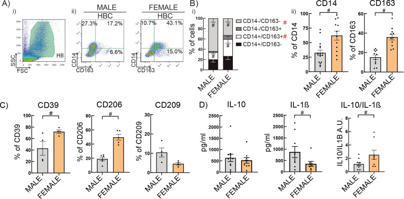
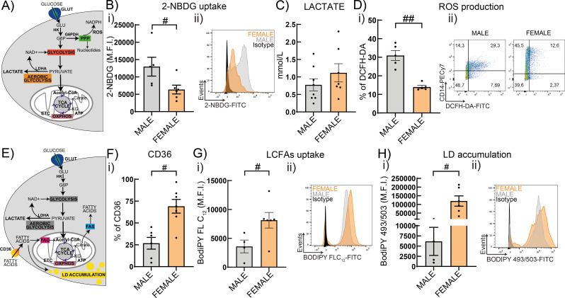
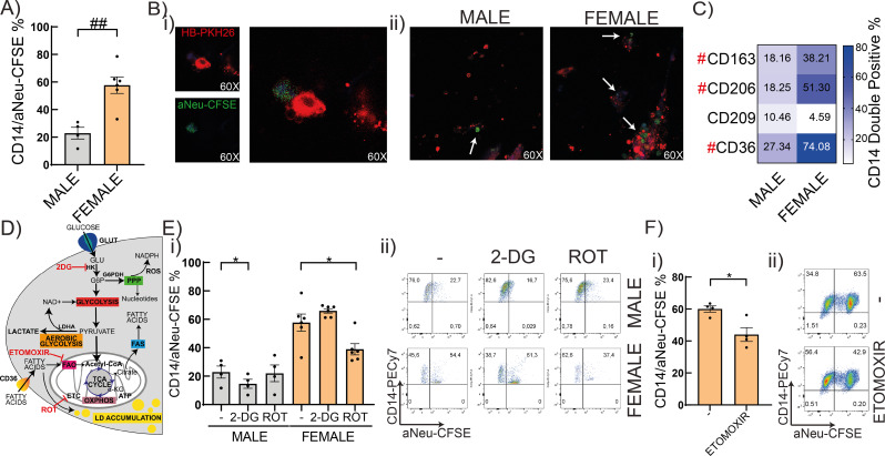
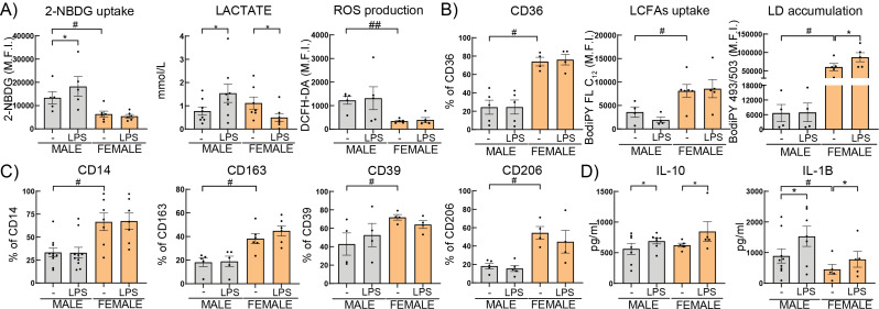
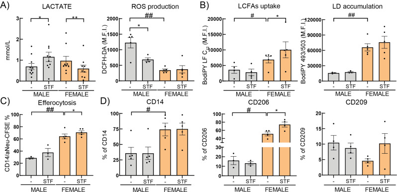
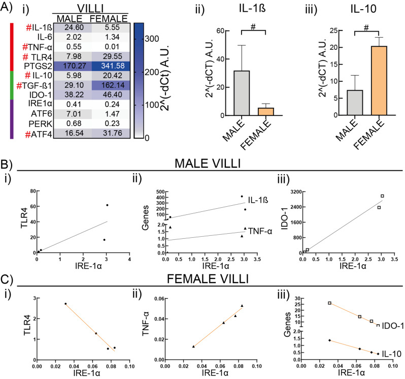
Results: HBC from female placentas displayed a stronger M2 phenotype accompanied by high rates of efferocytosis majorly sustained on lipid metabolism. On the other hand, male HBC expressed a weaker M2 phenotype with higher glycolytic metabolism. LPS stimulation reinforced the glycolytic metabolism in male but not in female HBC. Physiological endoplasmic reticulum stress activates IRE-1 differently, since its pharmacological inhibition increased lipid mobilization, accumulation and efferocytosis only in female HBC. Moreover, differential sex-associated pathways accompanying the phenotypic and functional profiles of HBC appeared related to the placental villi environment.

Conclusions: These results support sex-associated effects on the immunometabolism of the HBC and adds another layer of complexity to the intricate maternal-fetal immune interaction.

Keywords: Metabolism; Placental-macrophages; Sex-associated differences.

Plain language summary

Placental macrophages are the only fetal immune cell population within the stroma of healthy placenta along pregnancy and play a central role in contributing to the correct functioning of the placenta for the development of the fetus. Alterations in their metabolism lead to failures in their functions which are associated to pregnancy complications. Although, sex-specific differences were found in placental adaptation to pregnancy complications and outcomes, but the metabolism associated to their functions of placenta macrophages and whether they are associated to the sex of the placenta have not been explored so far. Here we studied human term placenta macrophages with special focus on their metabolism associated with their functions. We found out that macrophages from female placenta got energy from fatty acids whereas male macrophages used glucose. Furthermore, when female macrophages were exposed to a bacterial component, their metabolism or cellular function did not change towards one associated with a classic profile, but male macrophages did. These results might contribute to gain more insight into the immune-placental interactions at term human pregnancy and provide new clues to begin personalizing the pregnancy according to the sex of the fetus in physiological or pregnancy complications of inflammatory nature.


Conflict of interest statement

The authors declare no competing interests.

Figures

Fig. 1
Fig. 1
Female Hofbauer cells are more prone to an alternative-like profile. HBC were isolated from 31 placentas (16 male and 15 female newborn) by enzymatic digestion and cultured with RPMI 2% FCS ON. Cells were collected and analyzed by flow cytometry. A) Representative dot plots of (i) HBC gate and (ii) CD14-CD163% of positive cells. B) CD14 and CD163 proportion and C) CD39, CD206, CD209% of positive cells were shown. D) IL-10 and IL-1β secretion (pg/ml) were measured by ELISA in HBC supernatants. Results are expressed as Mean ± SEM. Statistically significant differences by placental sex are represented as in B) # (red) P < 0.05. Two-way ANOVA, Tukey’s multiple comparisons test [Interaction: F(3) = 8.628, P-value < 0.0001; Sex: F(1) = 5.99e-008, P-value = 0.998; Cell population: F(3) = 20.64, P < 0.0001]. B-D) # P < 0.05 (Mann-Whitney test) when only 2 conditions were analyzed. Each dot represents an independent experiment
Fig. 2
Fig. 2
Differential metabolic preferences in male and female HBC. A) and E) Simplified schematic overview of glucose and lipid metabolism, respectively. HBC cells were cultured with RPMI 2% FCS ON and collected to perform metabolic assays to study B) the glucose analogue 2-NBDG uptake, D) ROS production, F) CD36 expression, G) LCFAs uptake and H) Lipid Droplets (LD) accumulation by flow cytometry. Supernatants were used to study C) Lactate secretion with a colorimetric assay. Results are expressed as Mean ± SEM and in ii) representative histogram or dot plots are shown. Statistically significant differences between sex are represented as # P < 0.05 (Mann-Whitney test). Each dot represents an independent HBC sample. Abbreviations: Glucose (GLU), hexokinase (HK), glucose-6-phospate (G6P), G6P dehydrogenase (G6PDH), pentose phosphate pathway (PPP), reactive oxygen species (ROS), Lactate dehydrogenase A (LDHA), Tri-carboxylic acid (TCA), electron transport chain (ETC), oxidative phosphorylation (OXPHOS), fatty acid oxidation (FAO), fatty acid synthesis (FAS)
Fig. 3
Fig. 3
Sex-associated metabolic pathways in efferocytosis of HBC. A) Male or female HBC were challenged with maternal apoptotic neutrophils (aNeu), stained with CFSE, in a 1:5 ratio during 1 h. Then, they were washed and incubated with CD14 antibody. Double positive cells were analyzed by flow cytometry. B) Representative microphotographs of efferocytosis process (i) and MALE and FEMALE efferocytosis (ii) were taken by Zeiss LSM980 confocal microscope of 3 independent experiment each. White arrows show apoptotic bodies (green) in HBC-PKH26 (red). C) Phagocytic receptors expression (CD163, CD206, CD209 and CD36) were analyzed by flow cytometry. D) Simplified schematic overview of metabolic pathways and pharmacological inhibitors. E), F) HBC cells were cultured with metabolic inhibitors 2-DG (10 mM), ROT (100 nM) or ETOMOXIR (10 µM), stimulated with apopototic neutrophils as in (A) and analyzed by flow cytometry. Results are expressed as Mean ± SEM. Statistically significant differences: A, C) between HBC sex are shown as # P < 0.05, ## P < 0.01 (Mann-Whitney test). E) * P < 0.05, ** P < 0.01. Two-way ANOVA, Holm-Sidak’s multiple comparisons test. [Interaction: F(2) = 11.62, P-value = 0.0006; Sex: F(1) = 70.85, P-value < 0.0001; Treatment: F(1) = 6.157, P = 0.0095]. F) * P < 0.05 (basal vs. ETOMOXIR, Wilcoxon matched-pairs signed rank test). Each dot represents an independent HBC sample
Fig. 4
Fig. 4
HBC immunometabolic profile modulated by LPS. Male or female HBC were cultured in RPMI 2% FCS ON. A) glucose uptake and ROS production, B) CD36, LCFAs uptake, lipid droplets (LD) accumulation and C) CD14, CD163, CD206 and CD39, were measured by flow cytometry. In the supernatants A) Lactate or D) IL-10 and IL-1Β secretion were quantified by enzymatic assay or ELISA, respectively. Results are expressed as Mean ± SEM. Statistically significant differences by the stimuli within each sex are represented as * P < 0.05 (Wilcoxon matched-pairs signed rank test) and those parameters that were detected as significantly different between basal male and female HBC in Figs. 1 and 2, are here marked with #. Each dot represents an independent HBC sample
Fig. 5
Fig. 5
Pharmacological inhibition of IRE-1 and its association to HBC metabolic profile. HBC were cultured without or with 10μg/ml IRE1α inhibitor, STF-083010 (STF) in RPMI 2% FCS ON. A) ROS production, B) LCFA uptake and LD accumulation, C) Efferocytosis and D) CD14, CD206 and CD209 positive cells were evaluated by flow cytometry. In the supernatant A) Lactate was quantified by enzymatic assay. Results are expressed as Mean ± SEM. Statistically significant differences by the stimuli within each sex are represented as * P < 0.05 (Wilcoxon matched-pairs signed rank test) and those parameters that were detected as significantly different between basal male and female HBC in Figs. 1, 2 and 3, are here marked with #. Each dot represents an independent HBC sample
Fig. 6
Fig. 6
Sex associated transcriptionally differences in the placental microenvironment. Placentas (from 4 male and 4 female births) were processed to study gene expression in villi explants by RT-qPCR. A) Male or female gene expression is shown as their Mean 2^(-dCt). Statistically significant differences between sex are shown by # P < 0.05 (Mann-Whitney test). Red, green and purple lines mark pro, antiinflammatory and UPR genes, respectively. B) Male or C) Female gene correlations were evaluated by Pearson’s r. The r value was positive (blue) or negative (red) when the gene correlation was positive or negative, respectively. All correlations that are shown have P-values < 0.05

Similar articles

Cited by

References

    1. Mitchell AM, Palettas M, Christian LM. Fetal sex is associated with maternal stimulated cytokine production, but not serum cytokine levels, in human pregnancy. Brain Behav Immun. 2017;60:32–7. - PMC - PubMed
    1. O’Callaghan JL, Clifton VL, Prentis P, Ewing A, Saif Z, Pelzer ES. Sex-dependent differential transcript expression in the placenta of growth restricted infants. Placenta. 2022;128:1–8. - PubMed
    1. Eriksson JG, Kajantie E, Osmond C, Thornburg K, Barker DJP. Boys live dangerously in the womb. Am J Hum Biology. 2010;22:330–5. - PMC - PubMed
    1. Sun T, Gonzalez TL, Deng N, DiPentino R, Clark EL, Lee B et al. Sexually Dimorphic Crosstalk at the Maternal-Fetal Interface. J Clin Endocrinol Metab [Internet]. 2020;105:e4831-47. http://www.ncbi.nlm.nih.gov/pubmed/32772088 - PMC - PubMed
    1. Braun AE, Mitchel OR, Gonzalez TL, Sun T, Flowers AE, Pisarska MD, et al. Sex at the interface: the origin and impact of sex differences in the developing human placenta. Biol Sex Differ. BioMed Central Ltd; 2022. - PMC - PubMed

LinkOut - more resources