Quantum Dots-caused Retinal Degeneration in Zebrafish Regulated by Ferroptosis and Mitophagy in Retinal Pigment Epithelial Cells through Inhibiting Spliceosome
- PMID: 39420512
- PMCID: PMC11633537
- DOI: 10.1002/advs.202406343
Quantum Dots-caused Retinal Degeneration in Zebrafish Regulated by Ferroptosis and Mitophagy in Retinal Pigment Epithelial Cells through Inhibiting Spliceosome
Abstract
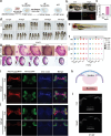
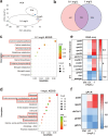
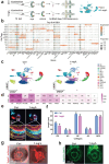
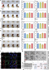
Quantum dots (QDs) are widely used, but their health impact on the visual system is little known. This study aims to elucidate the effects and mechanisms of typical metallic QDs on retinas using zebrafish. Comprehensive histology, imaging, and bulk RNA sequencing reveal that InP/ZnS QDs cause retinal degeneration. Furthermore, single-cell RNA-seq reveals a reduction in the number of retinal pigment epithelial cells (RPE) and short-wave cone UV photoreceptor cells (PR(UV)), accompanied by an increase in middle- and long-wave cone red, green, and blue photoreceptor cells [PR(RGB)]. Mechanistically, after endocytosis by RPE, InP/ZnS QDs inhibit the expression of splicing factor prpf8, resulting in gpx4b mRNA unsplicing, which finally decrease glutathione and induce ferroptosis and mitophagy. The decrease of RPE fails to engulf the damaged outer segments of PR, possibly promoting the differentiation of PR(UV) to PR(RGB). Knockout prpf8 or gpx4b with CRISPR/Cas9 system, the retinal damage is also observed. Whereas, overexpression of prpf8 or gpx4b, or supplement of glutathione can rescue the retinal degenerative damage caused by InP/ZnS QDs. In conclusion, this study illustrates the potential health risks of InP/ZnS QDs on eye development and provides valuable insights into the underlying mechanisms of InP/ZnS QDs-caused retinal degeneration.
Keywords: Ferroptosis; Quantum dots; Retinal degeneration; Single‐cell RNA sequencing; Spliceosome.
© 2024 The Author(s). Advanced Science published by Wiley‐VCH GmbH.
Conflict of interest statement
The authors declare no conflict of interest.
Figures


Similar articles
-
Involvement of FSP1-CoQ10-NADH and GSH-GPx-4 pathways in retinal pigment epithelium ferroptosis.Cell Death Dis. 2022 May 18;13(5):468. doi: 10.1038/s41419-022-04924-4. Cell Death Dis. 2022. PMID: 35585057 Free PMC article.
-
Conditional Loss of the Exocyst Component Exoc5 in Retinal Pigment Epithelium (RPE) Results in RPE Dysfunction, Photoreceptor Cell Degeneration, and Decreased Visual Function.Int J Mol Sci. 2021 May 11;22(10):5083. doi: 10.3390/ijms22105083. Int J Mol Sci. 2021. PMID: 34064901 Free PMC article.
-
Melanin-Like Nanomedicine Functions as a Novel RPE Ferroptosis Inhibitor to Ameliorate Retinal Degeneration and Visual Impairment in Dry Age-Related Macular Degeneration.Adv Healthc Mater. 2024 Dec;13(30):e2401613. doi: 10.1002/adhm.202401613. Epub 2024 Aug 11. Adv Healthc Mater. 2024. PMID: 39129350
-
Double cone dystrophy and RPE degeneration in the retina of the zebrafish gnn mutant.Invest Ophthalmol Vis Sci. 2003 Mar;44(3):1287-98. doi: 10.1167/iovs.02-0363. Invest Ophthalmol Vis Sci. 2003. PMID: 12601061
-
The pigmented epithelium, a bright partner against photoreceptor degeneration.J Neurogenet. 2017 Dec;31(4):203-215. doi: 10.1080/01677063.2017.1395876. Epub 2017 Nov 7. J Neurogenet. 2017. PMID: 29113536 Review.
Cited by
-
Ferroptosis: An Energetic Villain of Age-Related Macular Degeneration.Biomedicines. 2025 Apr 17;13(4):986. doi: 10.3390/biomedicines13040986. Biomedicines. 2025. PMID: 40299661 Free PMC article. Review.
References
-
- Sobhanan J., Rival J. V., Anas A., Shibu E. S., Takano Y., Biju V., Adv. Drug Deliv. Rev. 2023, 197, 114830. - PubMed
MeSH terms
Grants and funding
LinkOut - more resources
Full Text Sources
Molecular Biology Databases
Research Materials
