. 2024 Oct 16;57(2):286-294.
doi: 10.3724/abbs.2024172.

G6PC3 is involved in spermatogenesis by maintaining meiotic sex chromosome inactivation

G6PC3 is involved in spermatogenesis by maintaining meiotic sex chromosome inactivation

Yuming Cao et al. Acta Biochim Biophys Sin (Shanghai). .

Abstract

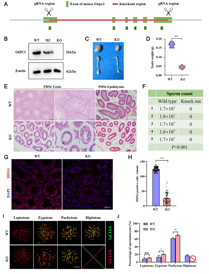
Meiosis, a process unique to germ cells, involves formation and repair of double-stranded nicks in DNA, pairing and segregation of homologous chromosomes, which ultimately achieves recombination of homologous chromosomes. Genetic abnormalities resulted from defects in meiosis are leading causes of infertility in humans. Meiotic sex chromosome inactivation (MSCI) plays a crucial role in the development of male germ cells in mammals, yet its underlying mechanisms remain poorly understood. In this study, we illustrate the predominant presence of a protein known as glucose 6 phosphatase catalyzed 3 (G6PC3) in pachytene spermatocytes, with a high concentration in the sex body (XY body), suggesting its significant involvement in male germ cell development. By employing CRISPR-Cas9 technology, we generate mice deficient in the G6pc3 gene, resulting in complete meiotic arrest at the pachytene stage in spermatocytes and are completely sterile. Additionally, we observe abnormal XY body formation and impaired MSCI in G6pc3-knockout spermatocytes. These findings underscore G6pc3 as a new essential regulator that is essential for meiotic progression. G6PC3 is involved in spermatocyte during male spermatogenesis development by the maintenance of meiosis chromosome silencing.

Keywords: glucose 6 phosphatase catalytic 3 (G6PC3); meiotic sex chromosome inactivation; pachytene arrest; sex chromosomes; spermatogenesis.


Conflict of interest statement

The authors declare that they have no conflict of interest.

Figures

None
Figure 1
G6PC3 is a testis-specific protein that accumulates on the XY body in spermatocytes (A) NCBI database analysis of the expression profile of G6pc3 mRNA in mouse tissues. (B) qRT-PCR verification of G6pc3 mRNA levels in multiple mouse tissues. Gapdh was used as a housekeeping gene, and the results were processed via the 2–ΔΔCT method. Data are presented as the mean ± SD, n = 3. (C) G6PC3 expression profile in adult (P56) mouse tissues detected by western blot analysis. β-Actin served as a loading control. (D) Immunofluorescence staining analysis of G6PC3 in testis sections. γH2AX was used as a marker for spermatocytes. The nuclei were stained with DAPI. The inset is an enlarged view of a pachytene spermatocyte with the XY body indicated. Scale bar: 50 μm. (E) Immunostaining of G6PC3 (red) and SYCP3 (green) on chromosome spreads of spermatocytes from P21 G6pc3 + / +  testes; the white arrow indicates the XY body, n = 3 mice for each group. Scale bar: 10 μm. (F) The expression pattern of G6pc3 in the mouse germline atlas was analyzed via a single-cell sequencing database (http://malehealthatlas.cn/).
None
Figure 2
G6PC3 is essential for spermatocyte development (A) Schematic diagram of the generation of G6pc3-deficient mice with the CRISPR-Cas9 genome editing system. (B) Western blot analysis of G6PC3 protein expression in testis extracts from G6pc3 + / +  and G6pc3‒/‒ testes at P56. β-Actin served as a loading control. (C,D) Litter size (right) and weight (left) of G6pc3 + / +  and G6PC3‒/‒ testes at P56. n = 13. (E) Hematoxylin and eosin (H&E) staining of histological sections of testes from G6pc3 + / +  and G6pc3‒/‒ mice at P56. Scale bar, left: 100 μm, right: 50 μm. (F) Sperm count in the tails of the epididymides of G6pc3 + / +  and G6pc3‒/‒ mice. P < 0.001, n = 5. (G) Frozen section staining of G6PC3 and DDX4 on the meiotic chromosomes in G6pc3 + / +  and G6pc3‒/‒ testicular tissue. Scale bar: 50 μm. (H) Statistical analysis of the number of seminiferous tubules without DDX4. Data are presented as the average percentage; n = 3 mice for each group, and 100 tubules were counted for each mouse. (G) Immunostaining of SYCP1 (green) and SYCP3 (red) on chromosome spreads of spermatocytes from P35 G6pc3 + / +  and G6pc3‒/‒ testes. n = 3 mice per group. Scale bar: 10 μm. (H) Frequency of meiotic prophase I stages. n = 3 mice per group, and 50 spermatocytes from each mouse were examined. Data are presented as the mean ± SD, *P < 0.05, **P < 0.01, ***P < 0.001 by two-tailed Student’s t test. WT, wild type; KO, knockout; HZ, heterozygote.
None
Figure 3
G6PC3 is essential for the formation of XY bodies (A) Immunostaining of SYCP3 (red) and H1T (green) on chromosome spreads of spermatocytes from P35 G6pc3 + / +  and G6PC3‒/‒ testes. n = 3 mice for each group. Scale bar: 10 μm. (B) Statistical analysis of the percentage of H1T-positive pachytene spermatocytes at different stages. Data are presented as the average percentage; n = 3 mice per group, and 100 pachytene spermatocytes were counted per mouse. (C) Immunostaining of γH2AX and DAPI in P56 G6pc3 + / +  and G6pc3‒/‒ testis frozen sections. Scale bar: 50 μm. (D) The number of abnormal XY bodies in each tubule of P56 G6pc3 + / +  and G6pc3‒/‒ mice. n = 3 mice for each group, and 20 tubules from each mouse were analyzed. (E) Immunostaining of SYCP3 (red) and γH2AX (green) in G6pc3 + / +  and G6pc3‒/‒ pachytene spermatocytes. Arrowhead: example of an extended XY pair. Scale bar, 10 μm. (F) Statistical results of (E). Data are presented as the average percentage; n = 3 mice for each group, and 50 pachytene spermatocytes were counted for each mouse. (G) TUNEL assays of testes sections prepared from 3-week-old G6pc3 + / +  and G6pc3‒/‒ mice. Scale bar, above: 100 μm, under: 50 μm. (H) Quantification of the number of TUNEL-positive seminiferous tubules. n = 3 mice for each group, and 50 spermatocytes were counted for each mouse. (I) Quantification of the number of TUNEL-positive cells per tubule. Twenty tubules per mouse were counted, and three mice from each genotype were analyzed. Data are presented as the mean ± SD. **P < 0.001, ***P < 0.001 by two-tailed Student’s t test.
None
Figure 4
Abnormal XY bodies undergo MSCI defects in G6pc3 ‒/‒ mice (A) Immunostaining of meiotic sex chromosome silencing in G6pc3 + / +  and G6pc3‒/‒ testis sections (γ-H2AX: red; RNA-Pol II: green). Scale bar: 50 μm. (B) Immunostaining of meiotic sex chromosome silencing in G6pc3 + / +  and G6pc3‒/‒ spermatocytes at the pachytene stage (SYCP3: red; RNA Pol II: green). Scale bar: 10 μm. (C) Immunostaining of H3K4me3 revealed active transcription of the XY chromosomes in G6pc3 + / +  and G6pc3‒/‒ testis sections (γ-H2AX: red; H3K4me3: green). Scale bar: 50 μm. (D) Immunostaining of H3K4me3 revealed active transcription of the XY chromosomes in G6pc3 + / +  and G6pc3‒/‒ spermatocytes at the pachytene stage (SYCP3: red; H3K4me3: green). Scale bar: 10 μm. (E) Statistical results of A and C. Data are presented as the average percentage; n = 3 mice for each group, and 50 pachytene spermatocytes were counted for each mouse. (F) Statistical results of B and D. Data are presented as the average percentage; n = 3 mice for each group, and 50 pachytene spermatocytes were counted for each mouse. (G–H) RNA-seq analysis of differential gene expression in testicular tissues of G6pc3 + / +  and G6pc3‒/‒ mice. (I) Functional annotation of DEGs between G6pc3 + / +  and G6pc3‒/‒ spermatocytes on the basis of RNA-seq data. ***P < 0.001.

References

    1. Farini D, De Felici M. The beginning of meiosis in mammalian female germ cells: a never-ending story of intrinsic and extrinsic factors. Int J Mol Sci. . 2022;23:12571. doi: 10.3390/ijms232012571. - DOI - PMC - PubMed
    1. MacKenzie A, Vicory V, Lacefield S, Lichten M. Meiotic cells escape prolonged spindle checkpoint activity through kinetochore silencing and slippage. PLoS Genet. . 2023;19:e1010707. doi: 10.1371/journal.pgen.1010707. - DOI - PMC - PubMed
    1. Alavattam KG, Maezawa S, Andreassen PR, Namekawa SH. Meiotic sex chromosome inactivation and the XY body: a phase separation hypothesis. Cell Mol Life Sci. . 2021;79:18. doi: 10.1007/s00018-021-04075-3. - DOI - PMC - PubMed
    1. Abe H, Yeh YH, Munakata Y, Ishiguro KI, Andreassen PR, Namekawa SH. Active DNA damage response signaling initiates and maintains meiotic sex chromosome inactivation. Nat Commun. . 2022;13:7212. doi: 10.1038/s41467-022-34295-5. - DOI - PMC - PubMed
    1. Hirota T, Blakeley P, Sangrithi MN, Mahadevaiah SK, Encheva V, Snijders AP, ElInati E, et al. SETDB1 links the meiotic DNA damage response to sex chromosome silencing in mice. Dev Cell. . 2018;47:645–659.e6. doi: 10.1016/j.devcel.2018.10.004. - DOI - PMC - PubMed

LinkOut - more resources