. 2024 Sep 12;7(10):3056-3070.
doi: 10.1021/acsptsci.4c00265. eCollection 2024 Oct 11.

Identification of a Compound Inhibiting Both the Enzymatic and Nonenzymatic Functions of Indoleamine 2,3-Dioxygenase 1

Affiliations

Identification of a Compound Inhibiting Both the Enzymatic and Nonenzymatic Functions of Indoleamine 2,3-Dioxygenase 1

Eleonora Panfili et al. ACS Pharmacol Transl Sci. .

Abstract

Indoleamine 2,3-dioxygenase 1 (IDO1) plays a key role in tumor immune escape. Besides being a metabolic enzyme that catalyzes the first step of tryptophan catabolism, it also acts as a signal-transducing protein, whose partnering with tyrosine phosphatase Src homology 2 (SH2) domain-containing protein tyrosine phosphatase substrate (SHPs) and phosphatidylinositol-3-kinase (PI3K) regulatory subunit p85 promotes the establishment of a sustained immunosuppressive phenotype. While IDO1 inhibitors typically interfere with its enzymatic activity, we aimed to discover a more effective modulator capable of blocking not only the enzymatic but also the signaling-mediated functions of IDO1. By virtual screening, we identified the compound VS-15, which selectively binds the heme-free form of IDO1, inhibits its enzymatic activity, and reduces the IDO1-mediated signaling pathway by negatively interfering with its partnership with SHPs and PI3K regulatory subunit p85 as well as with the IDO1 anchoring to the early endosomes in tumor cells. Moreover, VS-15 counteracts the TGF-β-mediated immunosuppressive phenotype in dendritic cells and reduces the level of inhibition of T cell proliferation by suppressive monocytes isolated from patients affected by pancreatic cancer. Herein, we describe the discovery and characterization of a small molecule with an unprecedented mechanism of action, capable of inhibiting both the enzymatic and nonenzymatic activities of IDO1 by binding to its apo-form. These results pave the way for the development of next-generation IDO1 inhibitors with a unique competitive advantage over the currently available modulators, thereby opening therapeutic opportunities in cancer immunotherapy.


Conflict of interest statement

The authors declare no competing financial interest.

Figures
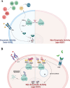
Figure 1
Figure 1
Enzymatic (A) and nonenzymatic (B) activities of IDO1.
Scheme 1
Scheme 1. Synthesis of VS-15
Reaction conditions: (a) dry THF, 0°C; (b) toluene, TEA, reflux; (c) TEA, HOBt, EDC, dry DCM, rt.
Figure 2
Figure 2
Identification and characterization of a novel compound binding to IDO1. (A) Cellular IC50 value (μM) of VS-15 obtained from a dose–response curve in P1 tumor cells stably transfected with human (left) or mouse (right) IDO1. (B) Kyn fold change in the supernatants from P1 tumor cells stably transfected with mouse IDO1 or TDO2 incubated with three different dilutions of VS-15 relative to the same cells treated with only the vehicle alone. (C) Residual activity of the iNOS enzyme expressed as a percentage of free enzyme activity that was defined as 100. (D) IDO1 MST binding curve in the presence of VS-15. The recombinant protein was mixed with a serial dilution of the compound starting at 250 μM to evaluate the binding of VS-15 to IDO1. In (A–D), results are the mean of three independent experiments, each performed in triplicate. Statistical analysis was performed using two-tailed paired Student’s t test (B) or ANOVA followed by posthoc Bonferroni’s test (C). ****p < 0.0001.
Figure 3
Figure 3
VS-15 does not affect IDO1 protein degradation. (A) Cycloheximide-chase assay followed by immunoblot analysis of IDO1 protein expression in lysates from P1.IDO1 cells pretreated with cycloheximide for 1 h and then exposed to VS-15 (1 μM) for the indicated time (from 0 to 8 h). For each time point, vehicle-treated cells were used as a control (Ctrl). β-tubulin expression was used as a normalizer. One representative experiment is shown. (B) Quantitative analysis of three independent experiments (mean ± SD) of Western blot analysis is represented as the fold change of normalized IDO1 protein at each time point over time 0. No statistically significant differences were determined between VS-15-treated and untreated samples.
Figure 4
Figure 4
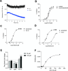
VS-15 is an inhibitor of apo-IDO1. (A) Absorbance-based tracking of the displacement of heme from IDO1. The loss of absorbance at the IDO1 λmax of the Soret peak (404 nm) has been evaluated as a function of time in the absence (black) and the presence (blue) of 10 μM VS-15 inhibitor. (B) Inhibition profile of VS-15 in A375 cells stimulated with IFN-γ in the presence or absence of succinylacetone, tryptophan, and increasing concentrations of VS-15 for 48 h. Each experiment included the corresponding controls: IFN-γ treatment alone for the standard assay and IFN-γ plus succinylacetone treatment. The IC50 value under physiological conditions (control) was 0.48 μM, while with succinylacetone treatment it was 0.27 μM. (C) Inhibition profile of VS-15 and epacadostat (epac.) (relative to the DMSO control) in the hIDO1 biochemical assay performed under standard conditions. The epacadostat IC50 value was 0.01 μM. (D) Inhibition profile of VS-15 and epacadostat (epac.) (relative to the DMSO control) in the hIDO1 biochemical assay performed at increased preincubation time and temperature. Under these conditions, the IC50 value was 0.05 μM for epacadostat and 4.6 μM for VS-15. (E) Free heme release after incubation of hIDO1 with two different concentrations of VS-15, epacadostat, or DMSO as a control (Ctrl). Data are presented relative to the untreated sample (dotted line, 1-fold). ****p < 0.0001 (paired Student’s t test). (F) Binding affinity of VS-15 defined as a result of the heme dissociation rate at different inhibitor concentrations (Kd = 63 μM). In (B–E), data are the mean ± SD of three independent experiments, each performed in triplicate.
Figure 5
Figure 5
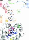
Docking pose of VS-15 in apo-IDO1. Location of the binding region on the IDO1 crystal structure (PDB id: 6MQ6) and docking pose of compound VS-15, depicted as blue sticks. The central aromatic ring of VS-15 is situated within pocket A (red) (Tyr126, Cys129, Val130, Phe163, Phe164, Ser167, and Ala264). The indole group interacts with Phe163 with a π–π interaction and is also located in a hydrophobic subpocket in the proximity of Phe226 in pocket B (green) (Phe226, Arg231, Ile232, Leu234, and Ser235). Furthermore, hydrogen bonding interactions occur between a carbonyl oxygen of the bicarbonyl moiety and Ser167 and also between another carbonyl oxygen and Ala264. Pocket C (blue) is not exploited, except for hydrophobic contact with Ser263. The other indole group is located in pocket D (yellow) (Val170, Ser267, Val269, Phe270, Leu359, Leu342, Arg343, and His346), where it forms a hydrogen bonding interaction with Ser267 and engages in hydrophobic interactions with the side chains of surrounding other amino acids (Val170, Leu342, and Phe270).
Figure 6
Figure 6
VS-15 negatively regulates IDO1-mediated signaling events in P1 tumor cells overexpressing IDO1. (A) Immunoprecipitation of IDO1 from P1.IDO1 cells treated with VS-15 (1 μM) or the vehicle alone (Ctrl) and detection of IDO1 or p85 PI3K by sequential immunoblotting. (B) Immunoblot analysis of endosome (EEs) and cytosol isolated from VS-15-treated P1.IDO1 cells. Cells incubated with the vehicle DMSO were used as a control (Ctrl). (C) Quantitative analysis of three independent immunoblot experiments, one of which is represented in (B). The IDO1:Rab5 ratio was calculated by densitometric quantification of the specific bands. (D) Immunoblot analysis of coimmunoprecipitates from P1.IDO1 cells treated as in (A). (E) Phosphatase activity produced by IDO1 immunoprecipitated from P1.IDO1 cells treated as in (A). Levels of free phosphate (pmol) generated during the incubation of a tyrosine-phosphorylated peptide with the coimmunoprecipitated IDO1 proteins from each condition were presented as a fold change relative to untreated cells (dotted line, 1-fold). In (A, B, D), one experiment representative of three is shown. In (C, E), results are representative of three independent experiments (mean ± SD), each performed in triplicate. In (C, E), **p < 0.01 (paired Student’s t test).
Figure 7
Figure 7
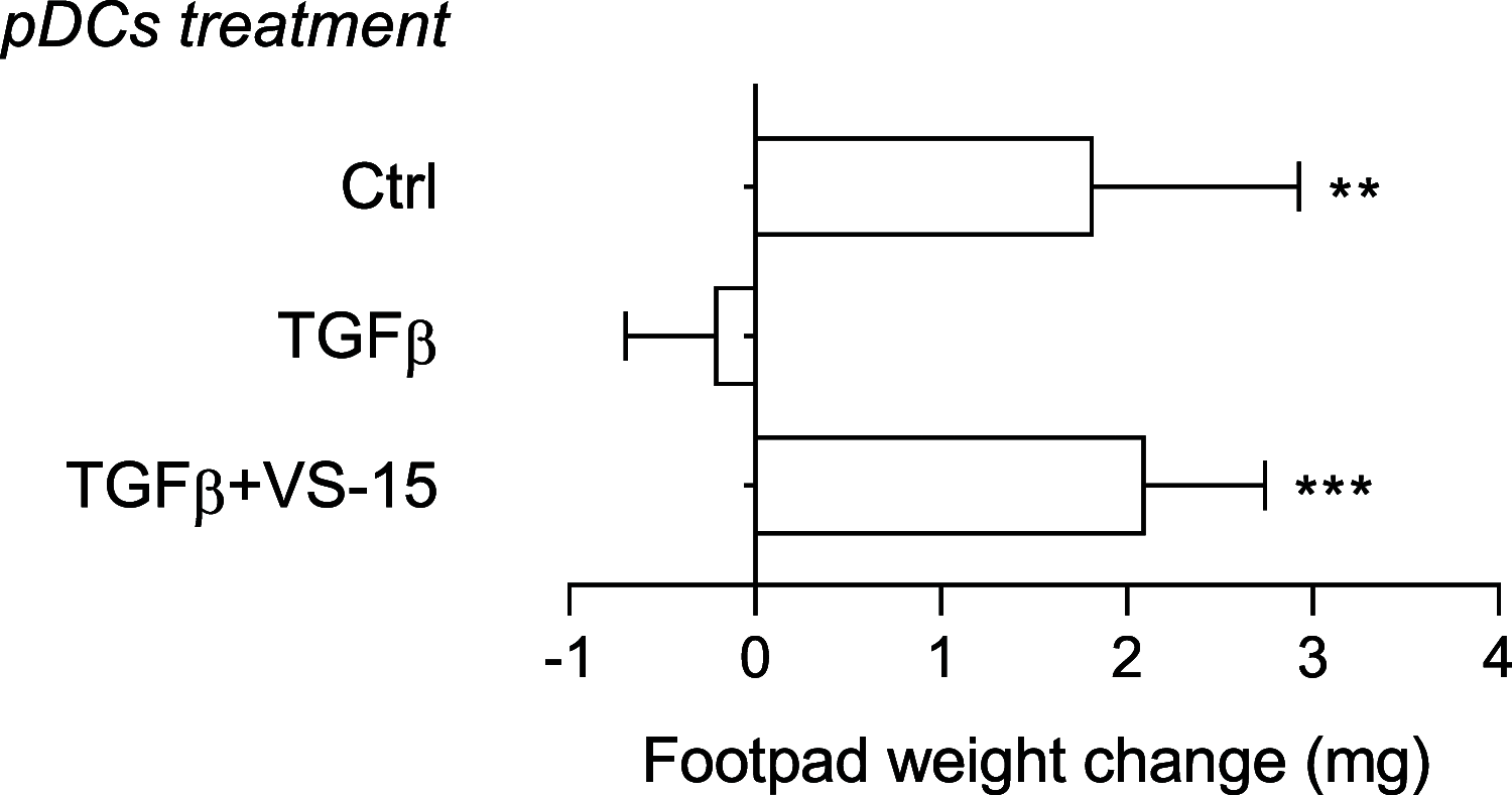
VS-15 abrogates the TGF-β-dependent tolerogenic response in pDCs in vivo. Skin test reactivity of mice sensitized with splenic HY-pulsed immunostimulatory cDCs combined with a minority fraction (5%, indicated) of pDCs. pDCs were conditioned in vitro with the vehicle DMSO (Ctrl) or TGF-β, in the presence or absence of VS-15 (1 μM), before being HY-pulsed together with the cDC majority fraction and i.p. transferred into syngeneic C57BL/6 recipient female mice. Analysis of skin reactivity of recipient mice to the eliciting peptide at 15 days is presented as a change in footpad weight (experimental versus control footpads). **p < 0.01 and ***p < 0.001 (Tukey’s multiple comparison test).
Figure 8
Figure 8
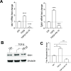
VS-15 negatively regulates IDO1-mediated signaling events in pDCs. (A) Real-time PCR analysis of Ido1 and Tgfb1 transcripts in pDCs treated with TGF-β alone or in combination with VS-15 (1 μM) or the vehicle as a control (Ctrl). Transcript expression was normalized to the expression of Gapdh and presented relative to results in untreated cells (dotted line, 1-fold). Results are representative of three independent experiments (mean ± SD), each performed in triplicate. (B) Immunoblot analysis of IDO1 protein expression in lysates from pDCs cells treated as in (A). β-Tubulin expression was used as a normalizer. One representative immunoblot of three is shown. (C) Phosphatase activity produced by IDO1 immunoprecipitated from pDCs treated as in (A). Levels of free phosphate (pmol) generated during the incubation of a tyrosine-phosphorylated peptide with the coimmunoprecipitated IDO1 proteins from each condition were presented as a fold change relative to untreated cells (dotted line, 1-fold). In (A, C), results are representative of three independent experiments (mean ± SD), each performed in triplicate. *p < 0.05, **p < 0.01, ***p < 0.001, and ****p < 0.0001. In (A) paired Student’s t test and in (C) one-way ANOVA followed by posthoc Bonferroni’s test.
Figure 9
Figure 9
VS-15 abrogates the immunosuppressive function of circulating monocytes isolated from tumor patients in controlling T cell proliferation. (A) Representative gating strategy to evaluate the percentage of circulating IDO1+ monocytes in the healthy donor (HD), pancreatic cancer (PC), breast cancer (BC), and lung cancer (LC) patients by flow cytometry. (B) Percentage of circulating IDO1+ monocytes (out of total monocytes) in HD (n = 9), PC (n = 30), BC (n = 20), and LC (n = 45) patients. (C) Correlation between the percentage of PC- and BC-derived circulating IDO1+ monocytes and the proliferation of activated CD3+ T lymphocytes following the coculture with PC- and BC-derived monocytes (n = 36). Representative percentage of IDO1+ monocytes analyzed by flow cytometry related to nonsuppressive and suppressive total monocytes derived from tumor patients is shown in the right panel. (D) Viability of HD-derived monocytes (n = 3) after 24 h treatment with different concentrations of VS-15 and DMSO as a control. (E) Viability of BC- and PC-derived monocytes (n = 5) after 24 h treatment with VS-15 at 1 μM. All values are normalized on cells treated with DMSO as a vehicle. (F) Relative proliferation percentage of CD3+ T cells after 4 days of coculture with either suppressive (PC, n = 3; BC, n = 5) or nonsuppressive (PC, n = 6; BC, n = 6) monocytes pretreated with 1 μM of VS-15 and DMSO as a control. All values are normalized on activated T cells in the absence of myeloid cells. Data are expressed as mean ± SEM. **p < 0.01, ***p < 0.001, and ****p < 0.0001. In (B) unpaired Student’s t test and in (F) Wilcoxon’s signed-rank test.

References

    1. Pallotta M. T.; Rossini S.; Suvieri C.; Coletti A.; Orabona C.; Macchiarulo A.; Volpi C.; Grohmann U. Indoleamine 2,3-dioxygenase 1 (IDO1): an up-to-date overview of an eclectic immunoregulatory enzyme. FEBS journal 2022, 289 (20), 6099–6118. 10.1111/febs.16086. - DOI - PMC - PubMed
    1. Schwarcz R. The kynurenine pathway of tryptophan degradation as a drug target. Current opinion in pharmacology 2004, 4 (1), 12–17. 10.1016/j.coph.2003.10.006. - DOI - PubMed
    1. Mellor A. L.; Munn D. H. IDO expression by dendritic cells: tolerance and tryptophan catabolism. Nature reviews. Immunology 2004, 4 (10), 762–774. 10.1038/nri1457. - DOI - PubMed
    1. Bessede A.; Gargaro M.; Pallotta M. T.; Matino D.; Servillo G.; Brunacci C.; Bicciato S.; Mazza E. M.; Macchiarulo A.; Vacca C.; Iannitti R.; Tissi L.; Volpi C.; Belladonna M. L.; Orabona C.; Bianchi R.; Lanz T. V.; Platten M.; Della Fazia M. A.; Piobbico D.; Puccetti P. Aryl hydrocarbon receptor control of a disease tolerance defence pathway. Nature 2014, 511 (7508), 184–190. 10.1038/nature13323. - DOI - PMC - PubMed
    1. Gutiérrez-Vázquez C.; Quintana F. J. Regulation of the Immune Response by the Aryl Hydrocarbon Receptor. Immunity 2018, 48 (1), 19–33. 10.1016/j.immuni.2017.12.012. - DOI - PMC - PubMed

LinkOut - more resources