. 2024 Oct 3:15:1466720.
doi: 10.3389/fimmu.2024.1466720. eCollection 2024.

Expanding the VEXAS diagnostic workup: the role of peripheral blood cytological analysis

Affiliations

Expanding the VEXAS diagnostic workup: the role of peripheral blood cytological analysis

Chiara Baggio et al. Front Immunol. .

Abstract

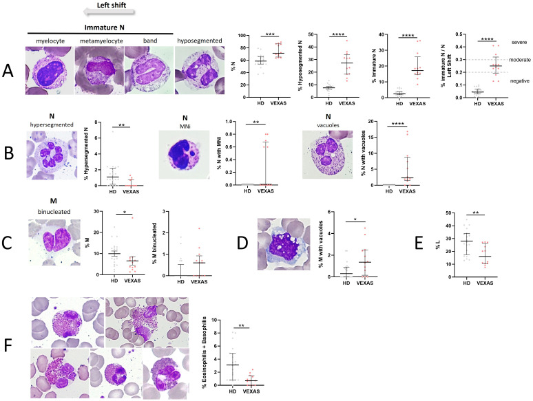
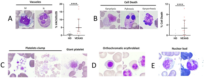
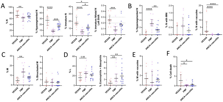
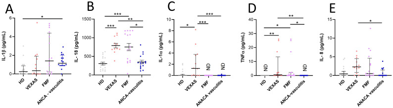
VEXAS syndrome is a newly described autoinflammatory entity characterized by somatic mutations in the UBA1 X-linked gene in hematopoietic progenitor cells. Several studies have demonstrated that the presence of vacuoles in progenitor cells from bone marrow aspirates is a hallmark finding for this syndrome. Therefore, this study aimed to characterize leukocytes from VEXAS patients versus patients with ANCA-associated vasculitis (AAV), familial Mediterranean fever (FMF), and healthy donors (HD) to define a specific cytological pattern that can support VEXAS diagnosis. Twelve VEXAS patients were included in the study. Blood samples from FMF (n = 16), AAV (n = 16) and HDs (n = 20) acted as controls. May-Grünwald Giemsa (MGG) staining was used for studying cellular morphology, including cytoplasm, granules, and vacuoles and to perform a cytogenic evaluation of leucocytes. Plasma IL-1β, IL-1α, TNFα, IL-18 and IL-8 were measured using ELISA assay. The cytological analysis from blood smears confirmed the presence of immature neutrophils in VEXAS patients. We found a greater number of vacuoles in VEXAS patients vs. FMF, AAV and HD. Micronuclei (MNi) and cell death rate were higher in VEXAS patients vs. HD. Cell death correlated with IL-1β and IL-8 levels. MNi were positively associated with IL-8 and IL-1β levels, and with the percentage of immature neutrophils and vacuoles. In conclusion, our findings suggested that cytological test may be supportive for VEXAS diagnosis, despite genetical analysis is mandatory for confirming the disease. Finally, we identified several cytological hallmarks that may distinguish the VEXAS "cytotype" not only from HD but also from other inflammatory diseases.

Keywords: VEXAS; cytokines; cytology; hematology; inflammation; vacuoles.


Conflict of interest statement

AD received consultancy fees from GSK, Eli Lilly, AstraZeneca, Otsuka. PS received grants from Novartis and Sobi. Authors DC, BP and GP were employed by R&I Genetics srl. The remaining authors declare that the research was conducted in the absence of any commercial or financial relationships that could be construed as a potential conflict of interest. The author(s) declared that they were an editorial board member of Frontiers, at the time of submission. This had no impact on the peer review process and the final decision.

Figures

Figure 1
Figure 1
Cytogenic and cellular morphology evaluation of leukocytes in blood smear from VEXAS patients. Blood smears from VEXAS patients (n = 12) and HD (n = 20) were stained using MGG staining as described in Material and Methods. (A) Left panel: representative image of immature N and hyposegmented N stained with MGG. Right panel: rate of N, hyposegmented N and immature N in VEXAS patients and HD. (B) Left panel: representative image of hypersegmented N, N with MNi and vacuoles stained with MGG. Right panel: rate of hypersegmented N, N with MNi and vacuoles in VEXAS patients and HD. (C) Left panel: representative image of binucleated M stained with MGG. Right panel: rate of M and binucleated M in VEXAS patients and HD. (D) Left panel: representative image of M with vacuoles stained with MGG. Right panel: rate of M with vacuoles in VEXAS patients and HD. (E) Rate of L in VEXAS patients and HD. (F) Left panel: representative image of eosinophils stained with MGG. Right panel: rate of granulocytes (eosinophils and basophils) in VEXAS patients and HD. Data are expressed as medians and IQR. p calculated using the Mann-Whitney test: *p<0.05, **p<0.01, ****p<0.0001. MGG, May-Grünwald Giemsa; N, neutrophils; MNi, micronuclei; M, monocytes; L, lymphocytes; HD, healthy donors.
Figure 2
Figure 2
Cell death rate of leukocytes and vacuoles evaluation in blood smear from VEXAS patients. Blood smears from VEXAS patients (n = 12) and HD (n = 20) were stained using MGG staining as described in Materials and Methods. (A) Left panel: representative image of M and N with vacuoles stained with MGG. Right panel: rate of total vacuoles in VEXAS patients and HD. (B) Left panel: representative image of karyorrhexis, karyolysis, and pyknosis in blood smears stained with MGG. Right panel: rate of cell death in VEXAS patients and HD. (C) Platelet morphology and platelet clumps. (D) Orthochromatic erythroblast. (E) Nuclear bud. Data are expressed as medians and IQR. p calculated using the Mann-Whitney test: ****p<0.0001. MGG, May-Grünwald Giemsa; M, monocytes; N, neutrophils; HD, healthy donors.
Figure 3
Figure 3
Cytogenic and cellular morphology evaluation of leukocytes in blood smear from VEXAS, FMF patients and patients with ANCA-associated vasculitis. Blood smears from VEXAS patients (n = 12), FMF (n = 16) and ANCA-associated vasculitis patients (n = 16) were stained using MGG staining as described in Materials and Methods. (A) rate of N, hyposegmented N and immature N in VEXAS, FMF patients ANCA-associated vasculitis patients. (B) Rate of hypersegmented N, N with MNi and vacuoles in VEXAS, FMF patients, ANCA-associated vasculitis patients. (C) Rate of M and binucleated M in VEXAS, FMF patients ANCA-associated vasculitis patients. (D) Rate of L and granulocytes (eosinophils and basophils) in VEXAS, FMF patients ANCA-associated vasculitis patients. (E) Rate of M with vacuoles in VEXAS, FMF patients, ANCA-associated vasculitis patients. (F) Rate of cell death in VEXAS, FMF patients, ANCA-associated vasculitis patients. Data are expressed as medians and IQR. p calculated using the Kruskal-Wallis test. Dunn’s post hoc test: *p<0.05, **p<0.01, ***p<0.001, ****p<0.0001. MGG, May-Grünwald Giemsa; N, neutrophils; MNi, micronuclei; M, monocytes; HD, healthy donors.
Figure 4
Figure 4
Cytokines and chemokines levels in plasma from VEXAS, FMF and AAV patients and correlation with cell death and I/T index. Cytokine and chemokines levels in plasma from VEXAS (n = 12), FMF (n = 16), AAV (n = 16) and HD (n = 20) were analyzed by ELISA as described in Materials and Methods. (A) IL-1β levels, (B) IL-18 levels, (C) IL-1α levels, (D) TNFα levels and (E) IL-8 levels. Data are expressed as medians and IQR. p calculated according using the Kruskal-Wallis test. Dunn’s post hoc test: *p<0.05, **p<0.01, ***p<0.001. HD, healthy donors.

Similar articles

References

    1. Vitale A, Caggiano V, Bimonte A, Caroni F, Tosi GM, Fabbiani A, et al. . VEXAS syndrome: a new paradigm for adult-onset monogenic autoinflammatory diseases. Intern Emerg Med. (2023) 18:711–22. doi: 10.1007/s11739-023-03193-z - DOI - PMC - PubMed
    1. Lacombe V, Hadjadj J, Georgin-Lavialle S, Lavigne C, Geneviève F, Kosmider O. Vacuoles in bone marrow progenitors: VEXAS syndrome and beyond. Lancet Haematol. (2024) 11:e160–7. doi: 10.1016/S2352-3026(23)00375-7 - DOI - PubMed
    1. Ferrada MA, Savic S, Cardona DO, Collins JC, Alessi H, Gutierrez-Rodrigues F, et al. . Translation of cytoplasmic UBA1 contributes to VEXAS syndrome pathogenesis. Blood. (2022) 140:1496–506. doi: 10.1182/blood.2022016985 - DOI - PMC - PubMed
    1. Al-Hakim A, Cull A, Topping J, Nadat F, Milek J, Alhefzi R, et al. . Recovery of bone marrow function in VEXAS syndrome-potential role for romiplostim. Hemasphere. (2023) 7:e934. doi: 10.1097/HS9.0000000000000934 - DOI - PMC - PubMed
    1. Chabrun F, Lacombe V, Dieu X, Geneviève F, Urbanski G. Accurate stratification between VEXAS syndrome and differential diagnoses by deep learning analysis of peripheral blood smears. Clin Chem Lab Med. (2023) 61:1275–9. doi: 10.1515/cclm-2022-1283 - DOI - PubMed

MeSH terms

LinkOut - more resources