. 2024 Nov 8;135(11):1033-1047.
doi: 10.1161/CIRCRESAHA.124.324248. Epub 2024 Oct 18.

Shear-Sensing by C-Reactive Protein: Linking Aortic Stenosis and Inflammation

Affiliations

Shear-Sensing by C-Reactive Protein: Linking Aortic Stenosis and Inflammation

Johannes Zeller et al. Circ Res. .

Abstract

Background: CRP (C-reactive protein) is a prototypical acute phase reactant. Upon dissociation of the pentameric isoform (pCRP [pentameric CRP]) into its monomeric subunits (mCRP [monomeric CRP]), it exhibits prothrombotic and proinflammatory activity. Pathophysiological shear rates as observed in aortic valve stenosis (AS) can influence protein conformation and function as observed with vWF (von Willebrand factor). Given the proinflammatory function of dissociated CRP and the important role of inflammation in the pathogenesis of AS, we investigated whether shear stress can modify CRP conformation and induce inflammatory effects relevant to AS.

Methods: To determine the effects of pathological shear rates on the function of human CRP, pCRP was subjected to pathophysiologically relevant shear rates and analyzed using biophysical and biochemical methods. To investigate the effect of shear on CRP conformation in vivo, we used a mouse model of arterial stenosis. Levels of mCRP and pCRP were measured in patients with severe AS pre- and post-transcatheter aortic valve implantation, and the presence of CRP was investigated on excised valves from patients undergoing aortic valve replacement surgery for severe AS. Microfluidic models of AS were then used to recapitulate the shear rates of patients with AS and to investigate this shear-dependent dissociation of pCRP and its inflammatory function.

Results: Exposed to high shear rates, pCRP dissociates into its proinflammatory monomers (mCRP) and aggregates into large particles. Our in vitro findings were further confirmed in a mouse carotid artery stenosis model, where the administration of human pCRP led to the deposition of mCRP poststenosis. Patients undergoing transcatheter aortic valve implantation demonstrated significantly higher mCRP bound to circulating microvesicles pre-transcatheter aortic valve implantation compared with post-transcatheter aortic valve implantation. Excised human stenotic aortic valves display mCRP deposition. pCRP dissociated in a microfluidic model of AS and induces endothelial cell activation as measured by increased ICAM-1 (intercellular adhesion molecule 1) and P-selectin expression. mCRP also induces platelet activation and TGF-β (transforming growth factor beta) expression on platelets.

Conclusions: We identify a novel mechanism of shear-induced pCRP dissociation, which results in the activation of cells central to the development of AS. This novel mechanosensing mechanism of pCRP dissociation to mCRP is likely also relevant to other pathologies involving increased shear rates, such as in atherosclerotic and injured arteries.

Keywords: C-reactive protein; aortic valve stenosis; inflammation; proteolysis; thrombosis.


Conflict of interest statement

None.

Figures
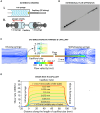
Figure 1.
Figure 1.
Extensional flow apparatus and computational fluid dynamics (CFD) calculations of the flow field generated. A, Schematic drawing of the utilized extensional flow apparatus. B, Image of extensional flow device. C, Flow streamlines in the middle cross-section of the infusing syringe, capillary tube, and withdrawing syringe colored according to flow velocity. Results were obtained using CFD simulation with a plunger velocity of 8 mm s−1 corresponding to a flow rate of 8 mL min−1. D, Variations in shear rate along the length of the capillary tube at different distances to the central axis of the capillary tube obtained using CFD simulation (0 mm, central axis; 0.17 mm, closest to the capillary wall). Results were obtained by setting the flow rate to 8 mL min−1. PE indicates polyethylene.
Figure 2.
Figure 2.
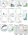
Shear dissociates pCRP (pentameric C-reactive protein) into mCRP (monomeric C-reactive protein), resulting in aggregate formation. A, Human pCRP subjected to shear dissociates into its monomeric subunits, as demonstrated by native Coomassie sodium dodecyl-sulfate polyacrylamide gel electrophoresis (SDS-PAGE). Human pCRP (2.5 mg/mL) was subjected to extensional shear stress for 200 passes and subsequently separated on a native SDS-PAGE. Unsheared pCRP (pCRP) did not show any staining at the size of mCRP (25 kDa, indicated in the protein ladder by the black arrowhead). However, pCRP sheared for 200 passes (sheared CRP [C-reactive protein]) showed a second band at the same size as the mCRP control (mCRP). The sheared CRP solution was separated into a pellet with decreased solubility (Pellet) and supernatant (Super). The pellet showed only minor staining at the size of the pCRP control (red arrowhead) and a strong band at ≈23 kDa. The soluble portion of the sheared sample (Super) showed up exclusively at the size of the control pCRP. The black arrow located above the 250-kDa marker indicates the border between the stacking and the separating gel. B, Further identification of mCRP was made by Western blotting using a conformation-specific mouse antihuman mCRP antibody clone 8C10. C, The shear-induced dissociation of pCRP increased with the number of passes (50, 200, and 1000 passes) as demonstrated by native SDS-PAGE. D, mCRP was further identified by Western blotting using the mCRP-specific antibody clone 8C10. E, A dot blot was used to detect a neoepitope only expressed in unfolded mCRP (inset shows a monomeric subunit of pCRP with the 8C10 epitope highlighted in red, PDB ID: 7TBA). F, pCRP subjected to shear and a static control were imaged by transmission electron microscopy. Images of pCRP (left) under control conditions showed the typical pentameric structure and size. However, pCRP subjected to shear (right, 1000 passes) showed large CRP aggregates. G, pCRP was subjected to increasing passes of shear and analyzed by mass photometry. Compared with the static pCRP control, increasing numbers of passes through the flow apparatus correlated with an increasing mass of CRP aggregates.
Figure 3.
Figure 3.
Biophysical measurements verified pCRP (pentameric C-reactive protein) dissociation and monomer formation under shear. A, Tryptophan (TRP) fluorescence was used to distinguish between different CRP (C-reactive protein) isoforms. The inset shows a globular monomeric subunit of pCRP with TRP residues highlighted in orange (PDB ID: 7TBA). With increasing numbers of passes (p) through the extensional flow field, TRP emission spectra decreased from pCRP toward mCRP (monomeric C-reactive protein). B, TRP fluorescence intensity at peak wavelength (λ=370 nm) was measured and compared for the different treatments by ANOVA and Tukey post hoc test (n=4), biological replicates. C and D, 8-anilino-1-naphthalenesulfonic acid (ANS) binding was used to further verify the conformational change in CRP. Shear-dissociated CRP demonstrated an increase in ANS fluorescence and, finally, (1000 passes) reached similar emission spectra and peak values to those of the control mCRP. ANS fluorescence intensity at peak wavelength (λ=500 nm) was measured and compared for the different treatments by ANOVA and Dunnett T3 test (n=5–6), biological replicates. E through G, Dynamic light scattering (DLS) size distribution plots showed a distinct shift of the unsheared pCRP (purple), with a single peak at Rh=12.26±0.63 nm (mean±SD), to multiple peaks for pCRP subjected to shear (200 passes and 1000 passes), with 200 passes still expressing a peak at the size of the unsheared pCRP and an additional peak at Rh=677.87±100.2 nm, while 1000 passes showed only minor peaks at Rh of the unsheared pCRP. This shift was also reflected in the mean hydrodynamic diameters (Z-averages) of the unsheared pCRP and sheared CRP in F and in all the sample correlograms in G, indicating both the size (via the decay time) and the particle size uniformity (via the consistency of the correlation plots). n=6, biological replicates, 1-way ANOVA, and Dunnett T3 test were applied. H, The ability of the different CRP isoforms to bind the ligand phosphocholine (PC) was evaluated using a sandwich ELISA. PC-coupled KLH (keyhole limpet hemocyanin) was coated to microtiter plates and incubated with different CRP isoforms generated by shear stress. Untreated pCRP and recombinant mCRP served as controls. While the unsheared pCRP harbored 5 functional PC binding sites (1 per monomeric subunit), the unfolded and disordered mCRP lacked a PC binding site. I, Aggregates produced from the sheared pCRP did not bind human C1q (complement component 1q). Equal amounts of protein were loaded into C1q-coated wells. Only the solubilized mCRP control was found to bind C1q significantly. n=3, biological replicates, 1-way ANOVA, and Tukey post hoc tests were used. J, The synoptical interpretation of the results presented in A through I. Shear-dissociated pCRP into monomeric subunits. The globular structure of mCRP was swiftly disrupted and became highly disordered, exposing C1q binding sites. As mCRP aggregated into larger fibrillar structures, to reduce exposure to the aqueous solvent, the C1q binding sites of each mCRP molecule were buried within the aggregate, thereby preventing access to C1q.
Figure 4.
Figure 4.
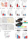
Human CRP (C-reactive protein) exposed to high shear was deposited as mCRP (monomeric C-reactive protein) in a mouse model of arterial stenosis. Graphical representation (A) and intraoperative images (B) of the carotid artery stenosis model, as described previously. B, The stenosis model with the right coronary artery (RCA) exposed. A consistent 80% stenosis was induced by applying a 6-0 braided polyester fiber suture circumferentially (black arrowhead) around a 150-µm needle. The direction of blood flow is indicated by the black arrow throughout B through D. C, Computational fluid dynamics (CFD) calculated wall shear rates in the artery stenosis model. The color scheme and scale indicate the respective wall shear rate. D and F, Representative immunohistochemistry of longitudinal sections of the carotid artery. The stenosis-inducing thread (black arrowhead) and the flow direction (black arrow) are indicated. Slides were incubated with either antihuman mCRP antibody (9C9; D) or antihuman pCRP (pentameric C-reactive protein) antibody (1D6; F) and visualized with secondary antibody conjugated to HRP, followed by incubation with 3,3’-diaminobenzidine peroxidase substrate. The scale bar indicates 20 µm. A secondary antibody only was used as a control. E and G, The planimetric analysis of 9C9 staining in stenosed carotid arteries showed significantly higher integrated density values in arteries with stenosis compared with vessels without stenosis (E). The planimetric analysis of 1D6 staining in stenosed and controlled carotid arteries, respectively, showed no significant difference (G). n=5 each; dots represent biological replicates. For statistical analysis, the Kruskal-Wallis test and the Dunn multiple comparison test were applied.
Figure 5.
Figure 5.
Circulating mCRP (monomeric C-reactive protein) is significantly reduced after transcatheter aortic valve implantation (TAVI). A, Schematic drawing of stenotic aortic valve with an illustration of the associated high shear stress. B through H, Clinical and experimental parameters of patients with TAVI before (red) and 1 month after TAVI (blue) are shown. B, Aortic mean gradients were significantly reduced after TAVI (n=37). P values were calculated using 2-tailed, matched-pairs signed rank tests (Wilcoxon). Dots represent biological replicates. C, Shear rates were calculated as described in detail in the methods section for patients before and after TAVI, demonstrating a significant reduction in mean shear rate (n=37, biological replicates). Paired 2-tailed t-tests were applied. D, High-sensitivity (hs) CRP (C-reactive protein) from patients before and 1 month after TAVI. pCRP (pentameric C-reactive protein) in patient blood was measured for 22 patients (n=22, biological replicates) by hsCRP assay, demonstrating no significant difference before and after TAVI, using paired nonparametric Wilcoxon matched-pairs signed rank text. E and F, Flow cytometry of microvesicles (MVs) isolated and analyzed as previously described from patients with TAVI (n=41) demonstrated a significant decrease in mCRP bound to MVs by both the ratio of mCRP-positive MV/all MV (E) and mean fluorescence intensity (MFI; F). Flow cytometry with mouse antihuman mCRP (9C9) and goat antimouse-Alexa Fluor 488 revealed a significantly reduced mCRP level in patients after TAVI (F). Data were analyzed using a matched 2-tailed t test (E; n=29, biological replicates) and paired nonparametric Wilcoxon matched-pairs signed rand test (F). G and H, Consistent with the hsCRP assay, flow cytometry analysis with mouse antihuman pCRP (clone 1D6) and goat antimouse-Alexa Fluor 488 did show a minor reduction in the MFI of the low level of pCRP-positive MVs (G) and no significant difference for the percentage of pCRP-positive MVs (H) before and after TAVI (n=41, biological replicates). Paired nonparametric Wilcoxon matched-pairs signed rank test was applied. I and J, CRP was further detected deposited as mCRP (I) but less so as pCRP (J), using immunohistochemistry on the cusps of stenotic aortic valves excised from patients undergoing surgical aortic valve replacement (representative example of stained valve sections shown). K, Immunohistochemical quantification of mCRP (9C9), pCRP (1D6), and isotype staining in valves of 8 patients (n=8, biological replicates) with severe aortic valve stenosis. P values were calculated using the Friedman nonparametric test and the Dunn multiple comparisons test, biological replicates. Respective antibody controls were used. L and M, Computational fluid dynamics calculations show the shear rates at the site of the stenosis in the microfluidic device, which approximates the situation before TAVI (upper half) and after TAVI (lower half; L). pCRP samples sheared in the microfluidic assay (n=4) showed dissociation as demonstrated by Western blots. M, Detection of pCRP (left) decreasing with increasing shear; in contrast, mCRP (right) was found to increase with increasing shear. Peak shear rates mimicking patients after TAVI mimic are given in blue (850 s−1), and peak shear rates mimicking stenotic aortic valves are given in red (3500 and 5000 s−1). n=4; dots represent biological replicates; and 1-way ANOVA and Tukey test were applied.
Figure 6.
Figure 6.
Shear-dissociated CRP (C-reactive protein) exhibits proinflammatory effects on endothelial cells and platelets. A, schematic drawing of the microfluidic model that recapitulates the shear conditions of aortic valve stenosis with direct flow into µ-slide channels covered with monolayers of human aortic valve endothelial cells (HAoVECs). B, Representative immunofluorescence images of human aortic valve endothelial cell monolayers incubated under static conditions (16 h) with 50-µg/mL pCRP (pentameric C-reactive protein; static pCRP) or mCRP (monomeric C-reactive protein; static mCRP). In addition, 50-µg/mL pCRP was subjected to flow for 16 h through an 80% stenosis microfluidic chip (5000 s−1) and subsequent perfusion over a monolayer of human aortic valve endothelial cells (sheared CRP). Representative expressions of the adhesion molecules, ICAM-1 (intercellular adhesion molecule 1) (green) and P-selectin (red), are shown (static control [no CRP], static pCRP, static mCRP, and sheared pCRP; n=16), and each is merged with DAPI staining. C and D, Quantification of B. mCRP increased the expression of ICAM-1 (C) and P-selectin (D) compared with static pCRP and static control (no CRP added). Shear-dissociated CRP induced a significant increase in expression of both ICAM-1 and P-selectin compared with flow with pCRP (shear control). n=16; dots represent biological replicates; and 1-way ANOVA and Tukey post hoc were used. E, PAC-1 binding and CD62P expression in washed human platelets (n=5) stimulated with ADP or increasing concentrations of pCRP and mCRP, with no treatment serving as resting platelet control. Cells were stained with PAC-1-FITC and antihuman CD62P-PE (cloneAC1.2) and analyzed by flow cytometry. pCRP failed to increase PAC-1 binding (left graph) and CD62P expression (right graph) compared with the resting control even at the highest concentration tested. In contrast, mCRP showed a concentration-dependent increase, with the lowest concentration tested (50 µg/mL) increasing both PAC-1 binding and CD62P expression significantly compared with resting control. n=5; dots represent biological replicates; and nonmatching Welch ANOVA and the Dunnett T3 test were used. F, TGF-β (transforming growth factor beta) surface expression on human platelets. Platelet-rich plasma from healthy donors (n=5) was stimulated with 50-µg/mL pCRP and mCRP, 100-nmol/L phorbol 12-myristate 13-acetate (PMA; positive control), or remained unstimulated for 30 min at room temperature. Flow cytometry was performed with a mouse antihuman TGF-β-AF488 (clone 1018746), mouse antihuman CD41a-APC (clone HIP8), and mouse antihuman CD62P-PE (clone AK-4), as previously described. mCRP, but not pCRP, increased the surface expression of TGF-β significantly compared with the unstimulated control. n=5; dots represent biological replicates; and ANOVA and the post hoc Tukey tests were applied.

References

    1. Baratchi S, Khoshmanesh K, Woodman OL, Potocnik S, Peter K, McIntyre P. Molecular sensors of blood flow in endothelial cells. Trends Mol Med. 2017;23:850–868. doi: 10.1016/j.molmed.2017.07.007 - PubMed
    1. Baratchi S, Zaldivia MTK, Wallert M, Loseff-Silver J, Al-Aryahi S, Zamani J, Thurgood P, Salim A, Htun NM, Stub D, et al. . Transcatheter aortic valve implantation represents an anti-inflammatory therapy via reduction of shear stress–induced, piezo-1–mediated monocyte activation. Circulation. 2020;142:1092–1105. doi: 10.1161/CIRCULATIONAHA.120.045536 - PubMed
    1. Malek AM, Alper SL, Izumo S. Hemodynamic shear stress and its role in atherosclerosis. JAMA. 1999;282:2035–2042. doi: 10.1001/jama.282.21.2035 - PubMed
    1. Ruggeri ZM, Platelets in atherothrombosis. Nat Med. 2002;8:1227–1234. doi: 10.1038/nm1102-1227 - PubMed
    1. Pareti FI, Lattuada A, Bressi C, Zanobini M, Sala A, Steffan A, Ruggeri ZM. Proteolysis of von Willebrand factor and shear stress–induced platelet aggregation in patients with aortic valve stenosis. Circulation. 2000;102:1290–1295. doi: 10.1161/01.cir.102.11.1290 - PubMed

Substances