. 2024 Oct;105(10):002032.
doi: 10.1099/jgv.0.002032.

A broadly reactive ultralong bovine antibody that can determine the integrity of foot-and-mouth disease virus capsids

Affiliations

A broadly reactive ultralong bovine antibody that can determine the integrity of foot-and-mouth disease virus capsids

John D Clarke et al. J Gen Virol. 2024 Oct.

Abstract

Foot-and-mouth disease vaccination using inactivated virus is suboptimal, as the icosahedral viral capsids often disassemble into antigenically distinct pentameric units during long-term storage, or exposure to elevated temperature or lowered pH, and thus raise a response that is no longer protective. Furthermore, as foot-and-mouth disease virus (FMDV)'s seven serotypes are antigenically diverse, cross-protection from a single serotype vaccine is limited, and most existing mouse and bovine antibodies and camelid single-domain heavy chain-only antibodies are serotype-specific. For quality control purposes, there is a real need for pan-serotype antibodies that clearly distinguish between pentamer (12S) and protective intact FMDV capsid. To date, few cross-serotype bovine-derived antibodies have been reported in the literature. We identify a bovine antibody with an ultralong CDR-H3, Ab117, whose structural analysis reveals that it binds to a deep, hydrophobic pocket on the interior surface of the capsid via the CDR-H3. Main-chain and hydrophobic interactions provide broad serotype specificity. ELISA analysis confirms that Ab117 is a novel pan-serotype and conformational epitope-specific 12S reagent, suitable for assessing capsid integrity.

Keywords: FMDV; pan-specific; single particle analysis; ultralong CDR antibody; vaccine quality assurance.


Conflict of interest statement

Some of the work presented in this study was presented within John D. Clarke’s doctoral thesis. The authors declare that no other conflicts of interest, direct or contrived, were identified in relation to this work. The funders had no role in the design of the study; in the collection, analyses or interpretation of data; in the writing of the manuscript; or in the decision to publish the results.

Figures

Fig. 1.
Fig. 1.. (a) Double sandwich homologous ELISA of Ab117 reactivity (the complex is not pre-made before application to the ELISA plate) with SAT2 topotypes. SAT2/ZIM/7/83 VLPs with S2093Y and SAT2/EGY/4/2012 inactivated virus. (b) Antigen titration ELISA (starting from 3.43 µg ml−1 as 1/3 dilution) utilizing M377, M311 and Ab117 as both capture and detection antibody for the SAT2/EGY/4/2012 inactivated virus without and with heat treatment (56 °C for 15 min) to disrupt the capsids.
Fig. 2.
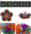
Fig. 2.. Fab117 binds to the interior surface of the dissociated SAT2/ZIM/7/83 pentamer. (a) Representative 2D class averages. (b) Final 3D reconstruction. (c, d) Close-up of the Fab117-SAT2/ZIM/7/83 complex density, viewed towards (c) the interior capsid surface and (d) from above the fivefold axis looking downward at the hydrophobic binding pocket. Density coloured by molecule: VP1 – blue, VP2 – turquoise, VP3 – orange and Fab117 CDR-H3 – purple.
Fig. 3.
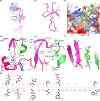
Fig. 3.. Details of Fab117 binding. For clarity, only interacting residues and bonds are shown. Residues are labelled with those from the virus given a preceding chain identifier for VP1, VP2 or VP3, respectively, such that they all have four digits. (a) AlphaFold model of Fab117 with HC in magenta and LC in cyan. (b) Cα trace of the ordered ‘knob’ domain of CDR-H3 in the SAT2 pentamer–Fab117 complex. Cαs of residues contacting the antigen (within 4.0 Å) are shown as grey spheres and disulphides as yellow sticks. The CDR-H3 is orientated as in (a). (c) Fab117 (rainbow backbone) engages the hydrophobic cleft of the SAT2 pentamer shown in surface representation and coloured by hydrophobicity. (d) The tip of the Fab117 CDR-H3 is shown in purple with W138 central and SAT2 residues within 4.0 Å of the latter represented as sticks. VP2 is coloured green, VP3 red and the antibody purple, with VP2 secondary structural elements labelled. (e, f) Zoomed perspective of the interactions between Fab117 and VP2. VP2 secondary structural elements are labelled. (g) LigPlot+v2.2.8 [38] of the interaction between the Fab117 CDR-H3 and interface residues of VP2 only (thus, for instance, the contacts of W138 to VP1 and VP3 are not shown).
Fig. 4.
Fig. 4.. Ab117 cross-specificity. (a) Sequence alignment of the representative FMDV serotypes [2830,52]. Residues with red background are conserved. Residues at the Fab117–SAT2/ZIM/7/83 interface are boxed. Black triangles indicate appreciable contribution to the interface, whilst red triangles indicate a hydrogen bond interaction (contributions determined using PISA). Residues directly making contact with W138 (≤4.0 Å) are indicated using an asterisk. The secondary structure of the SAT2/ZIM/7/83 proteins is indicated above the alignment. Alignment was performed using CLUSTALW [6061], and the figure was generated using ESPript [41]. (b) Heterologous sandwich ELISA shows pan-specific binding of Ab117 to thermally disrupted virus. Integrin αvβ6 was used as a universal capture for all intact viruses, whilst heated particles were trapped directly on the plates, with Ab117 applied for detection. SAT1/ZIM/22/89 inactivated virus was tested in an independent experiment. (c) Ab117 binding to SAT3/ZIM/4/81 [62] wild-type VLPs. Serum raised in animals infected with SAT3 was used as a positive control, whilst the lack of M3 binding to disrupted capsids showed the inability of this VHH to recognize SAT3-disrupted particles. Ab117 binds to untreated capsids, suggesting their instability. Heating at 56 °C increased the signal, consistent with increased disruption of particles, while heating at 65 °C exhibited a lower signal, which suggests the disruption of the conformational epitope recognized by Ab117 at higher temperatures. Cyan bars represent the signal using untreated capsids (4 °C), orange, and red bars represent the binding level using capsids heat treated for 15 min at 56 °C and 65 °C, respectively.

References

    1. The National Audit Office The 2001 Outbreak of Foot and Mouth Disease. Report. London: The National Audit Office, The Comptroller and Auditor General. 2002 102916454.
    1. Grubman MJ, Baxt B. Foot-and-mouth disease. Clin Microbiol Rev. 2004;17:465–493. doi: 10.1128/CMR.17.2.465-493.2004. - DOI - PMC - PubMed
    1. Knight-Jones TJD, Rushton J. The economic impacts of foot and mouth disease - what are they, how big are they and where do they occur? Prev Vet Med. 2013;112:161–173. doi: 10.1016/j.prevetmed.2013.07.013. - DOI - PMC - PubMed
    1. Gao Y, Sun S-Q, Guo H-C. Biological function of foot-and-mouth disease virus non-structural proteins and non-coding elements. Virol J. 2016;13:107. doi: 10.1186/s12985-016-0561-z. - DOI - PMC - PubMed
    1. Haydon DT, Samuel AR, Knowles NJ. The generation and persistence of genetic variation in foot-and-mouth disease virus. Prev Vet Med. 2001;51:111–124. doi: 10.1016/s0167-5877(01)00210-0. - DOI - PubMed

LinkOut - more resources