

Bone impairment in atypical hemolytic and uremic syndrome treated by long-term eculizumab
- PMID: 39422762
- PMCID: PMC11885323
- DOI: 10.1007/s00467-024-06564-6
Bone impairment in atypical hemolytic and uremic syndrome treated by long-term eculizumab
Abstract
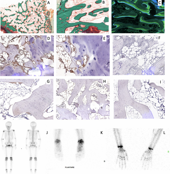
Atypical hemolytic uremic syndrome (aHUS) is a thrombotic microangiopathy, related to complement dysregulation, including Factor H deficiency (FH) treated by lifelong eculizumab therapy. Its long-term tolerance is not yet fully described. We report two patients with genetic FH deficiency receiving long-term eculizumab and displaying a peculiar bone phenotype. First case is a 13-year-old girl, presenting with bone pains, arthritis, and deformities, for which X-rays and MRI described multifocal osteochondritis. Bone biopsy revealed an active remodeling bone (many areas of bone formation and resorption) and C3c accumulation on immunohistochemical staining. The second patient is an 11-year-old girl, displaying mechanical bone pains, for which bone scintigraphy found hypofixation of wrists and ankles. These findings could be consistent with a side effect of eculizumab, as C3c accumulation may result from the downstream C5-blockade. Alternatively, bone alterations could be due to the absence of FH, as described in murine models. Further investigations are required to characterize bone disease in aHUS.
Keywords: AHUS; Bone; C3 deposits; Eculizumab.
© 2024. The Author(s).
Conflict of interest statement
Declarations. Conflict of interest: The authors declare no competing interests.
Figures
References
-
- Greenbaum LA, Fila M, Ardissino G, Al-Akash SI, Evans J, Henning P et al (2016) Eculizumab is a safe and effective treatment in pediatric patients with atypical hemolytic uremic syndrome. Kidney Int 89:701–711 - PubMed
-
- Daugan MV, Revel M, Lacroix L, Sautès-Fridman C, Fridman WH, Roumenina LT (2021) Complement detection in human tumors by immunohistochemistry and immunofluorescence. Methods Mol Biol 2227:191–203 - PubMed
-
- Mödinger Y, Löffler B, Huber-Lang M, Ignatius A (2018) Complement involvement in bone homeostasis and bone disorders. Semin Immunol 37:53–65 - PubMed
-
- Alexander JJ, Sankaran JS, Seldeen KL, Thiyagarajan R, Jacob A, Quigg RJ et al (2018) Absence of complement factor H alters bone architecture and dynamics. Immunobiology 223:761–771 - PubMed
Publication types
MeSH terms
Substances
Supplementary concepts
LinkOut - more resources
Full Text Sources
Miscellaneous
