. 2024 Oct 18;19(10):e0309845.
doi: 10.1371/journal.pone.0309845. eCollection 2024.

The curious case of the Dana platypus and what it can teach us about how lead shotgun pellets behave in fluid preserved museum specimens and may limit their scientific value

Affiliations

The curious case of the Dana platypus and what it can teach us about how lead shotgun pellets behave in fluid preserved museum specimens and may limit their scientific value

Henrik Lauridsen et al. PLoS One. .

Abstract

Fluid preserved animal specimens in the collections of natural history museums constitute an invaluable archive of past and present animal diversity. Well-preserved specimens have a shelf-life spanning centuries and are widely used for e.g. anatomical, taxonomical and genetic studies. The way specimens were collected depended on the type of animal and the historical setting. As many small mammals and birds were historically collected by shooting, large quantities of heavy metal residues, primarily lead, may have been introduced into the sample in the form of lead shot pellets. Over time, these pellets may react with tissue fluids and/or the fixation and preservation agents and corrode into lead salts. As these chemicals are toxic, they could constitute a health issue to collection staff. Additionally, heavy element chemicals interfere with several imaging technologies increasingly used for non-invasive studies, and may confound anatomical and pathological investigations on affected specimens. Here we present a case-study based on platypus (Ornithorhynchus anatinus) and other small mammals containing lead pellets from the collection of The Natural History Museum of Denmark.


Conflict of interest statement

I have read the journal’s policy and the author Michiel Krols of this manuscript has the following competing interests: Employed at TESCAN XRE, a manufacture of one of the micro-CT systems used in the study (UniTOM XL Spectral). The remaining authors have declared that no competing interests exist. This does not alter our adherence to PLOS ONE policies on sharing data and materials

Figures

Fig 1
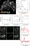
Fig 1. The Dana platypus (NHMD-M01-28) collected during The Carlsberg Foundation’s Oceanographical Expedition round the World 1928–30.
(a) Apart from a single puncture at the right side of the bill (black arrowhead) the specimen shows no superficial evidence of collection method; (b) Virtual coronal section from x-ray computed tomography (CT) showing hyperdense foreign objects (FO) embedded in the specimen; (c) Foreign object 16 dissected free from the thigh musculature appears as a centimetre sized nodule with great visual resemblance to dried chewing gum, but more brittle in nature; (d) Slice through FO 16 showing a whitish core and a brownish crust; (e) Virtual section of FO 16 from micro x-ray computed tomography (μCT) showing the crust is more radiodense than the core; (f) Virtual coronal section (similar to (b) from magnetic resonance imaging (MRI) of FO 11. In contrast to bony material that appears hypodense in the specific T2-weighted spin echo sequence, the foreign object appears hyperdense, but with a low level of image artifacts. This indicates that the foreign object does not consist of either bone mineral or paramagnetic substances; (g) Volume rending from CT of the Dana platypus allowing for the appreciation of the large number of foreign objects. At least 21 centimetre sized objects can be observed. The volume rending is calibrated to a 0–5000 Hounsfield unit (HU) calibration bar to demonstrate radiodensity of the skeleton and foreign objects. Skeletal components rarely exceed 1900 HU, but the average radiodensity of the foreign objects is 4320 HU indicating a non-biological origin.
Fig 2
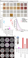
Fig 2. Chemical tests to reveal elemental components of foreign objects.
(a) Flame test of metal salts of most common biometals and pulverised core and crust of foreign object (FO) 12 and the whole of FO 11; (b) Calibration bar for lead (Pb) test paper 1, Tin (Sn) test paper, and arsenic (As) test kit. Additionally, test for interactions between As test kit and common physiological salts and Pb2+. No interactions were observed; (c) Serial dilution of PbCl2 on Pb test paper 1 and 2 to reveal lower detection limit of Pb on these test papers. Test paper 2 was most sensitive with a detection limit of 9.7 mg/l of PbCl2 equalling 34.9 μM Pb2+; (d) Analysis for the presence of Pb, antimony (Sb), Sn and As in double distilled water, 70% ethanol (EtOH), homogenized lead shot pellet, and homogenized core and crust of FO 14. No interaction was observed for any metal for water and ethanol whereas all four metals were detected in the lead shot pellet and in both core and crust of FO 14; (e) Salt solubility of FO 14 core material as a function of temperature in comparison with different lead salts (literature values); (f) Magnification of the graph above with a focus on FO 14 core solubility. The solubility of the FO 14 core material is much lower than would be expected for reaction products between ethanol and formaldehyde.
Fig 3
Fig 3. X-ray spectral analysis of foreign objects in the Dana platypus and in lead pellets.
(a) Overview of the Dana platypus and magnified lead pellets showing regions (circles) or points (crosshair) of interest; (b) Idealised x-ray attenuation spectra for lead (Pb) (primary second axis), cortical bone and calcium (Ca) (both on secondary second axis) at monochromatic x-ray photon energies from 60–200 keV. Lead has a sharp K-edge at 88 keV allowing for material identification in this range; (c) Polychromatic x-ray computed tomography (CT) attenuation spectra for peak energies between 80 and 135 keVp for the Dana platypus bone at regions of interest placed at the tibia, skull and a vertebra, respectively. All three spectra follow the expected decrease in attenuation at higher energies as predicted by the idealised spectrum for bone (b); (d) Polychromatic CT attenuation spectra for six foreign objects (FO) in the Dana platypus. The spectra are distinct from the bone spectra (c) showing an increase in attenuation at higher peak energies, but they are also not similar to the idealised lead spectrum (b) probably due to the polychromatic nature of conventional CT imaging; (e) Polychromatic CT attenuation spectra for the peripheral partial volume effected region of three lead pellets. In spite of the partial volume effect (necessary to avoid the Hounsfield unit threshold) and the polychromatic nature of conventional CT imaging, the lead pellet spectra are comparable to the idealised spectrum of lead (b) showing a pronounced increase in attenuation between 80 and 100 kVp; (f) Hyperspectral CT imaging of the neurocranium of the Dana platypus presented as coronal slices (top row) and maximum intensity projections (MIP, bottom row) of an averaged image of the 20–120 keV energy channels. K-edge subtraction at the K-edge of lead (88 keV) allows for material specific mapping of lead within the CT volume; (g) X-ray attenuation spectra (linear attenuation coefficient, μ, over x-ray energy) for five regions placed within FO 2 and FO 3 and five regions with skull bone. The sharp K-edge at 88 keV in the foreign objects confirms the presence of lead in foreign objects; (h) Derived curves of attenuation spectra in (g) showing maximum increase in attenuation at 88 keV.
Fig 4
Fig 4. Shot trajectory.
(a) Coronal (left) and oblique sagittal (right) virtual micro-CT sections in the head of the Dana platypus reveal a large foreign object in the right cerebral hemisphere and lead fragments and/or smears show the most likely shot trajectory; (b) Three-dimensional reconstructions of the head in a dorsal view (left) and viewed along the probable shot trajectory (right). Red and blue structures are hyperintense foreign objects; (c) Photos of the Dana platypus from the same viewpoints as in (b) and from a ventral viewpoint in addition (lower left). White arrowhead point to the exit wound on the dorsal portion of the bill. Magnifications show entry wounds. In the 70% ethanol soaked specimen (upper right) it is easy to overlook the small entry wound, whereas it is obvious after superficial preservation fluid has evaporated off (lower left and lower right).
Fig 5
Fig 5. Museum specimens collected with the shotgun.
(a) X-ray overview of small mammals in the fluid collection of The Natural History Museum of Denmark containing shotgun pellets. Oana = Ornithorhynchus anatinus, Pray = Pteropus rayneri, Pper = Pseudocheirus peregrinus, Cfin = Callosciurus finlaysonii. Six 3 mm lead pellets are shown (middle to the left) for comparison of size with foreign objects in the specimens; (b) Historical shotgun previously housed in the weapons collection of the The Natural History Museum of Denmark. This weapon and the custom made adapter sleeve (red arrow) was used at the Danish Noona Dan Expedition to the Pacific in 1961–62 and the Solomon Islands flying fox (Pray M05-CN2901) was most likely collected using this weapon.
Fig 6
Fig 6. Chemical analysis of preservation fluids for lead (Pb) and arsenic (As).
(a) Analysis for the presence of Pb in 30x concentrated samples of preservation fluids of mammal specimens (see Table 1 for full names and catalogue#) with (+) or without (-) hyperintense nodules on x-ray images. Some specimens were stored in the same jars as indicated with multiple + or -. Both the lipid (L) and the water (W) phase was analysed but the presence of Pb was not detected in any sample; (b) Analysis for the presence of As in 3.5x concentrated samples of preservation fluids (mixed lipid and water phase). In all but two samples As was detected; (c) Arsenic concentration in different samples of preservation fluids. There was no significant difference between [As] is samples with and without hyperintense nodules.
Fig 7
Fig 7. Corrosion test of lead pellets in different solutions.
(a) Analysis for the presence of lead (Pb), antimony (Sb), tin (Sn) and arsenic (As) after storing lead pellets for 22 days in 1 ml of either double distilled water, 70% ethanol (EtOH) with and without phosphate buffed saline (PBS), 100% EtOH, phosphate buffered 4% formaldehyde and unbuffered 4% and 40% formaldehyde. All but the 70% EtOH (PBS), 100% EtOH and the buffered 4% formaldehyde solutions tested positive for Pb. All solutions except the 100% EtOH tested positive for Sb. Tin was only detected in the unbuffered 40% formaldehyde solution. The water, the buffered 4% formaldehyde and in particular the 70% EtOH solution with PBS contained a detectable amount of As; (b) corrosion rate of lead pellets in different solutions. There was significant difference between the corrosion rate in different solutions (one way ANOVA with post hoc Tukey honest significance test). Significantly different groups are marked with different capital letters (A, B, C and D) i.e. a group labelled with A is significantly different from a groups labelled with B but not from one labelled with AC; (c) Lead pellet mass over time at the corrosion rate of different solutions. The point at which all pellet material has been corroded is marked with a circle in the respective line colours (or by an arrow in cases where circles are close); (d) Virtual cross sections of beef cubes previously hosting a lead pellet (top rows) or maximum intensity projections (MIP, bottom row). Varying degrees of corrosion products remain within the sample after 8.5 month of storage; (e) Volume (V) of corrosion products within beef samples preserved in different preservation fluids.
Fig 8
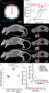
Fig 8. Determination of lead Hounsfield value and the effect on measurements of bone mineral content in animal specimens.
(a) Virtual section of 3 mm lead shot pellet from x-ray computed tomography (CT). Due to the high radiodensity of the lead alloy, the pellet core exceeds the maximum Hounsfield value on conventional 16-bit CT systems (from -32768 to +32767 HU) and actual radiodensity of lead pellets cannot be measured directly. However, peripheral voxels suffer from partial volume effects from the averaging of signal contribution from lead and surrounding air (-1000 HU); (b) By extrapolating the maximum slope of 12 longitudinal profile plots originating in air and terminating at the core of six lead pellets, the partial volume effect can be exploited to indirectly reveal the average radiodensity (43584 ± 4670 HU) of lead pellets allowing for original pellet size calculations of foreign objects in the Dana platypus; (c) Laboratory rat before (top) and after (middle) injection of six lead pellets, and after pellets have been digitally removed by subtracting all voxels with CT values > 1900 HU (bottom). To the left are maximum intensity projections, and to the right are virtual sagittal sections. Red boxes on the right are 2.5x magnifications of a pellet in the chest region; (d) Body mass adjusted bone mineral content (BMC) measured using quantitative CT on laboratory rats before and after injection of lead pellets and after digital removal of these. The presence of lead pellets increased body mass adjusted BMC significantly, and even after digital removal of lead pellets, the measurement was significantly affected due to the partial volume voxels with CT-values of a little less than 1900 HU that were still present in the images (see magnification in (c) lower right); (e) Percentage error (difference in estimated and actual value) of total BMC as a function of total BMC and the presence of either 1, 5 or 10 lead pellets. Due to the CT-value threshold of 32767 HU on most clinical CT systems, metallic lead alloys of lead shot pellets with CT values of 43584 ± 4670 HU do not express the maximum effect on BMC measurements. However, as lead pellets corrode and radiodensity of lead salts fall below the CT-value threshold, the effect of lead pellets is increased as these corrode. This is demonstrated using solid graph lines for the full effect of imbedded lead pellets and dashed lines for the limited effect when pellets are still in the metallic state.

References

    1. Birch, T. (1968). The history of the Royal Society of London for improving of natural knowledge from its first rise. Vol. 1. New York: Johnson reprint corporation. [Reprint of the 1756–1757 edition published for the Royal Society of London by A. Millar, London].
    1. Cole F.J. (1944). A history of comparative anatomy from Aristotle to the Eighteenth century. London: MacMillan.
    1. Simmons, J.E. (2014). Fluid preservation: A comprehensive reference. Lanham (MD, USA): Rowman & Littlefield.
    1. Blum F. (1893. a). Der formaldehyd als antisepticum. Münchener Medicinische Wochenschrift 40, 601–602.
    1. Blum F. (1893. b). Der formaldehyde als härtungsmittel. Zeitschrift der Wissenschaft für Mikroskopie 10, 314–315.