. 2024 Nov;300(11):107897.
doi: 10.1016/j.jbc.2024.107897. Epub 2024 Oct 17.

AMH regulates a mosaic population of AMHR2-positive cells in the ovarian surface epithelium

Affiliations

AMH regulates a mosaic population of AMHR2-positive cells in the ovarian surface epithelium

Elizabeth R Smith et al. J Biol Chem. 2024 Nov.

Abstract

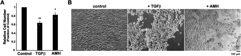
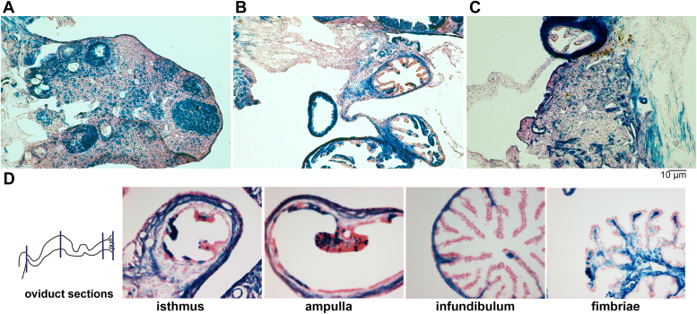
The function and homeostasis of the mammalian ovary depend on complex paracrine interactions between multiple cell types. Using primary mouse tissues and isolated cells, we showed in vitro that ovarian follicles secrete factor(s) that suppresses the growth of ovarian epithelial cells in culture. Most of the growth suppressive activity was accounted for by Anti-Mullerian Hormone/Mullerian Inhibitory Substance (AMH/MIS) secreted by granulosa cells of the follicles, as determined by immune depletion experiments. Additionally, conditioned medium from granulosa cells from wild-type control, but not AMH knockout, suppressed epithelial cell growth. Tracing of the AMH-regulated cells using AMHR2 (AMH receptor 2)-Cre:ROSA26 mutant mice indicated the presence of populations of AMHR2-positive epithelial cells on the ovarian surface and oviduct epithelia. Cells isolated from the mutant mice indicated that a subpopulation of cells marked by AMHR2-Cre:ROSA26 accounted for most cell growth and expansion in ovarian surface epithelial cells, and the AMHR2 lineage-derived cells were regulated by AMH in vitro; whereas, fewer AMHR2-Cre:ROSA26-marked cells accounted for oviduct epithelial cell outgrowth. The results reveal a paracrine pathway in maintaining follicle-epithelial homeostasis in the ovary and support a subpopulation of AMHR2 lineage marked epithelial cells as ovarian epithelial stem/progenitor cells with higher proliferative potential regulatable by follicle-secreted AMH.

Keywords: AMHR2; MIS/AMH; granulosa cell; ovarian cancer; ovarian epithelium; oviduct epithelium; stem cell.


Conflict of interest statement

Conflict of interest The authors declare that they have no conflicts of interest with the contents of this article.

Figures

Figure 1
Figure 1
Granulosa factor(s) suppress MOSE cell growth in culture.A, primary MOSE and OVD cells were isolated from wild-type mice and expanded in culture. Proliferation of cells was determined using a WST-1 assay, where cell number correlates with the absorbance measured at 450 nm. Relative cell numbers were measured in triplicate samples and are represented as the mean ± s.d. B, both MOSE and OVD have a characteristic cuboidal epithelial shape in culture. C, MOSE cells were co-cultured in transwell dishes with (+G) or without (control) granulosa cells (+G). Cell number was determined by the WST-1 proliferation assay kit. D, Conditioned medium was prepared from granulosa cells after 2 (2 days) or 4 (4 days) days in culture. MOSE cells were plated in triplicate in 96-well dishes and grown overnight, then incubated in granulosa cell conditioned medium (GCCM) mixed with normal culture medium at the percentages shown; cell number was assayed after 4 days in culture, when MOSE cells are actively proliferating. Data are expressed as mean ± s.d., with ∗p < 0.05, by Student’s unpaired two-tailed t test. E, representative images of MOSE cells co-cultured without (Control) or with (+Granulosa) granulosa cells.
Figure 2
Figure 2
AMH suppresses MOSE cell growth.A, MOSE cells were treated with TGF-β (0.1 μg/ml) or AMH (5 μg/ml) for 4 days, and cell growth was determined by WST-1 assay and expressed as the cell number of treated cells relative to control cells (fraction of control). The average of three experiments is shown as mean ± s.d., with ∗p < 0.05 or ∗∗p = 0.001, determined by an unpaired two-tailed Student’s t test. B, images of cells treated with TGF-β or AMH, as described.
Figure 3
Figure 3
AMH from granulosa cell conditioned medium suppresses MOSE growth in culture.A, AMH was immunodepleted from granulosa cell conditioned medium (GCCM) using an excess concentration of anti-AMH rabbit polyclonal antibody (Abcam ab84952) (Anti-AMH). Controls included normal culture medium (control) and granulosa cell conditioned medium that had no treatment (none), treated only with the affinity matrix used for IP (+ protein A/G), or non-specific pre-immune rabbit serum and Protein A/G (Pre-Ab). MOSE cultures were treated with the respective medium for 6 days, and relative cell number was determined by WST-1 assay, shown as mean ± s.d., with ∗p < 0.05, as determined by an unpaired two-tailed Student’s t test. The error bars indicate multiple and potentially overlapping measurements. There was no significant difference (N.S.) between cell proliferation in control medium and anti-AMH immunodepleted granulosa cell conditioned medium; however, a significant difference was determined between cells grown in untreated granulosa cell conditioned medium (none) and anti-AMH immunodepleted granulosa cell conditioned medium. B, granulosa cells were isolated from wild-type or AMH knockout mice and grown in culture for 6 days. The conditioned medium was collected and incubated with MOSE cells for an additional 6 days. The effect of granulosa cell conditioned medium from normal wild type or AMH knockout mouse ovaries was compared to normal culture medium. MOSE cell proliferation was significantly decreased by growth in WT GCCM, determined as ∗p < 0.05 by an unpaired two-tailed Student’s t test. C, MOSE cells were treated with TGFβ, EGF, or granulosa cell conditioned medium for 6 days, then collected in sample buffer, and analyzed by Western blot for the proteins indicated. D, images of cells treated with TGFβ, EGF, and conditioned medium collected from wild type (WT) or AMH KO granulosa cells grown in culture for 6 days.
Figure 4
Figure 4
LacZ staining of AMHR2-Cre+;Rosa ± ovaries and oviducts.A–B, the ovarian surface of adult mice is mosaic for AMHR2-Cre+;Rosa ± epithelial cells, as shown by positive LacZ (blue X-Gal) staining. Granulosa cells of the ovarian follicles also stain positive for LacZ. B, epithelial areas of the oviduct are heterogeneous for LacZ staining. C, X-Gal staining of an AMHR2-Cre+;Rosa+/−;Wv/Wv ovary; note the intra-ovarian proliferation of epithelial lesions. D, Cross sections of the oviduct represent areas from the isthmus, ampulla, infundibulum, and fimbriae, from left to right, as indicated in the diagram.
Figure 5
Figure 5
Expansion of AMHR2-Cre+;Rosa ± MOSE and OVD epithelial cells in vitro.A–B, primary cultures of MOSE and (C-D) OVD epithelial cells were established from 4-month-old AMHR2-Cre+;Rosa ± mice and allowed to expand in culture for 1 to 4 weeks. LacZ (X-Gal) staining of the cultures was performed. BD, the percentage of X-Gal positive cells was calculated from an average of at least three separate fields, expressed as mean ± s.d. Significant difference from the starting time point was calculated for each subsequent time point using an unpaired two-tailed Student’s t test, shown as ∗p < 0.05 and ∗∗p = 0.0001. Results are representative of three separate experiments. Images were taken at 63 × magnification.
Figure 6
Figure 6
AMHR2 protein is expressed in ovarian cancer cells and primary cells and tissues.A, AMHR2 was detected in lysates from ovarian cancer cell lines (OVCAR-3, -4, -5, -8, -10; A2780; OVSAHO); an immortalized human fallopian tube cell line (FTE); and a primary cell culture obtained a human ovarian surface epithelium (HOSE). AMHR2 was not detected in a human mammary fibroblast culture. B, protein lysates were obtained from normal wild type (WT) and Wv/Wv (Wv) mouse ovaries (Ov), oviducts (Od), uteri (Ut), adrenal glands (Ad), and testes (Te). AMHR2 was detected using a mouse mAb (Abcam) at 1:2000 dilution and normalized to β-actin.
Figure 7
Figure 7
AMH knockout ovaries experience early aging and depletion of follicles.A, phenotypes of wild type and AMH knockout mouse ovaries at 1 to 8 months. Ovaries from 3-month-old mice were immunostained for epithelial cytokeratin 8 (arrows). Images were taken at 40 × final magnification. B, one-year-old mouse ovaries from WT, AMH heterozygous (AMH Het), AMH knockout (AMH KO), and Wv/Wv homozygous mice. C, example of ovarian cyst (inset showing the whole ovary at 40 × ; panel at 100 × magnification) and overt aging morphology in 1-year-old AMH KO ovaries, compared to a Wv/Wv ovary. Samples were immunostained for cytokeratin 8.
Figure 8
Figure 8
A decrease in AMH contributes to ovarian aging and proliferative potential of the surface epithelium. Scheme: A, in the normal cycling ovary, AMH produced by follicular granulosa cells can feed back and regulate growth of the epithelial cells on the surface and potentially nearby oviduct epithelial cells expressing AMHR2. The presence of follicles also provides feedback regulation of FSH and LH levels. B, in the WvWv mouse ovary, the absence of follicles results in the absence of AMH and unrestricted LH and FSH levels, conditions that permit epithelial expansion of the ovarian surface into the ovary. C, in AMH knockout mice, the lack of AMH leads to a more rapid decline in follicle numbers, but follicle-derived factors, such as progesterone and estrogen, are significant enough to regulate LH and FSH levels.

References

    1. Hummitzsch K., Anderson R.A., Wilhelm D., Wu J., Telfer E.E., Russell D.L., et al. Stem cells, progenitor cells, and lineage decisions in the ovary. Endocr. Rev. 2015;36:65–91. - PMC - PubMed
    1. Lorenzo H.K., Teixeira J., Pahlavan N., Laurich V.M., Donahoe P.K., MacLaughlin D.T. New approaches for high-yield purification of Müllerian inhibiting substance improve its bioactivity. J. Chromatogr. B Analyt Technol. Biomed. Life Sci. 2002;766:89–98. - PubMed
    1. Eppig J.J. Reproduction: oocytes call, granulosa cells connect. Curr. Biol. 2018;28:R354–R356. - PubMed
    1. Schulz B.O., Krebs D., Diedrich K., Knoll H., Hobbel K., Hamerich U. Effects of granulosa cells and gonadotrophins on maturation of rabbit oocytes in vitro. Arch. Gynecol. 1985;236:135–143. - PubMed
    1. Matzuk M.M., Burns K.H., Viveiros M.M., Eppig J.J. Intercellular communication in the mammalian ovary: oocytes carry the conversation. Science. 2002;296:2178–2180. - PubMed

Publication types

MeSH terms

Substances

LinkOut - more resources