. 2024 Dec 11;44(50):e0438242024.
doi: 10.1523/JNEUROSCI.0438-24.2024.

Metabotropic NMDAR Signaling Contributes to Sex Differences in Synaptic Plasticity and Episodic Memory

Affiliations

Metabotropic NMDAR Signaling Contributes to Sex Differences in Synaptic Plasticity and Episodic Memory

Aliza A Le et al. J Neurosci. .

Abstract

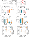
NMDA receptor (NMDAR)-mediated calcium influx triggers the induction and initial expression of long-term potentiation (LTP). Here we report that in male rodents, ion flux-independent (metabotropic) NMDAR signaling is critical for a third step in the production of enduring LTP, i.e., cytoskeletal changes that stabilize the activity-induced synaptic modifications. Surprisingly, females rely upon estrogen receptor alpha (ERα) for the metabotropic NMDAR operations used by males. Blocking NMDAR channels with MK-801 eliminated LTP expression in hippocampal field CA1 of both sexes but left intact theta burst stimulation (TBS)-induced actin polymerization within dendritic spines. A selective antagonist (Ro25-6981) of the NMDAR GluN2B subunit had minimal effects on synaptic responses but blocked actin polymerization and LTP consolidation in males only. Conversely, an ERα antagonist thoroughly disrupted TBS-induced actin polymerization and LTP in females while having no evident effect in males. In an episodic memory paradigm, Ro25-6981 prevented acquisition of spatial locations by males but not females, whereas an ERα antagonist blocked acquisition in females but not males. Sex differences in LTP consolidation were accompanied by pronounced differences in episodic memory in tasks involving minimal (for learning) cue sampling. Males did better on acquisition of spatial information whereas females had much higher scores than males on tests for acquisition of the identity of cues (episodic "what") and the order in which the cues were sampled (episodic "when"). We propose that sex differences in synaptic processes used to stabilize LTP result in differential encoding of the basic elements of episodic memory.

Keywords: NMDAR; actin; estrogen receptor; hippocampus; memory; metabotropic.


Conflict of interest statement

The authors declare no competing financial interests.

Figures

Figure 1.
Figure 1.
Theta burst stimulation (TBS) elicits nonionotropic NMDAR signaling and actin polymerization. Stimulation was applied to Schaffer-commissural (SC) projections to CA1 str. radiatum (SR) in slices from adult male rats. a, A 10-burst TBS train (arrow) elicited robust CA1 short- and long- term potentiation (STP and LTP, respectively) in vehicle (veh)-treated slices whereas MK-801 (30 µM, introduced 2 h before TBS) blocked this effect (p = 0.004 and p = 0.005 for STP and LTP, respectively; veh N = 8, MK-801 N = 5): Traces from before (solid) and 40 min after (dashed) TBS. b, Schematic showing placement of stimulating electrodes in CA1a (stim 1) and CA1c (stim 2); Gray box marks the CA1 SR field of F-actin and signaling protein analysis. c, Images show phalloidin labeling of F-actin (i.e., faint labeling of dendrites decorated with brighter punctate labeling of dendritic spines, arrows) in slices receiving low-frequency control (con) stimulation or TBS in the presence of veh or MK-801 (30 µM). d, Plot shows that TBS increased the proportion of phalloidin-positive puncta that were densely labeled normalized to control slice values (F(3,57) = 15.30, p < 0.0001; post hoc p = 0.0001); this effect was blocked by 100 µM APV (veh vs APV post hoc p = 0.0046) but not MK-801 (veh vs MK-801 p = 0.102, N = 8–24). e–g, Fluorescence deconvolution tomography was used to assess NMDAR contributions to TBS-induced synaptic signaling. Deconvolved images show immunolabeling for the kinase of interest and PSD95; the colors in the heading indicate the fluorophore for each protein; arrows indicate representative puncta with areas of double labeling that appear white (one element with double labeling is shown at higher magnification in an inset). e, TBS caused a rightward skew (toward greater densities) in the density–frequency histogram for synaptic pERK (i.e., colocalized with PSD95; F(38,608) = 18.50, p < 0.0001; con vs TBS post hoc: p = 0.0048); this was unaffected by MK-801. Inset, mean numbers of densely pERK-IR spines (≥100 density units) normalized to control slice values (F(2,32) = 10.33, p = 0.0003; N = 11–12/group) show that TBS increased pERK enriched postsynaptic elements in both veh-treated (p = 0.0007) and MK-801-treated (p = 0.0026) slices. f, TBS-induced increases of the proportion of synapses with dense pSrc immunoreactivity was blocked by APV (100 µM) but not MK-801 (F(3,62 = 15.11, p < 0.0001; veh vs MK-801 p = 0.4762; N = 7–32/group). g, TBS increased synaptic pCaMKII, and this effect was blocked by MK-801 (F(2,29) = 10.53, p = 0.0004; N = 8–16/group). Calibration: a, 1 mV, 10 ms; c, 5 µm; e–g, 2 µm; inset, 1 µm. Statistics: a, two-tailed unpaired t test; e, two-way repeated-measures ANOVA (interaction) with Bonferroni’s post hoc tests (d, e inset, f, g) and one-way ANOVA with Tukey post hoc tests. Asterisks inside bars denote significance versus con stimulation. Asterisks above bars denote significance between TBS groups. n.s., not significant, *p < 0.05, **p < 0.01, ***p < 0.001, ****p < 0.0001. Mean ± SEM values shown. See Extended Data Figure 1-1 for detailed statistics.
Figure 2.
Figure 2.
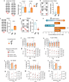
GluN2B-negative allosteric modulator Ro25-6981 (Ro25) blocks SC LTP in males but not females. a, b, In slices from male rats, the isolated NMDAR fEPSP response (i.e., 20 µM DNQX and 30 µM picrotoxin in bath) (a) was depressed by MK-801 (30 µM; N = 6) but (b) not by Ro25 (3 µM; N = 5). c, Voltage-clamp recordings from adult male mouse CA1 pyramidal cells held at +40 mV show that Ro25 infusion decreased NMDAR-EPSC amplitude (*p = 0.0313; veh N = 9, Ro25 N = 6). d, In adult male slices, TBS was applied to SC projections and CA1 fEPSP responses were evaluated 40 min after onset of vehicle (veh) or 3 µM Ro25 infusion. Traces at left show responses to the first three bursts in a 10-burst train (veh, black line; Ro25, dashed blue line; burst response area shaded gray). Graph shows that the area of individual burst responses (normalized to first burst) is not altered by Ro25 across the full theta train (F(9,126) = 0.268, p = 0.982. N = 7 veh, N = 9 Ro25). e, Measures within first TBS response (i.e., slope size of responses to pulses 2–4 normalized to that of pulse 1) show that the individual response profile was not affected by Ro25 (F(3,42 = 0.189, p = 0.903). f, g, In male rat slices, Ro25 (infused at horizontal bar) significantly reduced SC→CA1 LTP induced by (f) threshold-level TBS (4 TBS triplets, spaced by 90 s; p = 0.008; N = 5/group) or (g) a 10-burst TBS train (p = 0.0015, veh N = 7, Ro25 N = 9). h, i, In slices from adult female rats, TBS-induced SC LTP was fully blocked by 30 µM MK-801 (h; p = 0.0008; veh N = 5, MK-801 N = 4) but not by Ro25 (i; p = 0.972; veh N = 5, Ro25 N = 6). Traces in panels f–i are from before (solid) and 60 min after (dashed) TBS. Calibration: a, b, 100µV, 20 ms; c, 50pA, 50 ms; d–i, 1 mV, 10 ms. Statistics: two-tailed paired t test (a, b), unpaired Mann–Whitney U test (c, f), two-way repeated-measures ANOVA (d, e) and unpaired t test (g–i). Mean ± SEM values shown. See Extended Data Figure 1-1 for detailed statistics.
Figure 3.
Figure 3.
GluN2B-negative allosteric modulator Ro25-6981 (Ro25) blocks TBS-induced increases in spine F-actin in males but not females. Hippocampal slices received either control (con) low-frequency stimulation or 10 TBS of Schaffer-commissural (SC) afferents to CA1 in the presence of MK801 (30 µM) or Ro25 (3 µM), and then phalloidin labeling of F-actin or synaptic protein levels were evaluated in CA1 SR (blue and orange bars denote results from males and females, respectively). a, In males, TBS doubled the proportion of spines with dense phalloidin labeled F-actin in vehicle (veh)-treated slices and this effect was completely blocked by Ro25 applied alone (post hoc ****p < 0.0001) or in the presence of MK-801 (*p = 0.0304, N = 9–40/group; points denote individual slice measures). In a–d, bar graphs indicate values normalized to con mean; images show phalloidin labeling in CA1 SR in representative cases. b, In males, Ro25 blocked the TBS-induced increase in the proportion of PSD95-IR synapses with dense immunolabeling for pSrc and pCaMKII but not that for pERK (pSrc: F(2,27) = 6.517, p = 0.0049; N = 5–17/group; pERK, F(2,36) = 14.36; p < 0.0001, N = 11–17/group; pCaMKII: F(2,24) = 5.111, p = 0.0142; N = 7–12/group). c, In slices from females, TBS elicited comparable increases in punctate phalloidin labeled F-actin in the presence of veh, MK-801, and Ro25, but this effect was blocked by NMDAR antagonist APV (p < 0.0001; N = 5–33). d, TBS-induced increased numbers of densely phalloidin labeled puncta in CA1 SR are blocked by ERα antagonist MPP (3 µM) in females (F(2,29) = 16.02, p < 0.0001; N = 6–17) but not in males (p = 0.0003; N = 6–12). e, Deconvolved images show dual immunolabeling for NMDAR subunits (magenta) and PSD95 (green) in CA1 SR; arrows indicate double-labeled profiles. f, Quantification of immunofluorescence intensity (for puncta double-labeled for PSD95; female values normalized to male mean) shows that the proportion of postsynaptic elements with dense GluN1-immunoreactivity was greater in females than that in males (p = 0.0171) whereas levels of GluN2A- and GluN2B-immunoreactivity were comparable (N = 17–20/group). The proportion of PSD95-IR elements with dense pGluN2B Y1472 immunoreactivity was lower in females than that in males (p = 0.0041; N = 17–20/group). Scale bars: a, c, d, 5 μm; e, 2 μm. Statistics: a, c, d, male, Kruskal–Wallis with Dunn post hoc; b, d, female, one-way ANOVA with Tukey’s post hoc; f, two-tailed unpaired t test excepting NR2A (Welch's correction). Asterisks inside bars denote comparison with controls; n.s., not significant, *p < 0.05, **p < 0.01, ***p < 0.001, ****p < 0.00001. Mean ± SEM values shown. See Extended Data Figure 1-1 for detailed statistics.
Figure 4.
Figure 4.
Sex differences in the effects of GluN2B and ERα antagonists on episodic “Where” encoding. a, Mice received vehicle, Ro25, or MPP treatment before odor exposure in the four-corner episodic “Where” paradigm illustrated. b, c, Vehicle (veh)-treated males (blue) and females (orange; separate cohorts in panels b and c) preferentially sampled the cues moved to novel locations. b, Ro25 blocked discrimination of novel location cues in males (F(1,15) = 19.62, p = 0.0005; veh N = 5, Ro25 N = 4) but had no effect on female performance (N = 5/group). c, ERα antagonist MPP fully blocked discrimination of the moved cues in females but did not affect performance in males (F(1,24) = 8.001, p = 0.0093; N = 7/group). d–g, Measures of total (Σ) cue sampling times in seconds (d, e) and locomotor activity (distance traveled and velocity) (f, g) during both initial cue exposure and testing were not influenced by treatment (MPP, Ro25) in males or females (interaction, p > 0.05). Statistics: two-way ANOVA with post hoc Tukey. n.s., not significant, *p < 0.05, **p < 0.01, ***p < 0.001, ****p < 0.0001. Mean ± SEM values shown. See Extended Data Figure 1-1 for detailed statistics.
Figure 5.
Figure 5.
Sex differences in the acquisition of the What, When, and Where components of episodic memory. a, Left, Schematic of the serial odor “What” task using initial exposure (black) to three and four odors in series; in this and other schematics the novel cue or cue in novel location is indicated in red. Right: Bar graphs show that, in the three-odor task, both males (blue) and females (orange) preferentially explored the novel (D) versus the familiar (A) odor at testing (p = 0.94; male N = 6, female N = 10), whereas with initial exposure to a series of four odors, females preferentially explored the novel odor at testing but males did not (p = 0.0003; male vs female, N = 10/group). b, Left, Schematic of the Simultaneous “What” task. Right, At testing, females distinguished the novel from previously experienced odors but males did not (p = 0.0007; male N = 4 vs female N = 5). c, Left, Schematic of the “When” task. Right, Females preferentially sampled the least recently exposed odor but males did not (p = 0.001; male N = 5, female N = 6). d, Left, Simultaneous “Where” task schematic. Right, At testing males preferentially explored the novel location odors but females did not (p = 0.0022, N = 4/group). e, Female performance in the What, When, and Where tasks expressed as a z-score difference from the male group discrimination index mean. The female performance in the Simultaneous (sim) “What” task was greater than for the other tasks (F(3,21) = 49.11, p < 0.0001; post hoc *p ≤ 0.15; N = 4–10/group) and results on the “Where” tasks differed from the other three scores (# p < 0.0001). f–o, Top bar graphs show group means of total (Σ) sampling times in the initial exposure trial(s) leading to the test trial in a particular memory task. Bottom plots show the total distance traveled (DT, squares) and movement velocity (circles) during the same sessions. Analysis of interaction (sex*trial) via two-way ANOVA revealed no significant difference in the total time spent sampling odors during initial exposure and test trials for both serial “What” tasks (f, h), simultaneous “What” (j), “When” (l), and “Where” (n) (p > 0.05). g, i, k, m, o, The significance of interaction was also absent in measures of distance traveled and velocity across the same tasks (p > 0.05). Statistics: a–d, two-tailed unpaired t test; e, one-way ANOVA, post hoc Tukey; f–o, two-way ANOVA; n.s., not significant, *p < 0.05, **p < 0.01, ***p < 0.001. Mean ± SEM values shown. See Extended Data Figure 1-1 for detailed statistics.
Figure 6.
Figure 6.
Schematic illustration of mechanisms proposed to underlie sex differences in ion flux-independent NMDAR contributions to memory-related SC LTP. Observed effects of APV and MK-801 indicate that both males and females use ionotropic NMDAR functions to activate CaMKII and associated processes (e.g., AMPAR insertion) required for the induction of field CA1 LTP (indicated in red font). The present results show that both sexes also rely upon nonionotropic NMDAR functions (i.e., those blocked by APV but not by MK-801) for TBS-induced postsynaptic actin polymerization, Src phosphorylation, and LTP. Sex-specific effects of Ro25-6981 described here indicate GluN2B subserves these nonionotropic NMDAR functions in males, presumably via the subunit's cytoplasmic terminal domain (CTD) that acts as a docking site for signaling proteins including Src family kinases, SynGAP and CAMKII. Females do not use the Ro25-sensitive GluN2B mechanism for TBS-induced increases in postsynaptic F-actin or SC LTP but instead rely upon synaptic ERα to engage the same effectors as used by males. We propose that in females estrogen receptor alpha (ERα) may tonically suppress GluN2B activities by reducing phosphorylation of its CTD Y1472 site, thereby modulating CaMKII binding (Tullis and Bayer, 2023). However, results showing that, in females, APV blocks the TBS-induced increase in F-actin whereas MK-801 does not suggests that females do have m-NMDAR contributions to the LTP consolidation machinery; we speculate that the GluN2A CTD may contribute to these functions in females.

Update of

References

    1. Akashi K, Kakizaki T, Kamiya H, Fukaya M, Yamasaki M, Abe M, Natsume R, Watanabe M, Sakimura K (2009) NMDA receptor GluN2B (GluR epsilon 2/NR2B) subunit is crucial for channel function, postsynaptic macromolecular organization, and actin cytoskeleton at hippocampal CA3 synapses. J Neurosci 29:10869–10882. 10.1523/JNEUROSCI.5531-08.2009 - DOI - PMC - PubMed
    1. Amani M, Lauterborn JC, Le AA, Cox BM, Wang W, Quintanilla J, Cox CD, Gall CM, Lynch G (2021) Rapid aging in the perforant path projections to the rodent dentate gyrus. J Neurosci 41:2301–2312. 10.1523/JNEUROSCI.2376-20.2021 - DOI - PMC - PubMed
    1. Andreano JM, Cahill L (2009) Sex influences on the neurobiology of learning and memory. Learn Mem 16:248–266. 10.1101/lm.918309 - DOI - PubMed
    1. Asperholm M, Hogman N, Rafi J, Herlitz A (2019) What did you do yesterday? A meta-analysis of sex differences in episodic memory. Psychol Bull 145:785–821. 10.1037/bul0000197 - DOI - PubMed
    1. Babayan AH, et al. (2012) Integrin dynamics produce a delayed stage of long-term potentiation and memory consolidation. J Neurosci 32:12854–12861. 10.1523/JNEUROSCI.2024-12.2012 - DOI - PMC - PubMed

MeSH terms

LinkOut - more resources