. 2024 Oct 18;15(10):759.
doi: 10.1038/s41419-024-07146-y.

NLRP10 maintains epidermal homeostasis by promoting keratinocyte survival and P63-dependent differentiation and barrier function

Affiliations

NLRP10 maintains epidermal homeostasis by promoting keratinocyte survival and P63-dependent differentiation and barrier function

Yeonhee Cho et al. Cell Death Dis. .

Abstract

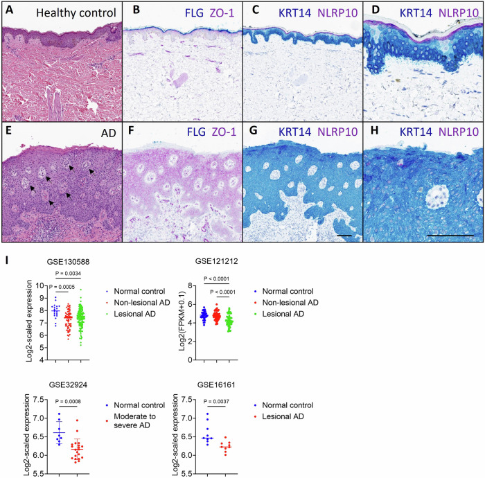
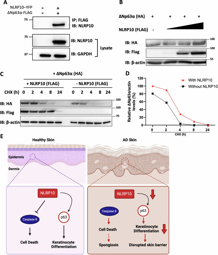
Atopic dermatitis (AD) is a common chronic inflammatory skin disorder characterized by disrupted epidermal barrier function and aberrant immune responses. Despite recent developments in new therapeutics for AD, there is still a large unmet medical need for disease management due to the complex and multifactorial nature of AD. Recent genome-wide association studies (GWAS) have identified NLRP10 as a susceptible gene for AD but the physiological role of NLRP10 in skin homeostasis and AD remains unknown. Here we show that NLRP10 is downregulated in AD skin samples. Using an air-lift human skin equivalent culture, we demonstrate that NLRP10 promotes keratinocyte survival and is required for epidermal differentiation and barrier function. Mechanistically, NLRP10 limits cell death by preventing the recruitment of caspase-8 to the death inducing signaling complex (DISC) and by inhibiting its subsequent activation. NLRP10 also stabilizes p63, the master regulator of keratinocyte differentiation, to drive proper keratinocyte differentiation and to reinforce the barrier function. Our findings underscore NLRP10 as a key player in atopic dermatitis pathogenesis, highlighting NLRP10 as a potential target for therapeutic intervention to restore skin barrier function and homeostasis in AD.


Conflict of interest statement

All authors are employees of Amgen Inc., a for-profit organization.

Figures

Fig. 1
Fig. 1. NLRP10 is downregulated in AD.
Representative images of healthy skin (AD) and severe atopic dermatitis (EH). Samples stained with H&E (A, E): Compared to healthy skin (A), AD skin (E) demonstrates epidermal thickening (acanthosis) and multiple foci of epidermal edema (spongiosis) as exemplified by arrows. Samples stained with ZO-1 and Filaggrin (B, F): In normal skin (B), ZO-1 is expressed at tight junctions of the stratum granulosum (and upper stratum spinosum) and Filaggrin is expressed in terminally differentiated keratinocytes (cytoplasmic expression within the stratum corneum and lucidum). In AD (F), ZO-1 expression extends throughout the stratum spinosum, diffusely within the cytoplasm (not limited to cell junctions). Filaggrin expression is lost with severe AD. Samples stained with NLRP10 and Cytokeratin14 (C, D and G, H): In control skin (C, D), NLRP10 is cytoplasmic within the stratum granulosum. With severe AD (G, H), its staining is reduced. D and H Are higher magnification images of (C) and (G) respectively. Scale bar (AC, EG): 100 µm; scale bar (D, H): 50 µm. I Gene expression profile of NLRP10 in normal and AD samples from four different study cohorts showing reduced NLRP10 expression in AD. Data represent mean ± S.D. and statistical significance was determined using one-way ANOVA for GSE130588 and GSE121212 or two tailed student t-test for GSE32924 and GSE16161. GSE130588 (n = 20 for normal control, n = 64 for non-lesional AD; n = 124 for lesional AD); GSE121212 (n = 37 for normal control, n = 54 for non-lesional AD; n = 55 for lesional AD); GSE32924 (n = 8 for normal control, n = 20 for moderate to severe AD); GSE16161 (n = 9 for normal control, n = 9 for lesional AD).
Fig. 2
Fig. 2. NLRP10 promotes epidermal differentiation.
A, B Representative images of H&E staining (A) and quantification of epidermal thickness (B) of human skin equivalent (HSE) developed from WT or NLRP10 KO NHEKs (n = 4). C, D Representative images (C) and quantification of thickness (D) of Keratin 10+ and Keratin 14+ layers in HSE (n = 3). Yellow arrow lines highlight Keratin 14+ layers and white arrow lines indicates Keratin 10+ layers. E, F Representative images (E) and quantification of relative fluorescence intensity (F) of β-catenin in WT and NLRP10 KO HSEs (n = 3 for WT, n = 4 for KO). G Filaggrin and caspase-14 (Casp-14) from WT and NLRP10 KO undifferentiated NHEKs and HSEs were analyzed by Western blotting. Data represent mean ± S.D. and statistical significance was determined using two tailed student t-test. Dashed lines highlight the epidermis region within the HSEs. Scale bar: 20 µm.
Fig. 3
Fig. 3. NLRP10 augments epidermal barrier function.
A Volcano plot of lipid species differentially detected in NLRP10 KO compared to WT HSEs. B Boxplot of log2 fold change (logFC) of different lipid classes in NLRP10 KO compared to WT HSEs. Red color highlights statistically significantly changed lipid classes (P < 0.005). C Heatmaps from WT and NLRP10 KO HSE (n = 3) lipidomics analysis of the ceramide subclasses (acylceramides, N-acylsphinganines, N-acylsphingosines) included in the Cer category in the lipid set enrichment analysis in (B). Heatmap intensity is colored by z-Score for each lipid species. D Representative images and quantification of biotin signal intensity in HSEs with topical biotin treatment (n = 3). Biotin was added on top of the HSEs on the 9th day of air-lift for 45 min and washed with PBS. Frozen sections of HSE were stained with streptavidin conjugated with Alexa Fluor 594. Dashed lines highlight the epidermis region within the HSEs. Scale bar: 20 µm. Data are representative of two independent experiments and data represent mean ± S.D. and statistical significance was determined using two tailed student t-test.
Fig. 4
Fig. 4. NLRP10 prevents keratinocyte cell death.
Cell death monitored by LDH release assay (A) and immunoblotting (BD). A LDH release from WT or NLRP10 KO NHEKs was measured (n = 6). Data represent mean ± S.D. and statistical significance was determined using two tailed student t-test. B WT and NLRP10 KO NHEKs treated with staurosporine (SS, 1 µM, 4 h), TNFα (20 ng/ml, 7 h)/SM-164 (100 µM, 7 h)/zVAD (20 µM, 7.5 h) (TSZ), amino acids deprivation (AA-, 1 h), VbP (2 µM, 4 h) or nigericin (Nig, 6.7 µM, 4 h) were analyzed by western blot with indicated antibodies. Clv. Casp8, cleaved caspase-8; Clv. Casp3, cleaved caspase-3; Clv. GSDMD, cleaved Gasdermin D. C WT and NLRP10 KO NHEKs incubated for 6 h in NHEK complete medium after UVB exposure (250 mJ/cm2 or 400 mJ/cm2) were analyzed by western blot with indicated antibodies. WT NHEKs were stimulated with staurosporine (SS, 1 µM, 4 h) as a positive control. D WT and NLRP10 KO NHEKs treated with TRAIL (300 ng/ml, 2.5 h) were analyzed by western blot with indicated antibodies. GAPDH was used as a loading control and all data are representative of more than three independent experiments.
Fig. 5
Fig. 5. NLRP10 inhibits caspase-8 activation.
A Co-immunoprecipitation (co-IP) of NLRP10-Flag with caspase-1-HA and caspase-8-HA in 293 T cells. Co-IP elution and cell lysate was analyzed by Western blotting using anti-HA and anti-NLRP10 antibodies. GAPDH was used as a loading control. B Construct design of different caspase-8 plasmids. C Caspase-8 constructs shown in (B) were co-transfected with NLRP10-FLAG in 293 T cells, and protein complex was pulled down using anti-FLAG resin and analyzed by Western blotting. D Western blot analysis of DR5 DISC IP carried out in WT and NLRP10 KO NHEK cells showing increased cleaved caspase-8 p43/p41 form in the DISC of NLRP10 KO NHEKs. E Western blot analysis of DR5 DISC IP carried out in Jurkat cells without or with NLRP10 overexpression showing decreased full length and cleaved caspase-8 in the DISC of NLRP10-overexpressing cells. The DR5 DISC IP was performed 1 h after addition of beads coupled with an agonist anti-DR5 antibody. β-actin was used as a loading control. Black arrowheads indicate full length caspase-8, and blue arrowheads represent cleaved caspase-8 in the DISC. DISC: death inducing signaling complex. F Illustration of DR5 DISC in the absence or presence of NLRP10. Agonistic anti-DR5 antibody induces the assembly of DISC and recruitment of caspase-8 to the complex. NLRP10 interacts with the DED domain of caspase-8, reduces caspase-8 density recruited to the DISC and thereby suppresses caspase-8 activation. Caspase-8 is color-coded to highlight the tandem DED (tDED) domain in green and catalytic domain in red. DR5, death receptor 5; FADD, Fas-Associated Death Domain Protein; DED, death effector domain.
Fig. 6
Fig. 6. NLRP10 enhances p63 signaling in keratinocytes.
A Dot plot of enriched reactome pathways downregulated and upregulated in NLRP10 KO HSEs. B, C Gene set enrichment plots for top reactome pathways are shown: Formation of the cornified envelope (B) and keratinization (C). D scatter plot showing common genes significantly changed (adjusted P < 0.05) between NLRP10 KO (this study) and p63 knockdown (Truong et al.) [42]. Correlation coefficient (R) and p-value are shown. E Immunohistochemistry staining of p63 in WT and NLRP10 KO HSEs. Dashed lines highlight the boundary between the epidermis and the dermis within the HSEs. F, G Western blot of p63α in WT and NLRP10 KO NHEK (n = 5). Quantification was performed with densitometry normalized to housekeeping gene GAPDH.
Fig. 7
Fig. 7. NLRP10 stabilizes p63 in keratinocytes.
A Co-immunoprecipitation of NLRP10-YFP and Np63α-FLAG in 293 T cells. GAPDH was used as a loading control. B COS-7 cells transfected with Np63α-HA and increasing doses of NLRP10-FLAG were subjected to cell fractionation and immunoblotted with antibodies to the indicated proteins. β-actin was used as a loading control. Samples were prepared 48 h after transfection. C COS-7 cells transfected with Np63α-HA with or without NLRP10-FLAG for 48 h were incubated with cycloheximide (CHX) for up to 24 h, and analyzed by immunoblotting for Np63α-HA, NLRP10-FLAG and β-actin (loading control) levels. D Relative level of the ratio of Np63α-HA to β-actin in (C). The immunoblot band was analyzed by ImageJ software. E Proposed working model of NLRP10. NLRP10 inhibits caspase-8-mediated cell death and stabilizes p63 to promote keratinocyte differentiation and formation of skin barrier in healthy skin. In AD skin, NLRP10 expression is reduced. With decreased NLRP10 level, keratinocytes are more susceptible to cell death and have reduced differentiation, which altogether lead to spongiosis and disrupted skin barrier.

References

    1. Langan SM, Irvine AD, Weidinger S. Atopic dermatitis. Lancet. 2020;396:345–60. - PubMed
    1. Bieber T. Atopic dermatitis: an expanding therapeutic pipeline for a complex disease. Nat Rev Drug Discov. 2021;21:21–40. - PMC - PubMed
    1. Cork MJ, Danby SG, Vasilopoulos Y, Hadgraft J, Lane ME, Moustafa M, et al. Epidermal barrier dysfunction in atopic dermatitis. J Invest Dermatol. 2009;129:1892–908. - PubMed
    1. Boguniewicz M, Fonacier L, Guttman-Yassky E, Ong PY, Silverberg JI. Atopic dermatitis yardstick update. Ann Allergy Asthma Immunol. 2023;130:811–20. - PubMed
    1. Weidinger S, Beck LA, Bieber T, Kabashima K, Irvine AD. Atopic dermatitis. Nat Rev Dis Prim. 2018;4:1. - PubMed

MeSH terms

Substances